. 2010 Feb;84(4):1989-2000.
doi: 10.1128/JVI.01902-09. Epub 2009 Dec 9.

Fine mapping of pre-S sequence requirements for hepatitis B virus large envelope protein-mediated receptor interaction

Affiliations

Fine mapping of pre-S sequence requirements for hepatitis B virus large envelope protein-mediated receptor interaction

Andreas Schulze et al. J Virol. 2010 Feb.

Abstract

Previous studies showed that the N-terminal 75 amino acids of the pre-S1 domain of the hepatitis B virus (HBV) L protein are essential for HBV and hepatitis delta virus (HDV) infectivity. Consistently, synthetic lipopeptides encompassing this sequence or only parts of it efficiently block HBV and HDV infection, presumably through specific interference with a cellular receptor. Crucial for both virus infectivity and the inhibitory activity of the peptides are N-terminal myristoylation and a highly conserved motif within the N-terminal 48 amino acids. To refine the sequence requirements, we synthesized a series of HBV pre-S1 peptides containing deletions, point mutations, d-amino acid exchanges, or genotype-specific sequence permutations. Using the HepaRG cell line and a genotype D-derived virus, we determined the specific inhibitory activities of the peptides and found that (i) lipopeptides with an artificial consensus sequence inhibit HBV genotype D infection more potently than the corresponding genotype D peptides; (ii) point mutations, d-amino acid exchanges, or deletions introduced into the highly conserved part of the pre-S1 domain result in an almost complete loss of activity; and (iii) the flanking sequences comprising amino acids 2 to 8, 16 to 20, and, to a less pronounced extent, 34 to 48 gradually increase the inhibitory activity, while amino acids 21 to 33 behave indifferently. Taken together, our data suggest that HBV pre-S1-mediated receptor interference and, thus, HBV receptor recognition form a highly specific process. It requires an N-terminal acyl moiety and a highly conserved sequence that is present in primate but not rodent or avian hepadnaviruses, indicating different entry pathways for the different family members.


Figures
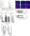
FIG. 1.
FIG. 1.
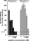
HBVpreS/2-48stearoyl efficiently inhibits HBV infection of HepaRG cells, requires N-terminal acylation, and does not obstruct HBV attachment at concentrations up to 1 μM. (A) Elution profiles of analytical HPLC runs of HBVpreS/2-48stearoyl, HBVpreS/2-48scrambledstearoyl, and HBVpreS/2-48. Note that, due to size limitations, all peptide names have been shortened and are represented by their amino acid sequences (e.g., 2-48 for HBVpreS/2-48). Peptide bonds were detected by UV absorbance at 214 nm and are given as arbitrary units. (B) At left, data are shown for HBV infection inhibition using increasing concentrations (0 to 1,000 nM) of HBVpreS/2-48stearoyl, HBVpreS/2-48scrambledstearoyl, and HBVpreS/1-48. At right, data are shown for HBV infection inhibition using increasing concentrations (0 to 10,000 nM) of WHVpreS/2-81myr. As control, 1 μM HBVpreS/2-48myr was used in parallel. The amount of HBsAg secreted from days 7 to 11 p.i. was determined by ELISA and is given as a percentage of the control infection. The sensitivity of the ELISA (cutoff) is indicated by the dotted line. (C) Quantification of HBV binding/uptake (4 h at 37°C) to HepaRG cells in the absence (left) and presence of 200 μg/ml heparin (middle) or 1 μM HBVpreS/2-48myr (right). Cell-associated HBV genome equivalents were quantified by real-time PCR. (D) Effect of 10 μM HBVpreS/2-48stearoyl on HBV infection when added together with virus (middle) or 16 h after the start of virus inoculation (right). HBsAg amounts from days 7 to 11 p.i. were determined by ELISA. (E) Analysis of the inhibitory activities of HBVpreS/2-48stearoyl (upper row) and HBVpreS/2-48scrambledstearoyl (lower row) by HBcAg-specific IF. At 30 min prior to infection HepaRG cells were incubated with buffer (left) or 250 nM (middle) or 2.5 μM (right) of the respective peptides and subsequently infected with HBV. At 11 days p.i. cells were analyzed for HBcAg (red) and nuclei (DAPI; blue). (F) Quantification of the IF analyses shown in panel E. In total 61,304 DAPI-positive cells were counted for HBVpreS/2-48scrambledstearoyl; for HBVpreS/2-48stearoyl all HBcAg-positive cells in the corresponding well were counted. (G) Measurement of the secreted HBsAg (days 7 to 11 p.i.) of the infection shown in panel E. The results are depicted as a percentage of the control infection. The sensitivity of the assay (cutoff) is indicated by the dotted line.
FIG. 2.
FIG. 2.
Infection inhibition activities of C-terminally shortened and antigenically altered HBV pre-S1 lipopeptides. HBV infection competition experiments on HepaRG cells using increasing concentrations (0 to 1,000 nM) of the indicated peptides. All peptide names have been shortened due to size limitation. The amount of secreted HBsAg (days 7 to 11 p.i.) was determined by ELISA. Values are presented as a percentage of the control infection. The detection limit of the assay is indicated by the dotted line. Uncomp, uncompeted.
FIG. 3.
FIG. 3.
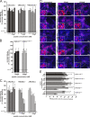
Infection inhibition activity of N-terminally shortened or modified HBVpre-S1 lipopeptides. (A to C) HBV infection inhibition using increasing concentrations (0 to 1 μM or 0 to 10 μM) of HBVpreS/5-48stearoyl and HBVpreS/9-48stearoyl (A); HBVpreS/5-48stearoyl, HBVpreS/6-48stearoyl, HBVpreS/7-48stearoyl, and HBVpreS/9-48stearoyl (B); or HBVpreS/2-48(A2-8)stearoyl, HBVpreS/2-48(A2-9)stearoyl, and HBVpreS/9-48stearoyl (C). All peptide designations have been shortened due to size limitations. The amount of HBsAg or HBeAg secreted from days 7 to 11 p.i. was determined by ELISA. The values are given as a percentage of the untreated control infection. The sensitivity of the ELISA (cutoff) is indicated by a dotted line.
FIG. 4.
FIG. 4.
Infection inhibition activity of combined N- and C-terminally deleted HBV pre-S1 lipopeptides. HBV infection competition using 0, 1, 5, 25, 100, and 1,000 nM HBVpreS/5-33stearoyl or HBVpreS/9-33stearoyl. All peptide names have been shortened due to size limitations. The amount of HBsAg secreted between days 7 to 11 p.i. was determined by ELISA. The sensitivity of the ELISA is indicated by the dotted line.
FIG. 5.
FIG. 5.
Infection inhibition activity of HBV pre-S1 lipopeptides carrying mutations in the genotypically highly conserved region 11-LGFFP-15. (A to C) HBV infection competition experiments using 0 to 1,000 nM HBVpreS/2-48(Δ11-15)stearoyl, HBVpreS/2-48(A11-15)stearoyl, HBVpreS/2-48(d-11/13-15)stearoyl (A), HBVpreS/2-48(d-11/13-15)stearoyl and HBVpreS/2-48(d-11/13)stearoyl (B), or HBVpreS/2-48(L11R)stearoyl, HBVpreS/2-48(G12E)stearoyl, and HBVpreS/2-48(F13S)stearoyl (C). All peptide names have been shortened due to size limitation. The amount of secreted HBsAg of days 7 to 11 p.i. was quantified by ELISA and is shown as a percentage of the control infection. (D) HBcAg-specific IF analyses (red) and nuclei staining (DAPI; blue) of infection competition experiments on HepaRG cells using a 250 nM or 2.5 μM concentration of the above-mentioned peptides. (E) Quantification of the IF analyses shown in panel D. In total, 143,678 DAPI-positive cells were counted. The number of HBcAg-positive cells is presented as a percentage of the untreated control.
FIG. 6.
FIG. 6.
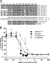
Infection inhibition activity of myristoylated and stearoylated HBVpre-S1-derived peptides comprising the complete or a shortened consensus sequence. (A) Sequence alignment of the human HBV genotypes A to H and the chimpanzee, gorilla, and woolly monkey hepatitis B viruses. The short (residues 2 to 48) and long (residues −11 to 48) consensus sequences derived from HBV genotypes A to H are depicted in the middle. Identical amino acids are marked by dark-gray boxes. Nonidentical amino acids used for the consensus sequences are marked by light-gray boxes. (B) HBV infection competition using increasing concentrations (0 to 1,000 nM) of the aforementioned consensus peptides. All peptide names have been shortened due to size limitations. The amount of secreted HBsAg between days 7 to 11 p.i. was measured by ELISA and is presented as a percentage of the control infection.

References

    1. Abou-Jaoude, G., and C. Sureau. 2007. Entry of hepatitis delta virus requires the conserved cysteine residues of the hepatitis B virus envelope protein antigenic loop and is blocked by inhibitors of thiol-disulfide exchange. J. Virol. 81:13057-13066. - PMC - PubMed
    1. Barrera, A., B. Guerra, L. Notvall, and R. E. Lanford. 2005. Mapping of the hepatitis B virus pre-S1 domain involved in receptor recognition. J. Virol. 79:9786-9798. - PMC - PubMed
    1. Blanchet, M., and C. Sureau. 2007. Infectivity determinants of the hepatitis B virus pre-S domain are confined to the N-terminal 75 amino acid residues. J. Virol. 81:5841-5849. - PMC - PubMed
    1. Bruss, V. 2007. Hepatitis B virus morphogenesis. World J. Gastroenterol. 13:65-73. - PMC - PubMed
    1. Bruss, V., and D. Ganem. 1991. The role of envelope proteins in hepatitis B virus assembly. Proc. Natl. Acad. Sci. U. S. A. 88:1059-1063. - PMC - PubMed

Publication types

MeSH terms

Substances

LinkOut - more resources