. 2010 Feb;84(4):1934-44.
doi: 10.1128/JVI.01657-09. Epub 2009 Dec 9.

An N-terminal region of Lassa virus L protein plays a critical role in transcription but not replication of the virus genome

Affiliations

An N-terminal region of Lassa virus L protein plays a critical role in transcription but not replication of the virus genome

Michaela Lelke et al. J Virol. 2010 Feb.

Abstract

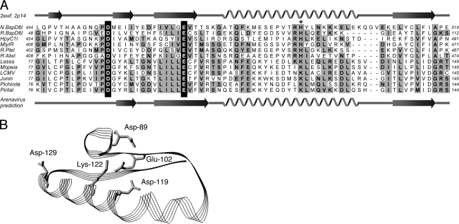
The central domain of the 200-kDa Lassa virus L protein is a putative RNA-dependent RNA polymerase. N- and C-terminal domains may harbor enzymatic functions important for viral mRNA synthesis, including capping enzymes or cap-snatching endoribonucleases. In the present study, we have employed a large-scale mutagenesis approach to map functionally relevant residues in these regions. The main targets were acidic (Asp and Glu) and basic residues (Lys and Arg) known to form catalytic and binding sites of capping enzymes and endoribonucleases. A total of 149 different mutants were generated and tested in the Lassa virus replicon system. Nearly 25% of evolutionarily highly conserved acidic and basic side chains were dispensable for function of L protein in the replicon context. The vast majority of the remaining mutants had defects in both transcription and replication. Seven residues (Asp-89, Glu-102, Asp-119, Lys-122, Asp-129, Glu-180, and Arg-185) were selectively important for mRNA synthesis. The phenotype was particularly pronounced for Asp-89, Glu-102, and Asp-129, which were indispensable for transcription but could be replaced by a variety of amino acid residues without affecting genome replication. Bioinformatics disclosed the remote similarity of this region to type IIs endonucleases. The mutagenesis was complemented by experiments with the RNA polymerase II inhibitor alpha-amanitin, demonstrating dependence of viral transcription from the cellular mRNA pool. In conclusion, this paper describes an N-terminal region in L protein being important for mRNA, but not genome synthesis. Bioinformatics and cell biological experiments lend support to the hypothesis that this region could be part of a cap-snatching enzyme.


Figures

FIG. 1.
FIG. 1.
Alanine mutations introduced in Lassa virus L protein. The activity of the respective mutants in the replicon system as measured by expression of Ren-Luc is indicated as follows: (i) white typeface on black background, inactive mutant; (ii) black typeface on gray background, mutant with reduced activity; (iii) black typeface on white background, mutant with wild-type activity. (Upper panel) Asp, Glu, Lys, and Arg mutants; (lower panel) Phe mutants. The localization of conserved domains L1 to L4 is shown at the bottom (47).
FIG. 2.
FIG. 2.
Immunoblot analysis of HA-tagged L protein mutants. BSR T7/5 cells were infected with MVA-T7 and transfected with PCR products expressing L protein mutants containing a C-terminal HA tag. L protein in cytoplasmic lysate was separated by SDS-PAGE, blotted, and detected with anti-HA antibody. A semiquantitative standard based on titration of lysate is shown in panel J. Negative (neg.) control cells were infected with MVA-T7 but not transfected.
FIG. 3.
FIG. 3.
Influence of L protein mutations on transcription and replication. RNA synthesized by L protein mutants in the context of the replicon system was analyzed by Northern blotting. Antigenomic RNA and Ren-Luc mRNA were detected using a radiolabeled riboprobe hybridizing to the Ren-Luc gene. The activity of the mutants in luciferase assay is indicated below the blot (−, inactive mutant; +, mutant with reduced activity; ‡, mutant with wild-type activity). Mutations in L protein are indicated above the blot. Negative (neg.) control cells expressed minigenome, NP, and an L protein mutant with a mutation in the catalytic site of the RdRp. The methylene blue-stained 28S rRNA is shown below the blots as a marker for gel loading and RNA transfer. Each panel represents an independent transfection experiment.
FIG. 4.
FIG. 4.
Level of Ren-Luc mRNA in relation to antigenome level. Signals on Northern blots in Fig. 3 were quantified via intensity profiles (for details and examples, see Fig. S2 in the supplemental material). All mutants with detectable signals are shown. Those with significant reduction of the mRNA/antigenome ratio compared to the wild type (<40%) are indicated by black bars. The wild-type mRNA/antigenome ratio was set at 1 for each experiment to render independent blots comparable. Careful examination of the intensity profiles revealed residual signals at the mRNA position (about 10% of the wild-type mRNA signal) for mutants completely negative in the Ren-Luc assay. These signals were not seen in the negative control lanes. The precise nature of this RNA is not clear, as the luciferase data indicate that functional mRNA is not expressed.
FIG. 5.
FIG. 5.
Amino acid sequence alignment of the N-terminal part of L protein (L1 domain) of Old and New World arenaviruses and summary of the mutagenesis experiments. Positions subjected to mutagenesis are marked above the sequence. The data from Northern blotting are coded as follows: (i) large inverted triangle, mutant with reduced mRNA expression but wild-type levels of antigenome; (ii) small inverted triangle, mutant with selective reduction of the mRNA level associated with overall reduction of RNA synthesis; (iii) circle, mutant with wild-type mRNA/antigenome ratio or no RNA signals at all. The Ren-Luc data are coded as follows: (i) black head, inactive mutant; (ii) gray head, mutant with reduced activity; (iii) white head, mutant with wild-type activity. The predicted secondary structure of the domain is shown below the alignment.
FIG. 6.
FIG. 6.
Effect of α-amanitin treatment on Lassa virus RNA synthesis. (A) Northern blot analysis. Vero cells were infected with Mopeia virus at an MOI of 3 (left) or with Lassa virus at different MOIs (right) and treated with 10 μg/ml α-amanitin or left untreated. Total RNA was prepared from Mopeia virus-infected cells at the indicated intervals and from Lassa virus-infected cells 24 h after infection and subjected to Northern blotting. Hybridization was performed with a 32P-labeled antisense probe of NP gene. The methylene blue-stained 28S rRNA is shown below the blot as a semiquantitative marker for gel loading and RNA transfer. (B) Representative intensity profiles from Northern blot lanes in panel A used for quantification of signals. (C) Quantitative evaluation of S RNA and NP mRNA steady-state levels. Signals on the Northern blot were quantified via intensity profiles as shown in panel B, and the NP mRNA/S RNA ratio was calculated for α-amanitin-treated and untreated cells. The NP mRNA/S RNA ratio in untreated cells was set at 100%. p.i., postinfection.
FIG. 7.
FIG. 7.
Effect of α-amanitin treatment on capping of Lassa virus mRNA. (A) IP of capped RNA. Vero cells were infected with Lassa virus at a MOI of 1 and treated with 10 μg/ml α-amanitin. After 24 h, total cellular RNA was prepared and capped RNA was immunoprecipitated in two successive rounds by using an antibody against m3G-cap/m7G-cap structures. An unrelated antibody (anti-NP) was used as a control. Precipitate and supernatant were subjected to Northern blotting, and virus RNA was detected by using an NP gene-specific probe. The methylene blue-stained 28S rRNA is shown below the blot as a semiquantitative marker for gel loading and RNA transfer. T, total RNA; P1, pellet after first round of IP; S1, supernatant after first round of IP; P2, pellet after second round of IP; S2, supernatant after second round of IP. (B) Cumulative fractions of NP mRNA and S RNA precipitated after the first and second rounds of IP. The fraction was calculated based on the signal intensities on the blot shown in panel A and the amount of material subjected to each round of IP.
FIG. 8.
FIG. 8.
Similarity between L1 fragment of Lassa virus L protein (positions 77 to 145) and type IIs or nicking endonucleases. (A) Threading analysis using mGenTHREADER (22) identified structural similarity of the L1 fragment with structure 2ewf of the nicking endonuclease N.BspD6I (23). The alignment was extended to related type IIs and nicking endonucleases as well as further Old and New World arenaviruses. Secondary structure elements in 2ewf (N.BspD6I) and 2p14 (R.BspD6I) and the predicted secondary structure of arenavirus L protein are shown, respectively, above and below the alignment. Key catalytic residues of the endonucleases (Asp and Glu) (20, 23, 49) align with Asp-89 and Glu-102 of the L1 domain (highlighted in white on black background). An additional putative catalytic residue (His; marked with an asterisk) (23) aligns with Lys-122 of L protein. (B) Three-dimensional model of the L1 fragment shown in panel A using the structure of N.BspD6I (23) as a template. Modeling and ribbon drawing were performed with Pdb Viewer 3.7 (12). Side chains of residues relevant for mRNA but not antigenome synthesis are shown.

References

    1. Auperin, D. D., V. Romanowski, M. Galinski, and D. H. Bishop. 1984. Sequencing studies of Pichinde arenavirus S RNA indicate a novel coding strategy, an ambisense viral S RNA. J. Virol. 52:897-904. - PMC - PubMed
    1. Buchholz, U. J., S. Finke, and K. K. Conzelmann. 1999. Generation of bovine respiratory syncytial virus (BRSV) from cDNA: BRSV NS2 is not essential for virus replication in tissue culture, and the human RSV leader region acts as a functional BRSV genome promoter. J. Virol. 73:251-259. - PMC - PubMed
    1. Bushnell, D. A., P. Cramer, and R. D. Kornberg. 2002. Structural basis of transcription: alpha-amanitin-RNA polymerase II cocrystal at 2.8 A resolution. Proc. Natl. Acad. Sci. U. S. A. 99:1218-1222. - PMC - PubMed
    1. Dias, A., D. Bouvier, T. Crepin, A. A. McCarthy, D. J. Hart, F. Baudin, S. Cusack, and R. W. Ruigrok. 2009. The cap-snatching endonuclease of influenza virus polymerase resides in the PA subunit. Nature 458:914-918. - PubMed
    1. Duijsings, D., R. Kormelink, and R. Goldbach. 2001. In vivo analysis of the TSWV cap-snatching mechanism: single base complementarity and primer length requirements. EMBO J. 20:2545-2552. - PMC - PubMed

Publication types

MeSH terms