. 2010 Feb;84(4):1920-33.
doi: 10.1128/JVI.01934-09. Epub 2009 Dec 9.

During lytic infections, herpes simplex virus type 1 DNA is in complexes with the properties of unstable nucleosomes

Affiliations

During lytic infections, herpes simplex virus type 1 DNA is in complexes with the properties of unstable nucleosomes

Jonathan J Lacasse et al. J Virol. 2010 Feb.

Abstract

The genomes of herpes simplex virus type 1 (HSV-1) are regularly chromatinized during latency such that their digestion with micrococcal nuclease (MCN) releases nucleosome-sized DNA fragments. In lytically infected cells, in contrast, MCN releases HSV-1 DNA in primarily heterogeneously sized fragments. Consistently, only a small percentage of this HSV-1 DNA coimmunoprecipitates with histones. Most current models propose that histones associate with HSV-1 DNA during lytic infections at low occupancy. However, histone modification or occupation is also proposed to regulate HSV-1 transcription. It remains unclear how the histones associated with a small percentage of HSV-1 DNA may regulate transcription globally. Moreover, the physical properties of the complexes containing histones and HSV-1 DNA are unknown. We evaluated the HSV-1 DNA-containing complexes at 5 h after (lytic) infection by biochemical fractionations. Nuclear HSV-1 DNA did not fractionate as protein-free HSV-1 DNA but as DNA in cellular nucleosomes. Moreover, MCN released HSV-1 DNA in complexes that fractionate as cellular mono- and dinucleosomes by centrifugation followed by sucrose gradients and size-exclusion chromatography. The HSV-1 DNA in such complexes was protected to heterogeneous sizes and was more accessible to MCN than DNA in most cellular chromatin. Using a modified MCN digestion to trap unstable digestion intermediates, HSV-1 DNA was quantitatively recovered in discrete mono- to polynucleosome sizes in complexes fractionating as cellular mono- to polynucleosomes. The HSV-1 DNAs in complexes fractionating as mono- to dinucleosomes were stabilized by cross-linking. Therefore, most HSV-1 DNA forms particularly unstable nucleosome-like complexes at 5 h of lytic infection.


Figures
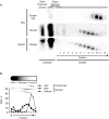
FIG. 1.
FIG. 1.
HSV-1 DNA in nuclei of lytically infected cells does not fractionate as protein-free DNA. Vero cells were mock infected or infected with HSV-1 (Nuclear) and harvested 5 h later. Half of the mock-infected nuclei were then spiked with protein-free HSV-1 DNA. Infected, mock-infected, and spiked mock-infected nuclei were digested with BamHI and lysed. Soluble and insoluble protein complexes were separated by differential centrifugation. The soluble DNA-protein complexes were then further resolved on sucrose gradients (Fraction). DNA in the pellet and each gradient fraction was analyzed by Southern blotting with HSV-1 or cellular probes. (A) Images of the hybridizations. Bottom fractions loaded to the left show different exposures under each condition. Exposures optimized for soluble fractions are also shown for insoluble fractions, which are therefore overexposed (Equal exposure). Lower exposures optimized for the insoluble fractions are also shown (Short Exposure). (B) Line graph presenting HSV-1 and cellular DNA in each fraction as a percentage of DNA in the gradient. The panels on the top are the same hybridizations shown in A, shown as a reference for the graph below and resized to fit the figure.
FIG. 2.
FIG. 2.
MCN digestion releases HSV-1 DNA in complexes that fractionate as cellular mono- to dinucleosomes. Nuclei of infected cells were digested for 150 s with 0.05 U of MCN per 1 × 10⁷ nuclei, and the soluble DNA-protein complexes were resolved on sucrose gradients (A and B). Following a similar experiment, fractions 10 and 11 from the gradients were further fractionated by size-exclusion chromatography (C). DNA from each fraction was analyzed by Southern blotting with HSV-1 or cellular probes. (A) Images of the membrane hybridized with HSV-1 or cellular probes. Bottom fractions are to the left. (B) Line graphs presenting HSV-1 and cellular DNA in each fraction as a percentage of DNA in the gradient. The panels on the top are the same as those shown in A, shown as reference for the graph below and resized to fit in the figure. (C) Images of the membranes hybridized with cellular or HSV-1-specific probes. Arrowheads indicate the migration of mono- or dinucleosomes.
FIG. 3.
FIG. 3.
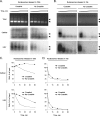
Nuclear HSV-1 DNA is more accessible to MCN than DNA in most cellular chromatin. Purified nuclei of infected cells were digested for 0.5, 2.5, 5, 15, 30, and 60 min with 0.005, 0.05, 0.5, or 5 U MCN per 1 × 10⁷ nuclei, and the DNA was analyzed by Southern blotting with HSV-1 or cellular probes. (A) Images of ethidium bromide-stained gels (Total) and membranes hybridized with cellular or HSV-1-specific probes. M, molecular mass marker (in basepairs). Normal exposures and overexposures (bottom), in which the nucleosome-sized and MCN-resistant HSV-1 DNAs are more clearly visible, are shown. To achieve comparable signal intensities, only 50% of the sample was loaded for 0.5 min at 0.005 to 5.0 U of MCN. (B and C) Line graphs presenting normalized levels of cellular (B) and HSV-1 (C) DNAs against digestion time (averages ± standard deviations) (n = 3).
FIG. 4.
FIG. 4.
Most HSV-1 DNA released by MCN as soluble chromatin is in complexes that fractionate as mono- to dinucleosomes. Nuclei of infected cells were digested for 15, 30, 150, or 300 s with 0.05 U of MCN per 1 × 10⁷ nuclei. The soluble DNA-protein complexes were resolved on sucrose gradients. DNA from each fraction was analyzed by Southern blotting with HSV-1- or cell-specific probes. Line graphs present cellular and HSV-1 DNAs in each fraction as a percentage of DNA in the gradient. Inserts on top show images of the membranes hybridized with HSV-1 or cellular probes, resized to fit the graph. Bottom fractions are loaded to the left.
FIG. 5.
FIG. 5.
HSV-1 DNA- or cellular DNA-containing complexes resolve to different fractions at low salt concentrations. Nuclei of infected cells were digested for 150 s with 0.05 U MCN per 1 × 10⁷ nuclei and then lysed. The soluble DNA-protein complexes were resolved on sucrose gradients containing 0, 80, 225, or 450 mM NaCl. DNA from each fraction was analyzed by Southern blotting with HSV-1- or cell-specific probes. Line graphs present cellular and HSV-1 DNA in each fraction as a percentage of DNA in the gradient. Insets on the top show images of the membrane hybridized with HSV-1 or cellular probes, resized to fit the graphs. Bottom fractions are loaded to the left.
FIG. 6.
FIG. 6.
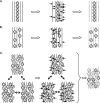
Potential types of HSV-1 nucleoprotein complexes. Cartoons represent three potential models of HSV-1 DNA nucleoprotein complexes in lytically infected cells. (A and B) A small percentage of HSV-1 genomes is either regularly chromatinized (A) or irregularly chromatinized with randomly positioned nucleosomes (B). Protein-free genomes are represented as straight lines without histones. Sites of MCN digestion are indicated by arrows. MCN first randomly cleaves the protein-free genomes and the linker region between nucleosomes. Protein-free genomes are completely digested by longer digestions, whereas chromatinized genomes are protected to mononucleosome sizes. As a result, only a small percentage of HSV-1 DNA is protected to nucleosome-sized fragments or coimmunoprecipitates with histones. (C) Most HSV-1 DNA is in unstable nucleosome-like complexes. Unstable nucleosomes are represented with dotted lines. MCN first cleaves the DNA within the unstable nucleosomes and the linker DNA. DNA within the unstable nucleosomes is then promptly degraded. As a result, only a minor fraction of HSV-1 is protected to nucleosome-sized fragments at any given time or coimmunoprecipitates with histones.
FIG. 7.
FIG. 7.
HSV-1 DNA fractionates as polynucleosomes after modified MCN digestion. (A) Cartoon representing the modified MCN digestion protocol designed to “trap” potential intermediate unstable HSV-1 nucleosome-like complexes. MCN digestion randomly cleaves the protein-free genomes and linker DNA as well as the DNA within the unstable nucleosomes. “Soluble chromatin” remains in the supernatant, unlike the “insoluble chromatin” pellets. Soluble chromatin is removed, and MCN is quenched. Insoluble chromatin is resuspended with fresh MCN digestion buffer. The process is repeated nine times. (B) Nuclei of infected cells were lysed, and soluble chromatin and insoluble chromatin were fractionated. Insoluble chromatin was resuspended in MCN digestion buffer (0.05 U MCN/ml) and subjected to modified MCN digestions. Supernatants were periodically removed and quenched, and the insoluble pellets were resuspended with fresh MCN. Soluble DNA-protein complexes were pooled and further resolved on sucrose gradients. DNA from each fraction was analyzed by Southern blotting with HSV-1- or cell-specific probes. Images of the membranes hybridized with cell- or HSV-1-specific probes are shown.
FIG. 8.
FIG. 8.
Unstable HSV-1 nucleosome-like complexes are partially stabilized by cross-linking. Nuclei of infected cells were digested with 0.05 U MCN per 1 × 10⁷ nuclei for 150 s or 15 s and lysed. Soluble DNA-protein complexes were resolved on sucrose gradients, and the relevant fractions from the sucrose gradients were then further fractionated by size-exclusion chromatography. Relevant fractions from the size-exclusion columns were either cross-linked or not for 1 h at 4°C, quenched with 125 mM glycine for 10 min, and redigested with 0.05 U/ml MCN for 0, 5, 15, 30, and 60 min. DNA was analyzed by Southern blotting with HSV-1 or cellular probes. (A and B) Images of ethidium bromide-stained gels (Total) and membranes hybridized with cell- or HSV-1-specific probes. Different exposures are shown for each hybridization. Arrowheads indicate the migration of mono- or dinucleosomes. (C and D) Line graphs presenting cellular and HSV-1 DNAs against digestion time, expressed as a percentage of DNA prior to digestion.

Similar articles

Cited by

References

    1. Alazard, N., H. Gruffat, E. Hiriart, A. Sergeant, and E. Manet. 2003. Differential hyperacetylation of histones H3 and H4 upon promoter-specific recruitment of EBNA2 in Epstein-Barr virus chromatin. J. Virol. 77:8166-8172. - PMC - PubMed
    1. Amelio, A. L., N. V. Giordani, N. J. Kubat, J. E. O'Neil, and D. C. Bloom. 2006. Deacetylation of the herpes simplex virus type 1 latency-associated transcript (LAT) enhancer and a decrease in LAT abundance precede an increase in ICP0 transcriptional permissiveness at early times postexplant. J. Virol. 80:2063-2068. - PMC - PubMed
    1. Amelio, A. L., P. K. McAnany, and D. C. Bloom. 2006. A chromatin insulator-like element in the herpes simplex virus type 1 latency-associated transcript region binds CCCTC-binding factor and displays enhancer-blocking and silencing activities. J. Virol. 80:2358-2368. - PMC - PubMed
    1. Ascoli, C. A., and G. G. Maul. 1991. Identification of a novel nuclear domain. J. Cell Biol. 112:785-795. - PMC - PubMed
    1. Ausio, J., F. Dong, and K. E. van Holde. 1989. Use of selectively trypsinized nucleosome core particles to analyze the role of the histone “tails” in the stabilization of the nucleosome. J. Mol. Biol. 206:451-463. - PubMed

Publication types

LinkOut - more resources