. 2010 Feb;84(4):1957-66.
doi: 10.1128/JVI.01627-09. Epub 2009 Dec 9.

Influenza C virus NS1 protein upregulates the splicing of viral mRNAs

Affiliations

Influenza C virus NS1 protein upregulates the splicing of viral mRNAs

Yasushi Muraki et al. J Virol. 2010 Feb.

Abstract

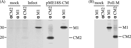
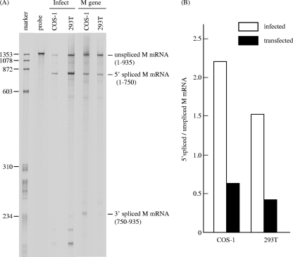
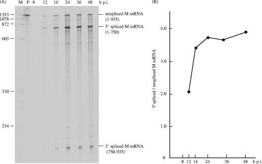
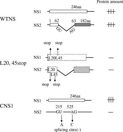
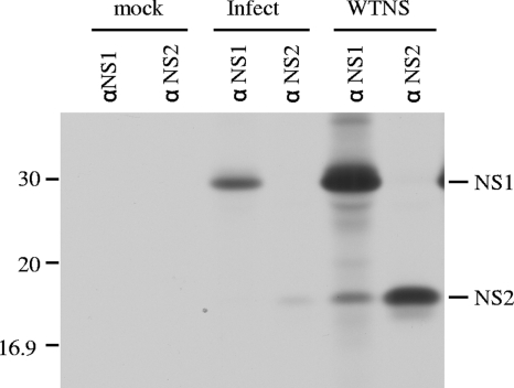
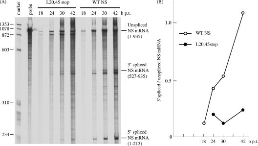
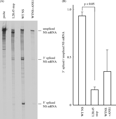
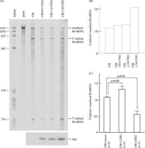
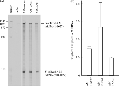
Pre-mRNAs of the influenza A virus M and NS genes are poorly spliced in virus-infected cells. By contrast, in influenza C virus-infected cells, the predominant transcript from the M gene is spliced mRNA. The present study was performed to investigate the mechanism by which influenza C virus M gene-specific mRNA (M mRNA) is readily spliced. The ratio of M1 encoded by a spliced M mRNA to CM2 encoded by an unspliced M mRNA in influenza C virus-infected cells was about 10 times larger than that in M gene-transfected cells, suggesting that a viral protein(s) other than M gene translational products facilitates viral mRNA splicing. RNase protection assays showed that the splicing of M mRNA in infected cells was much higher than that in M gene-transfected cells. The unspliced and spliced mRNAs of the influenza C virus NS gene encode two nonstructural (NS) proteins, NS1(C/NS1) and NS2(C/NS2), respectively. The introduction of premature translational termination into the NS gene, which blocked the synthesis of the C/NS1 and C/NS2 proteins, drastically reduced the splicing of NS mRNA, raising the possibility that C/NS1 or C/NS2 enhances viral mRNA splicing. The splicing of influenza C virus M mRNA was increased by coexpression of C/NS1, whereas it was reduced by coexpression of the influenza A virus NS1 protein (A/NS1). The splicing of influenza A virus M mRNA was also increased by coexpression of C/NS1, though it was inhibited by that of A/NS1. These results suggest that influenza C virus NS1, but not A/NS1, can upregulate viral mRNA splicing.


Figures

FIG. 1.
FIG. 1.
M gene translational products synthesized in infected and M gene-transfected cells. (A) COS-1 cells mock infected (lane mock), infected with C/Yamagata/1/88 (lane Infect) at an MOI of 10 PFU/cell, or transfected with the transient expression vector pME18S, containing the full-length cDNA of the influenza C virus M gene (lane pME18S-CM), were labeled with [35S]methionine for 60 min at 26 h p.i. or at 48 h p.t., and cell lysates were immunoprecipitated with anti-GST/CM2 serum (lane αCM2) or anti M1 MAb (L2) (lane αM1). The resulting immunoprecipitates were analyzed by SDS-PAGE on 17.5% gels containing 4 M urea under reducing conditions, followed by fluorography. The positions of molecular weight markers are shown to the left. (B) 293T cells mock transfected (lane mock) or transfected with an RNA polymerase I-driven vector containing influenza C virus M gene cDNA, pPolI/M, and plasmids expressing polymerases (PB2, PB1, and P3) and NP, pcDNA/PB2-AA, pcDNA/PB1-AA, pcDNA/P3-AA, and pCAGGS.MCS/NP-AA, respectively (20) (lane PolI-M), were labeled with [35S]methionine for 60 min at 48 h p.t., and cell lysates were immunoprecipitated with anti-GST/CM2 serum (lane αCM2) or anti M1 MAb (L2) (lane αM1).
FIG. 2.
FIG. 2.
RNase protection assay of influenza C virus M gene-derived mRNAs in infected and M gene-transfected cells. (A) Total RNA was extracted from COS-1 and 293T cells infected with C/Yamagata/1/88 at an MOI of 10 PFU/cell at 24 h p.i. (lane Infect) and from cells transfected with pME18S-CM at 48 h p.t. (lane M gene) and analyzed by RNase protection assay using a 33P-labeled influenza C virus RNA segment 6 (M gene)-specific RNA probe (vRNA sense). The protected fragments were separated on a 4% polyacrylamide gel containing 4 M urea. The HaeIII digest of ψX174 DNA was 5′ end labeled and used as a size marker (marker lane). The probe lane shows an undigested probe. (B) The ratios of 5′ spliced M mRNA to unspliced M mRNA in infected (open box) and transfected (closed box) COS-1 and 293T cells were calculated after densitometry of panel A.
FIG. 3.
FIG. 3.
Kinetics of M mRNA splicing in influenza C virus-infected cells. (A) RNA was extracted from C/Yamagata/1/88 virus-infected cells at various times postinfection and processed for RNase protection assay using an antisense M gene-specific RNA probe. Lanes M and P show the 5′ end-labeled HaeIII digest of ψX174 DNA as a size marker and an undigested probe, respectively. (B) The ratios of 5′ spliced M mRNA to unspliced M mRNA in each lane of panel A are shown by closed circles.
FIG. 4.
FIG. 4.
Indirect immunofluorescent staining of infected and transfected cells. HMV-II cells mock infected (mock) or infected with C/Yamagata/1/88 (upper panel) and COS-1 cells mock transfected (mock) or transfected with pME18S-CNS1 (lower panel) were fixed with carbon tetrachloride at 12, 24, 36, and 48 h after infection or transfection, respectively. The cells were then stained by an indirect method using anti-GST/NS1 serum.
FIG. 5.
FIG. 5.
Schematic representation of the NS gene construct. Boxes show open reading frames. (WTNS construct) A 246-amino-acid (aa) NS1 protein encoded by a colinear mRNA transcript of influenza C virus RNA segment 7 (NS gene) and a 182-amino-acid NS2 protein encoded by a spliced mRNA are expressed in cells transfected with pME18S-WTNS. (L20,45stop construct) Premature termination codons were introduced into Leu residues 20 and 45 of the N-terminal 62-amino-acid region, which is common to both the NS1 and NS2 coding regions, to eliminate the expression of both the NS1 and NS2 proteins. (CNS1 construct) Plasmid pME18S-CNS1, which expressed NS1 alone, was constructed by substituting the donor (214-GU-215) and acceptor (525-AG-526) for splicing into 214-GA-215 and 525-CG-526, respectively, thereby resulting in the abolition of transcription to the NS2 mRNA.
FIG. 6.
FIG. 6.
NS1 and NS2 synthesis in influenza C virus-infected and NS gene-transfected cells. COS-1 cells mock infected (lane mock), infected with C/Yamagata/1/88 (Infect lanes) at an MOI of 10 PFU/cell, or transfected with pME18S-WTNS (WTNS lanes) were labeled with [35S]methionine and immunoprecipitated with anti-GST/NS1 serum (αNS1 lanes) or anti-GST/NS2 serum (αNS2 lanes). The resulting immunoprecipitates were analyzed by SDS-PAGE, followed by fluorography.
FIG. 7.
FIG. 7.
Kinetics of NS mRNAs in transfected cells. (A) COS-1 cells were transfected with either pME18S-WTNS (WTNS lanes) or pME18S-L20,45stop (L20,45stop lanes) in which a premature termination of translation was introduced into the Leu residues at positions 20 and 45 of the influenza C virus NS coding region. The unspliced and spliced NS mRNAs at the indicated times posttransfection were quantitated by RNase protection assay using a 33P-labeled influenza C virus RNA segment 7 (NS gene)-specific RNA probe (vRNA sense). The probe lane contains the undigested probe. (B) Ratios of 3′ spliced NS mRNA to unspliced NS mRNA in cells transfected with pME18S-WTNS (open circles) or pME18S-L20,45stop (closed circles).
FIG. 8.
FIG. 8.
RNase protection assay of NS mRNAs in transfected cells. (A) Cytoplasmic RNA was isolated from COS-1 cells transfected with either pME18S-WTNS (WTNS lane) or pME18S-L20,45stop (L20,45stop lane) or cotransfected with pME18S-WTNS and pME18S-ANS1 that expressed the NS1 protein of influenza A/PR/8/34 (WTNS+ANS1 lane) at 48 h p.t. and analyzed by RNase protection assay as described in Materials and Methods. The representative data of two independent experiments are shown. (B) The splicing ratios of 3′ spliced to unspliced NS mRNAs in cells transfected with each NS construct in the two experiments whose results are shown in panel A were analyzed statistically.
FIG. 9.
FIG. 9.
RNase protection assay of M mRNAs in influenza C virus M gene-transfected cells coexpressing influenza C virus NS1 protein. (A) pME18S-CM (0.5 μg) was introduced into COS-1 cells either together with 0.5, 1.0, or 2.0 μg of pME18S-CNS1 (right three lanes) or without pME18S-CNS1 (CM lane) and analyzed by RNase protection assay using an antisense M-specific RNA probe. The probe lane contains the undigested probe (upper panel). The expression of the C/NS1 protein in cells transfected as described above was analyzed by RIP with anti-GST/NS1 serum (lower panel). (B) Ratios of 5′ spliced to unspliced M mRNA in the transfected cells shown in panel A. (C) The ratios of 5′ spliced to unspliced M mRNA in COS-1 cells cotransfected with pME18S-CM and the pME18S vector as a control (CM+vector bar) or pME18S-CM and pME18S-CNS1 (CM+CNS1 bar) in four independent RNase protection assay experiments and cotransfected with pME18S-CM and pME18S-ANS1 (CM+ANS1 bar) in three independent experiments were compared.
FIG. 10.
FIG. 10.
RNase protection assay of influenza A virus M mRNAs in influenza A virus M gene-transfected cells coexpressing either influenza C virus NS1 or influenza A virus NS1. (A) pME18S-AM (0.33 μg), which can express the M gene-derived mRNA of influenza A virus PR/8/34 strain, was introduced into COS-1 cells with either 0.67 μg pME18S alone (AM+vector lane), 0.67 μg of pME18S-CNS1 (AM+CNS1 lane), or 0.67 μg of pME18S-ANS1, which can express the NS1 protein of influenza A/PR/8/34 (AM+ANS1 lane). The influenza A virus M mRNAs were analyzed by RNase protection assay using an antisense probe specific for the influenza A virus M gene. The probe lane shows an undigested probe. Representative data from two independent experiments are shown. (B) The splicing ratios of 3′ spliced to unspliced AM mRNAs in the cells cotransfected with each construct in the two experiments whose results are shown in panel A were compared.

Similar articles

Cited by

References

    1. Alamgir, A. S. M., Y. Matsuzaki, S. Hongo, E. Tsuchiya, K. Sugawara, Y. Muraki, and K. Nakamura. 2000. Phylogenetic analysis of influenza C virus nonstructural (NS) protein genes and identification of the NS2 protein. J. Gen. Virol. 81:1933-1940. - PubMed
    1. Betakova, T., and A. J. Hay. 2007. Evidence that the CM2 protein of influenza C virus can modify the pH of the exocytic pathway of transfected cells. J. Gen. Virol. 88:2291-2296. - PubMed
    1. Bukrinskaya, A. G., N. K. Vorunova, G. V. Kornilayeva, R. A. Narmanbetova, and A. J. Hay. 1982. Influenza virus uncoating in infected cells and effect of rimantadine. J. Gen. Virol. 60:49-59. - PubMed
    1. Ciampor, F., C. A. Thompson, S. Grambas, and A. J. Hay. 1992. Regulation of pH by the M2 protein of influenza A viruses. Virus Res. 22:247-258. - PubMed
    1. Enami, M., G. Sharma, C. Benham, and P. Palese. 1991. An influenza virus containing nine different RNA segments. Virology 185:291-298. - PubMed

Publication types

MeSH terms

LinkOut - more resources