. 2010 Feb;84(4):1891-901.
doi: 10.1128/JVI.02458-09. Epub 2009 Dec 9.

A single tyrosine in the severe acute respiratory syndrome coronavirus membrane protein cytoplasmic tail is important for efficient interaction with spike protein

Affiliations

A single tyrosine in the severe acute respiratory syndrome coronavirus membrane protein cytoplasmic tail is important for efficient interaction with spike protein

Corrin E McBride et al. J Virol. 2010 Feb.

Abstract

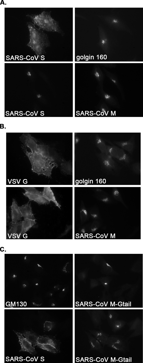
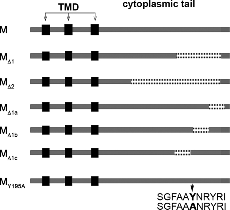
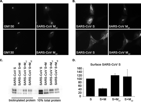
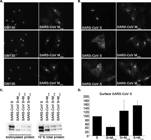
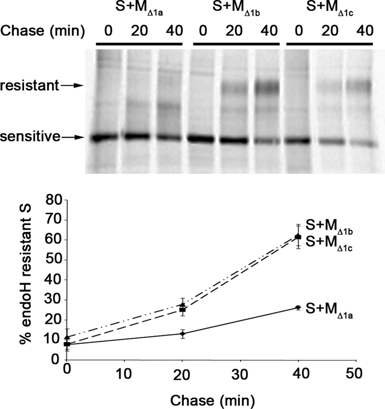
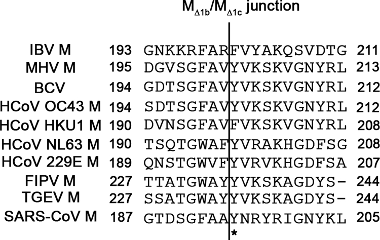
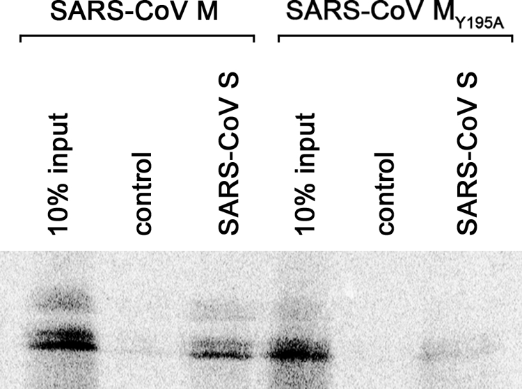
Severe acute respiratory syndrome coronavirus (SARS-CoV) encodes 3 major envelope proteins: spike (S), membrane (M), and envelope (E). Previous work identified a dibasic endoplasmic reticulum retrieval signal in the cytoplasmic tail of SARS-CoV S that promotes efficient interaction with SARS-CoV M. The dibasic signal was shown to be important for concentrating S near the virus assembly site rather than for direct interaction with M. Here, we investigated the sequence requirements of the SARS-CoV M protein that are necessary for interaction with SARS-CoV S. The SARS-CoV M tail was shown to be necessary for S localization in the Golgi region when the proteins were exogenously coexpressed in cells. This was specific, since SARS-CoV M did not retain an unrelated glycoprotein in the Golgi. Importantly, we found that an essential tyrosine residue in the SARS-CoV M cytoplasmic tail, Y(195), was important for S-M interaction. When Y(195) was mutated to alanine, M(Y195A) no longer retained S intracellularly at the Golgi. Unlike wild-type M, M(Y195A) did not reduce the amount of SARS-CoV S carbohydrate processing or surface levels when the two proteins were coexpressed. Mutating Y(195) also disrupted SARS-CoV S-M interaction in vitro. These results suggest that Y(195) is necessary for efficient SARS-CoV S-M interaction and, thus, has a significant involvement in assembly of infectious virus.


Figures

FIG. 1.
FIG. 1.
The cytoplasmic tail of SARS-CoV M specifically mediates interaction with SARS-CoV S. HeLa cells expressing SARS-CoV S in the presence or absence of SARS-CoV M (A), SARS-CoV M in the presence or absence of VSV G (B), or SARS-CoV M-Gtail in the presence or absence of SARS CoV-S (C) for 24 h were fixed, permeabilized, and stained with mouse anti-SARS-CoV S and rabbit anti-golgin-160 (a Golgi marker) or rabbit anti-SARS-CoV M (A), mouse anti-VSV G and rabbit anti-golgin-160 or rabbit anti-SARS-CoV M (B), or rabbit anti-VSV G and mouse anti-GM130 (a Golgi marker) or mouse anti-SARS-CoV S (C). The secondary antibodies were Alexa 488-conjugated donkey anti-mouse IgG and Texas red-conjugated donkey anti-rabbit IgG. The same field is shown for each horizontal set of panels.
FIG. 2.
FIG. 2.
Cartoon depicting wild-type SARS-CoV M and M mutants. Deletions or point mutations were introduced into the cytoplasmic tail of SARS-CoV M using site-directed mutagenesis. Deleted regions are marked with dotted lines. The Y195 mutation is in bold and marked with a black arrow in the sequence alignment below the cartoon. Gray arrows show transmembrane domains (TMD).
FIG. 3.
FIG. 3.
SARS-CoV MΔ1 and MΔ2 localize to the Golgi compartment but do not retain SARS-CoV S or reduce the amount of SARS-CoV S at the plasma membrane. (A) HeLa cells expressing SARS-CoV MΔ1 or SARS-CoV MΔ2 for 24 h were fixed, permeabilized, and stained with rabbit anti-SARS-CoV M and mouse anti-GM130. Secondary antibodies were Alexa 488-conjugated donkey anti-mouse IgG and Texas red-conjugated donkey anti-rabbit IgG. The same field is shown for each horizontal set of panels. (B) HeLa cells coexpressing SARS-CoV S and MΔ1 or MΔ2 for 24 h were fixed, permeabilized, and stained with mouse anti-SARS-CoV S and rabbit anti-SARS-CoV M. Secondary antibodies were Alexa 488-conjugated donkey anti-mouse IgG and Texas red-conjugated donkey anti-rabbit IgG. The same field is shown for each horizontal set of panels. (C and D) HEK293T cells expressing SARS-CoV S in the absence or presence of SARS-CoV M, MΔ1, or MΔ2 were surface biotinylated at 24 h posttransfection. After lysis, 10% of the lysate was saved for quantification of total S. Biotinylated proteins were collected from the remainder of the lysate by using streptavidin agarose resin, washed, and eluted in sample buffer. After SDS-PAGE and Western blotting with antibodies to SARS-CoV S (C), total biotinylated S protein was quantified for each sample (D). Values in panel D are normalized to the amount of biotinylated S when expressed alone. The averages of the results of three independent experiments ± standard errors of the means are shown.
FIG. 4.
FIG. 4.
SARS-CoV MΔ1 and MΔ2 are less effective at reducing SARS-CoV S trafficking through the Golgi compartment than wild-type SARS-CoV M. At 24 h posttransfection, HEK293T cells expressing SARS-CoV S in the absence or presence of SARS-CoV M, MΔ1, or MΔ2 were labeled with [35S]methionine-cysteine for 20 min and then chased for 0, 20, or 40 min. After lysis, S proteins were immunoprecipitated, denatured, and digested with endo H. Endo H-sensitive and -resistant forms are indicated. After electrophoresis and phosphorimaging, the percentage of endo H-resistant S was quantified. The averages of the results of five independent experiments ± standard errors of the means are shown.
FIG. 5.
FIG. 5.
SARS-CoV MΔ1a, MΔ1b, and MΔ1c localize to the Golgi compartment, but only SARS-CoV MΔ1a can retain SARS-CoV S and reduce the amount of SARS-CoV S at the plasma membrane. (A) HeLa cells expressing SARS-CoV MΔ1a, MΔ1b, or MΔ1c for 24 h were fixed, permeabilized, and stained with rabbit anti-SARS-CoV M and mouse anti-GM130. Secondary antibodies were Alexa 488-conjugated donkey anti-mouse IgG and Texas red-conjugated donkey anti-rabbit IgG. The same field is shown for each horizontal set of panels. (B) HeLa cells coexpressing SARS-CoV S and MΔ1a, MΔ1b, or MΔ1c for 24 h were fixed, permeabilized, and stained with mouse anti-SARS-CoV S and rabbit anti-SARS-CoV M. Secondary antibodies were Alexa 488-conjugated donkey anti-mouse IgG and Texas Red-conjugated donkey anti-rabbit IgG. The same field is shown for each horizontal set of panels. (C and D) HEK293T cells expressing SARS-CoV S in the absence or presence of SARS-CoV MΔ1a, MΔ1b, or MΔ1c were surface biotinylated. After lysis, biotinylated cell surface proteins were collected by using streptavidin agarose resin, washed, and eluted in sample buffer. After SDS-PAGE and Western blotting with rabbit anti-SARS-CoV S antibodies, total biotinylated S protein was quantified for each sample (D). Values in panel D are normalized to the amount of biotinylated S when expressed alone. The averages of the results of three independent experiments ± standard errors of the means are shown.
FIG. 6.
FIG. 6.
Neither SARS-CoV MΔ1b nor MΔ1c slows SARS-CoV S trafficking through the Golgi compartment. HEK293T cells expressing SARS-CoV S in the presence of SARS-CoV MΔ1a, MΔ1b, or MΔ1c were labeled with [35S]methionine-cysteine for 20 min and then chased for 0, 20, or 40 min. After lysis, S proteins were immunoprecipitated, denatured, and digested with endo H. Endo H-sensitive and -resistant forms are indicated. After electrophoresis and phosphorimaging, the percentage of endo H-resistant S was quantified. The averages of the results of three independent experiments ± standard errors of the means are shown.
FIG. 7.
FIG. 7.
Sequence alignment of CoV M protein cytoplasmic tails. The SARS-CoV MΔ1b/MΔ1c junction is marked with a vertical line. Y195 that is mutated in SARS-CoV MY195A is marked with an asterisk. IBV, avian infectious bronchitis virus; MHV, mouse hepatitis virus; BCV, bovine coronavirus; HCoV, human coronavirus; FIPV, feline infectious peritonitis virus; TGEV, porcine transmissible gastroenteritis coronavirus; SARS-CoV, severe acute respiratory syndrome coronavirus. The alignment was generated using MultAlin multiple sequence alignment with hierarchical clustering (8).
FIG. 8.
FIG. 8.
Tyrosine 195 is important for efficient S and M interaction. (A) SARS-CoV MY195A localizes to the Golgi compartment but does not retain SARS-CoV S intracellularly. HeLa cells expressing SARS-CoV MY195A in the absence or presence of SARS-CoV S were stained as described for Fig. 3. The same field is shown for each horizontal set of panels. (B) SARS-CoV MY195A does not reduce the amount of SARS-CoV S at the plasma membrane. HEK293T cells exogenously coexpressing SARS-CoV S in the absence or presence of SARS-CoV M, MΔ1, or MY195A were surface biotinylated as described for Fig. 3C. The averages of the results of three independent experiments ± standard errors of the means are shown. (C) SARS-CoV MY195A does not slow SARS-CoV S trafficking through the Golgi as well as wild-type SARS-CoV M does. HEK293T cells exogenously coexpressing SARS-CoV S and SARS-CoV M, MΔ1, or MY195A were pulse-labeled and chased, and immunoprecipitated S protein was treated with endo H as described for Fig. 4. The averages of the results of three independent experiments ± standard errors of the means are shown.
FIG. 9.
FIG. 9.
Mutating Y195 disrupts the SARS-CoV S-M interaction in vitro. Equal amounts of solubilized in vitro-transcribed and -translated SARS-CoV M or MY195A were incubated with His-tagged SARS-CoV S prebound to nickel beads or with nickel beads alone. Samples were washed, subjected to SDS-PAGE, and visualized and quantified by phosphorimaging. Equal loading of recombinant S protein was ensured by staining with Coomassie blue (not shown).

Similar articles

Cited by

References

    1. Armstrong, J., S. Patel, and P. Riddle. 1990. Lysosomal sorting mutants of coronavirus E1 protein, a Golgi membrane protein. J. Cell Sci. 95(Pt. 2):191-197. - PubMed
    1. Barcena, M., G. T. Oostergetel, W. Bartelink, F. G. Faas, A. Verkleij, P. J. Rottier, A. J. Koster, and B. J. Bosch. 2009. Cryo-electron tomography of mouse hepatitis virus: insights into the structure of the coronavirion. Proc. Natl. Acad. Sci. U. S. A. 106:582-587. - PMC - PubMed
    1. Baudoux, P., C. Carrat, L. Besnardeau, B. Charley, and H. Laude. 1998. Coronavirus pseudoparticles formed with recombinant M and E proteins induce alpha interferon synthesis by leukocytes. J. Virol. 72:8636-8643. - PMC - PubMed
    1. Bisht, H., A. Roberts, L. Vogel, A. Bukreyev, P. L. Collins, B. R. Murphy, K. Subbarao, and B. Moss. 2004. Severe acute respiratory syndrome coronavirus spike protein expressed by attenuated vaccinia virus protectively immunizes mice. Proc. Natl. Acad. Sci. U. S. A. 101:6641-6646. - PMC - PubMed
    1. Bonifacino, J. S., and L. M. Traub. 2003. Signals for sorting of transmembrane proteins to endosomes and lysosomes. Annu. Rev. Biochem. 72:395-447. - PubMed

Publication types

MeSH terms

LinkOut - more resources