. 2010 Feb 12;285(7):4715-24.
doi: 10.1074/jbc.M109.086660. Epub 2009 Dec 9.

Negative autoregulation of GTF2IRD1 in Williams-Beuren syndrome via a novel DNA binding mechanism

Affiliations

Negative autoregulation of GTF2IRD1 in Williams-Beuren syndrome via a novel DNA binding mechanism

Stephen J Palmer et al. J Biol Chem. .

Abstract

The GTF2IRD1 gene is of principal interest to the study of Williams-Beuren syndrome (WBS). This neurodevelopmental disorder results from the hemizygous deletion of a region of chromosome 7q11.23 containing 28 genes including GTF2IRD1. WBS is thought to be caused by haploinsufficiency of certain dosage-sensitive genes within the deleted region, and the feature of supravalvular aortic stenosis (SVAS) has been attributed to reduced elastin caused by deletion of ELN. Human genetic mapping data have implicated two related genes GTF2IRD1 and GTF2I in the cause of some the key features of WBS, including craniofacial dysmorphology, hypersociability, and visuospatial deficits. Mice with mutations of the Gtf2ird1 allele show evidence of craniofacial abnormalities and behavioral changes. Here we show the existence of a negative autoregulatory mechanism that controls the level of GTF2IRD1 transcription via direct binding of the GTF2IRD1 protein to a highly conserved region of the GTF2IRD1 promoter containing an array of three binding sites. The affinity for this protein-DNA interaction is critically dependent upon multiple interactions between separate domains of the protein and at least two of the DNA binding sites. This autoregulatory mechanism leads to dosage compensation of GTF2IRD1 transcription in WBS patients. The GTF2IRD1 promoter represents the first established in vivo gene target of the GTF2IRD1 protein, and we use it to model its DNA interaction capabilities.


Figures
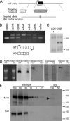
FIGURE 1.
FIGURE 1.
Generation of the Gtf2ird1tm1Hrd mutant allele and the consequences for transcription and translation. A, homologous recombination of the targeting construct with the wild-type (WT) allele using positive neomycin (Neo) selection and negative diphtheria toxin (DTA) selection. The Neo cassette was subsequently removed using the flanking LoxP sites (arrowheads). B, RTPCR analysis of Gtf2ird1 transcripts in brown adipose tissue from a litter of mice segregating the mutant allele using primers in exon 1 and exon 3 (shown in scheme). A novel exon 1–3 splice transcript is produced with equal efficiency to the wild-type transcript. C, Western blot of proteins immunoprecipitated (IP) from C2C12 cell extracts using the M19 antibody for IP and as probe. The immunoprecipitation was conducted in the presence (+) or absence (−) of a blocking peptide antigen to demonstrate specificity. D, analysis of transcription and translation from a wild-type Gtf2ird1 cDNA and the equivalent mutant cDNA lacking exon 2. Extracts from transfected COS-7 cells show that both transcripts are abundantly produced (Northern Gtf2ird1) but the GTF2IRD1 peptide is below detectable levels in cells expressing the mutant transcript (KO) using two antibodies, M19 and G21. Equal loading is shown by ethidium bromide staining of RNA and Coomassie Blue staining of protein in the M19 Western blot. E, Western blot analysis using sequential dilutions of protein extract from cells transfected with the wild-type construct compared with high concentration loadings of mutant extract (KO), probed with M19 and G21 antibodies. A faint band is visible at the highest concentration in the M19 analysis (arrowhead). Numbers represent μg of total protein.
FIGURE 2.
FIGURE 2.
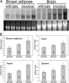
Gtf2ird1 transcript production approximately doubles in the tissues of Gtf2ird1tm1Hrd knock-out mice. A, Northern blot of total RNA extracted from brown adipose and brain tissue of three wild-type and three homozygous knock-out mice showing that the 3.5-kb Gtf2ird1 band is more intense in all three knock-out mice. Close scrutiny also shows an expected slight reduction in transcript length because of the loss of exon 2 (129 bp) in the knock-out samples. B, quantitative RT-PCR analysis of Gtf2ird1 levels. Error bars show S.D., and the numbers indicate the number of mice used. Unpaired two-tailed Student's t tests show a difference between wild-type (WT/WT) and homozygous null (KO/KO) genotypes with a probability of: p = 0.0011 for brown adipose, p = 0.012 for brain, p = 0.0019 for heart, and p = 0.022 for spleen.
FIGURE 3.
FIGURE 3.
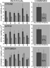
Quantitative RT-PCR analysis of CYLN2, GTF2I, and GTF2IRD1 expression in lymphoblastoid cell lines derived from Williams-Beuren syndrome patients and their relatives. Error bars show S.D., and the figure in the patient bar indicates the estimated mean expression level as a percentage of the relatives' mean. Paired two-tailed Student's t tests show differences between the means of genotype groups with a probability of: CYLN2 p = 0.004575, GTF2I p = 0.000342, and GTF2IRD1 p = 0.559668.
FIGURE 4.
FIGURE 4.
ClustalW alignment of the GUR in four vertebrate species. Colored boxes indicate identical base residues, and the arrows indicate the position and orientation of three highly conserved GTF2IRD1 recognition sequences. The core motifs are perfectly conserved, but conservation quickly diminishes either side of the flanking sites. The transition to bold lowercase indicates the start of the most 5′ ESTs found in data base searches. Human EST BM544769, Mouse EST CJ178886, X. tropicalis EST CN092749. No informative Fugu ESTs were found.
FIGURE 5.
FIGURE 5.
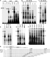
EMSA of GTF2IRD1 proteins showing affinity for the GUR and the tandem nature of the binding. A, binding of human GTF2IRD1 to synthetic multimers of the TNNI1-derived probe, B1, originally used as a yeast one-hybrid bait (10). GTF2IRD1 (IRD1) does not bind detectably to single copies, but binds well to B1 dimers (2xB1) and trimers (3xB1). A lower shift complex (LC) is present in both, but a higher shift complex (HC) appears with the trimer. Probes combined with unprogrammed in vitro translation mix (IVT) or with GTF2IRD1 and excess cold competitor oligonucleotide (COLD) showed no shift. An unknown endogenous shift complex (arrows) present in the in vitro translation mix shows only a minor affinity increase as a result of multimerization. B, GUR fragment is bound efficiently by the two most abundant mouse isoforms of GTF2IRD1, 3α7, and 3α5 and by human GTF2IRD1, but not by the mouse β isoform of TFII-I (GTF2I). No shift occurs in the IVT control or probe-only lane (PR). C, deletion of the leucine zipper domain of GTF2IRD1 (IRD1ΔLZ) intensifies affinity of the LC, but ablates the HC, demonstrating that the HC contains a GTF2IRD1 dimer. D and E, probes containing mutations in single GGATTA sites or in combinations were tested with human GTF2IRD1 protein to determine changes in affinity. Position of the mutation (M) is indicated on the scheme of the GUR (arrows). F, mutation of the middle site alone leads to absence of binding, but the shifts are restored by reducing the distance between the flanking sites, as illustrated using a series of probes with successive deletions (GURDEL1–5). G, sequences of the double-stranded probes used in the EMSA: binding sites are indicated in bold type, and mutations are indicated in lowercase bold type. In the GUR DEL series, the residues deleted in each successive probe are indicated by underlines. Gaps between images indicate where irrelevant lanes have been removed.
FIGURE 6.
FIGURE 6.
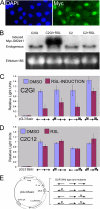
The GUR acts as an effective enhancer/promoter in cell culture assays and is negatively regulated by exogenously derived inducible GTF2IRD1. A, inducible Myc-GTF2IRD1 clonal cell line C2GI, with nuclei stained by DAPI and Myc-tagged GTF2IRD1 detected by anti-Myc immunofluorescence (Myc). B, Northern blot of C2GI and parental C2C12 (C2) cell RNA with or without the inducer RSL. C, C2GI inducible GTF2IRD1 cells with pGL3-Basic and pGL3-GUR constructs containing the wild-type GUR or with mutations (M) in the flanking recognition sites. Induction of GTF2IRD1 leads to a repression of the reporter containing the wild-type GUR. Mutations in the GTF2IRD1 binding sites result in attenuation of repression. Luciferase activity was set at 1 for the wild-type GUR, and measurements were normalized relative to this to combine experiments. Error bars indicate the S.D. of duplicate measurements in two separate experiments. D, C2C12 parental cell line control experiment showing that repression is not caused by the induction ligand RSL. A single experiment conducted in duplicate. Error bars indicate range. E, diagram of reporter constructs used in C and D. The wild-type GUR and permutations containing 3-bp mutations (M) in the GGATTA site were introduced into pGL3-Basic in the appropriate orientation.
FIGURE 7.
FIGURE 7.
Model of the proposed mechanism for GTF2IRD1 binding to the GUR. EMSA experiments suggest that the middle and proximal GGATTA sites, which are both in the same orientation, are bound in tandem by two RDs of GTF2IRD1. Binding to the middle and distal sites is also possible, but has a lower affinity. Formation of the dimer is dependent on the presence of the leucine zipper domain. The orientation of the protein on the DNA and identification of which binding site the RDs contact is speculative.

Similar articles

Cited by

References

    1. Strømme P., Bjørnstad P. G., Ramstad K. (2002) J. Child Neurol. 17, 269–271 - PubMed
    1. Osborne L. R., Mervis C. B. (2007) Expert. Rev. Mol. Med. 9, 1–16 - PMC - PubMed
    1. Valero M. C., de Luis O., Cruces J., Pérez Jurado L. A. (2000) Genomics 69, 1–13 - PubMed
    1. Curran M. E., Atkinson D. L., Ewart A. K., Morris C. A., Leppert M. F., Keating M. T. (1993) Cell 73, 159–168 - PubMed
    1. Hirota H., Matsuoka R., Chen X. N., Salandanan L. S., Lincoln A., Rose F. E., Sunahara M., Osawa M., Bellugi U., Korenberg J. R. (2003) Genet. Med. 5, 311–321 - PubMed

Publication types

MeSH terms

LinkOut - more resources