. 2010 Feb 12;285(7):4931-40.
doi: 10.1074/jbc.M109.048397. Epub 2009 Dec 9.

Stem-like cancer cells are inducible by increasing genomic instability in cancer cells

Affiliations

Stem-like cancer cells are inducible by increasing genomic instability in cancer cells

Yi Liang et al. J Biol Chem. .

Abstract

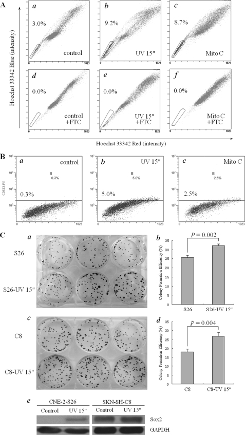
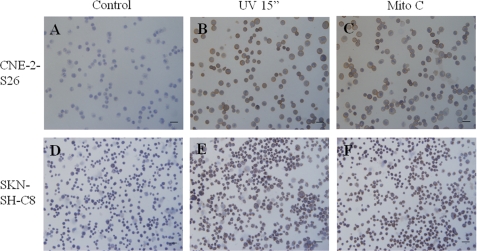
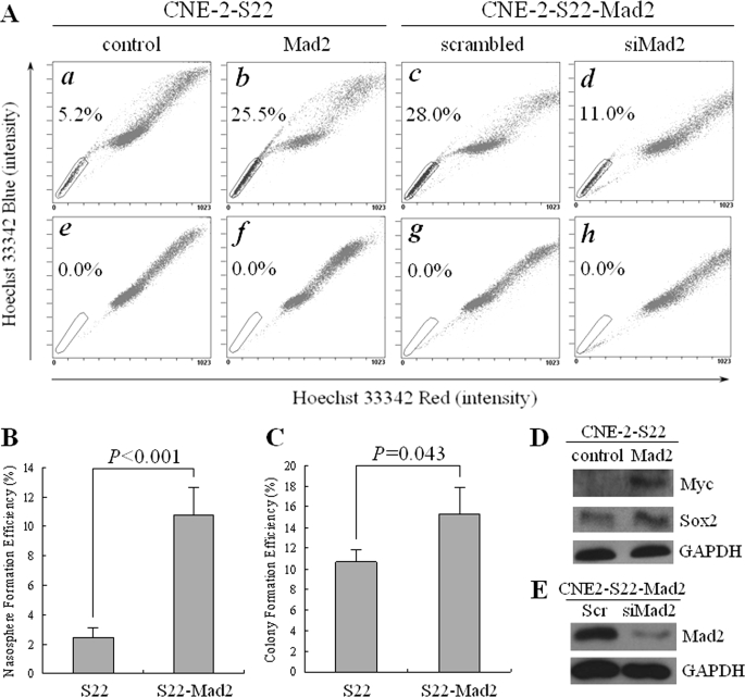
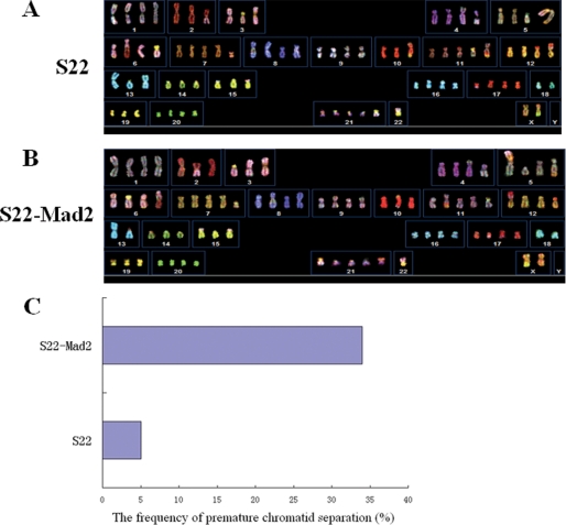
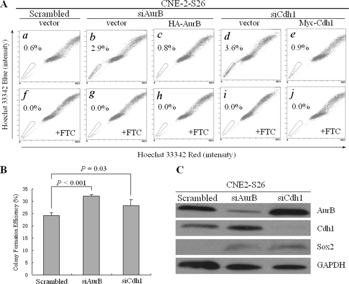
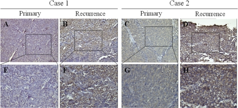
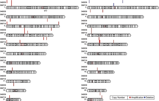
The existence of cancer stem cells (CSCs) or stem-like cancer cells (SLCCs) is regarded as the cause of tumor formation and recurrence. However, the origin of such cells remains controversial with two competing hypotheses: CSCs are either transformed from tissue adult stem cells or dedifferentiated from transformed progenitor cells. Compelling evidence has determined the chromosomal aneuploidy to be one of the hallmarks of cancer cells, indicating genome instability plays an important role in tumorigenesis, for which CSCs are believed to be the initiator. To gain direct evidence that genomic instability is involved in the induction of SLCCs, we utilized multiple approaches to enhance genomic instability and monitored the percentage of SLCC in cultured cancer cells. Using side population (SP) cells as a marker for SLCC in human nasopharyngeal carcinoma (NPC) and CD133 for human neuroblastoma cells, we found that DNA damage inducers, UV and mitomycin C were capable of increasing SP cells in NPC CNE-2 and neuroblastoma SKN-SH cells. Likewise, either overexpression of a key regulator of cell cycle, Mad2, or knock down of Aurora B, an important kinase in mitosis, or Cdh1, a key E3 ligase in cell cycle, resulted in a significant increase of SP cells in CNE-2. More interestingly, enrichment of SP cells was observed in recurrent tumor tissues as compared with the primary tumor in the same NPC patients. Our study thus suggested that, beside transformation of tissue stem cells leading to CSC generation, genomic instability could be another potential mechanism resulting in SLCC formation, especially at tumor recurrence stage.


Figures

FIGURE 1.
FIGURE 1.
SLCC induction in human NPC cells with DNA damage treatment. A, SP cells were induced by UV light or mitomycin C in human NPC cells. a–f, NPC CNE-2 cells were single cell cloned, and a single cell clone CNE-2-S26 was selected and expanded for UV light or mitomycin C treatment and Hoechst 33342 staining. a, without treatment; b, treated with UV light for 15 s and cultured for 24 h; c, treated with 2.5 μg/ml mitomycin C for 24 h; d–f, the same as a–c, except that FTC at a final concentration of 5 μm was added for 5 min before Hoechst 33342 staining. The percentage of SP cells is indicated. B, CD133-positive cells were induced with UV light or mitomycin C in human neuroblastoma cells. a–c, human neuroblastoma SKN-SH cells were single cell cloned, and a single cell clone SKN-SH-C8 was selected for treatment and CD133-positive cell counting. a, without treatment; b, treated with UV radiation for 15 s and cultured for 24 h; c, treated with 2.5 μg/ml mitomycin C for 24 h. The percentage of CD133-positive cells was indicated in the figures. C, the properties of SLCCs was enhanced by UV treatment in both NPC cells and neuroblastoma cells. a and b, colony formation assay of CNE-2-S26 cells before/after UV treatment. a, colony formation results; b, statistical analysis. c to d: Colony formation assay of SKN-SH-C8 cells before/after UV treatment. c, colony formation results; d, statistical analysis. The p value was indicated in the figures. e, the protein level of Sox2 in both CNE-2-S26 cells and SKN-SH-C8 cells before/after UV treatment.
FIGURE 2.
FIGURE 2.
The expression of ABCG2 or CD133 was induced by DNA damage treatment. A–C, the expression of ABCG2 of CNE-2-S26 was enhanced after UV light or mitomycin C treatment. A, without treatment; B, treated with UV radiation for 15 s and cultured for 24 h; C, treated with 2.5 μg/ml mitomycin C for 24 h. D–F, the expression of CD133 of SKN-SH-C8 was enhanced after UV light or mitomycin C treatment. D, without treatment; E, treated with UV radiation for 15 s and cultured for 24 h; F, treated with 2.5 μg/ml mitomycin C for 24 h.
FIGURE 3.
FIGURE 3.
SP cells induction with stable transfection of Mad2 in human NPC cells. A, Hoechst 33342 staining was performed in human NPC CNE-2-S22-Mad2 cells. a, parental CNE-2-S22 cells; b, Mad2-transfected CNE-2-S22 cells; c, CNE-2-S22-Mad2 cells transfected with scrambled siRNA; d, CNE-2-S22-Mad2 cells transfected with siMad2. e–h, the same as a–d, except that FTC at a final concentration of 5 μm was added before Hoechst 33342 staining. The percentage of SP cells is indicated. B, spheroid (nasosphere) formation assay of CNE-2-S22 and CNE-2-S22-Mad2 in suspension culture circumstance. C, colony formation assay of CNE-2-S22 and CNE-2-S22-Mad2 in adherent culture circumstance. The p value was indicated in the figures. D, Western blot analysis of exogenous Mad2 tagged with Myc and endogenous Sox2 in CNE-2-S22 and CNE-2-S22-Mad2 stable cell lines. E, Western blot analysis of endogenous Mad2. Glyceraldehyde-3-phosphate dehydrogenase (GAPDH) was used as a loading control.
FIGURE 4.
FIGURE 4.
Genomic instability induced by stable overexpression of Mad2 in human NPC cells. A and B, SKY of human NPC CNE-2-S22 cells before/after stable overexpression of Mad2. A, parental CNE-2-S22 cells; B, CNE-2-S22-Mad2 cells. C, the frequency of premature chromatid separation of CNE-2-S22 cells and CNE-2-S22-Mad2 cells.
FIGURE 5.
FIGURE 5.
Transient RNA interference of Aurora B or Cdh1 increased the percentage of SP cells in human NPC cells. A, Hoechst 33342 staining was performed in human NPC CNE-2-S26 cells after cotransfection of Aurora B/Cdh1 siRNA and rescue plasmids. a, scrambled siRNA and vector plasmids; b, Aurora B siRNA and vector plasmid; c, Aurora B siRNA and HA-Aurora B rescue plasmid; d, Cdh1 siRNA and vector plasmid; e, Cdh1 siRNA and Myc-Cdh1 rescue plasmid. f–j, the same as a–e, except that FTC at a final concentration of 5 μm was added before Hoechst 33342 staining. The percentage of SP cells is indicated. B, the colony formation efficiency after Aurora B/Cdh1 siRNA was transfected, compared with the scrambled siRNA. The p values are indicated. C, Western blot analysis of Aurora B, Cdh1, and Sox2 after Aurora B or Cdh1 was knocked down. Glyceraldehyde-3-phosphate dehydrogenase (GAPDH) was used as a loading control.
FIGURE 6.
FIGURE 6.
Spontaneous emergence of ABCG2-positive cells in recurrent NPC tissues. A, C, E, and G, representative immunohistochemistry staining of ABCG2 in primary tumors of two NPC patients. A, Case 1; C, Case 2; E and G, the magnification of A and C, respectively. B, D, F, and H, representative immunohistochemistry staining of ABCG2 in recurrent tumors of the same patients. B, Case 1; D, Case 2; F and H, the magnification of B and D, respectively.
FIGURE 7.
FIGURE 7.
The genome-wide CNV of two couples of primary and recurrent NPC tissues. Using the copy number of the primary tissues as controls, relative copy number of the matched recurrent tissues was calculated and is shown as red lines (amplification) or blue lines (deletion). The sample numbers of the two NPC cases are indicated.

Similar articles

Cited by

References

    1. Quintana E., Shackleton M., Sabel M. S., Fullen D. R., Johnson T. M., Morrison S. J. (2008) Nature 456, 593–598 - PMC - PubMed
    1. Reya T., Morrison S. J., Clarke M. F., Weissman I. L. (2001) Nature 414, 105–111 - PubMed
    1. Bapat S. A. (2007) Semin. Cancer Biol. 17, 204–213 - PubMed
    1. Houghton J., Morozov A., Smirnova I., Wang T. C. (2007) Semin. Cancer Biol. 17, 191–203 - PubMed
    1. Huntly B. J., Gilliland D. G. (2005) Nat. Rev. Cancer 5, 311–321 - PubMed

Publication types

MeSH terms

Substances