. 2009 Dec 22;106(51):21848-53.
doi: 10.1073/pnas.0911354106. Epub 2009 Dec 9.

Giant Marseillevirus highlights the role of amoebae as a melting pot in emergence of chimeric microorganisms

Affiliations

Giant Marseillevirus highlights the role of amoebae as a melting pot in emergence of chimeric microorganisms

Mickaël Boyer et al. Proc Natl Acad Sci U S A. .

Abstract

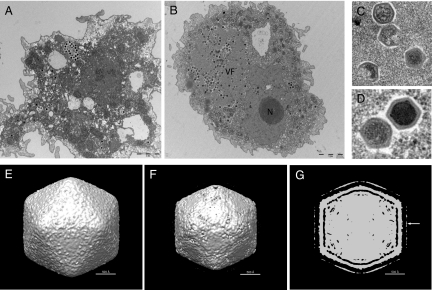
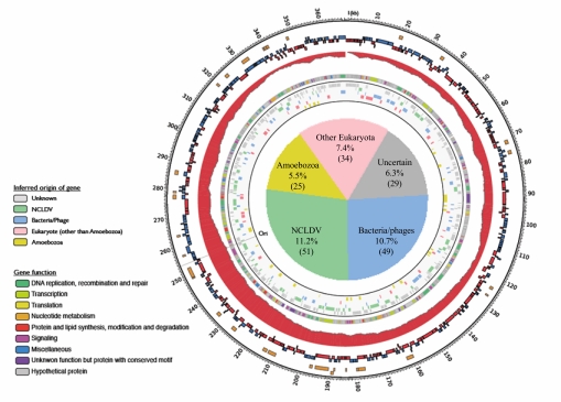
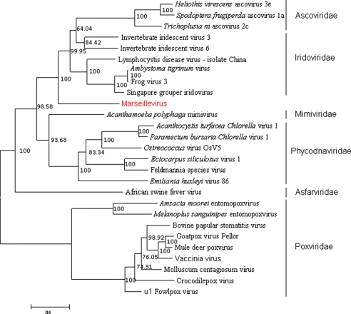
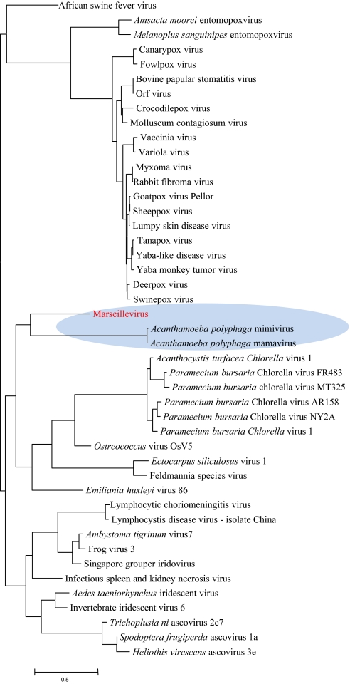
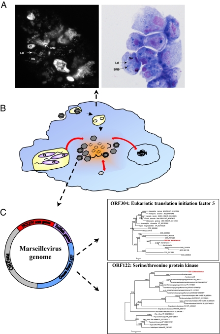
Giant viruses such as Mimivirus isolated from amoeba found in aquatic habitats show biological sophistication comparable to that of simple cellular life forms and seem to evolve by similar mechanisms, including extensive gene duplication and horizontal gene transfer (HGT), possibly in part through a viral parasite, the virophage. We report here the isolation of "Marseille" virus, a previously uncharacterized giant virus of amoeba. The virions of Marseillevirus encompass a 368-kb genome, a minimum of 49 proteins, and some messenger RNAs. Phylogenetic analysis of core genes indicates that Marseillevirus is the prototype of a family of nucleocytoplasmic large DNA viruses (NCLDV) of eukaryotes. The genome repertoire of the virus is composed of typical NCLDV core genes and genes apparently obtained from eukaryotic hosts and their parasites or symbionts, both bacterial and viral. We propose that amoebae are "melting pots" of microbial evolution where diverse forms emerge, including giant viruses with complex gene repertoires of various origins.


Conflict of interest statement

The authors declare no conflict of interest.

Figures

Fig. 1.
Fig. 1.
Ultrastructure of Marseillevirus. Transmission electron microscopy images were taken at 30 min p.i. (A) and at 6 h p.i. (B). (A) Marseillevirus particles being phagocytosed by an amoeba. (Scale bar: 2 μm.) (B) A virus factory (VF) developed through the cell cytoplasm, near the nucleus (N). (Scale bar: 2 μm.) (C) Different stages of Marseillevirus assembly. (D) Complete immature and mature virus particles. (E-G) Cryo-EM 3D reconstruction using images of purified Marseillevirus. (E) Shaded-surface representation of the Marseilles virus 3D density map at contour level σ = 0.5 viewed along an icosahedral twofold axis. (F) Same density map as (E) at a higher contour level (σ = 1.75). The density of the fibers is lower than that of the capsid and is not visible at this contour level. (G) A central sliced view of the Marseillevirus 3D density map at contour level σ = 1.2. Only the globular ends of the fibers are visible as an outer layer of density (white arrow). The stems of the fibers are not visible. However, the fibers can be seen in the original micrographs. The absence of the fibers in the reconstruction is a result of low resolution and/or the fibers being flexible.
Fig. 2.
Fig. 2.
Map of the Marseillevirus chromosome. Rings starting from outer to innermost correspond to (i) genome coordinates in kilobases; (ii) proteins identified through 2D mass spectrometry (orange); (iii) predicted protein-coding genes oriented in forward (blue) or reverse (red) strand; (iv) cumulative gene orientation skew; (v) predicted functions of proteins; and (vi) origin of each gene inferred from sequence comparison and phylogenetic analyses (light gray background). The pie chart inside the ring represents taxonomic breakdown of Marseillevirus genes by probable origins inferred by phylogenetic analysis or sequence conservation (Table S4). “Ori” indicates putative origin of replication deduced from the position of slope reversal (around position 253,000) of the cumulative gene orientation skew.
Fig. 3.
Fig. 3.
A maximum-likelihood tree based on concatenated alignments (1,849 positions) of five NCLDV core proteins: D5 type ATPase, DNA polymerase B, A32 ATPase, major capsid protein, and A1L/VLTF2 transcription factor. The tree was built using TreeFinder (WAG[,]:G[Optimum]:4, 1,000 replicates, Search Depth 2).
Fig. 4.
Fig. 4.
Neighbor-joining clustering of NCLDV by phyletic pattern. The phyletic patterns of the orthologous sets of NCLDV genes (8, 9) indicating the presence/absence of the respective gene in each virus were used for the construction of the neighbor-joining tree (phylip3.66) after adding the Marseillevirus orthologs.
Fig. 5.
Fig. 5.
(A) DAPI (Left) and Hemacolor (Right) staining of A. castellanii (nucleus, Nu) coinfected with Legionella drancourtii (Ld), Parachlamydia strain BN9 (BN9), and Marseillevirus (VF). Amoeba cells containing the three microorganisms were observed at 16 h and 24 h p.i. The DAPI and Hemacolor-stained microorganisms were controlled by performing amoeba infection with each microorganism alone. Marseillevirus was detected by the characteristic morphology of its VF. (B) Schematic representation of multiple intracellular microorganisms (bacteria in purple, Marseillevirus in dark gray, and its VF in orange, and other viruses in light gray) infecting amoeba. Lateral gene exchange (red arrow) could occur during microorganism multiplication. (C) Schematic representation of Marseillevirus genome with some examples of gene probably acquired by lateral HGT. *Marseillevirus homolog sequence was detected in Acanthamoeba castellanii Neff draft genome and included in the phylogenetic studies (Fig. S4). **Numbers in brackets indicate the position of Marseillevirus homolog sequence in Acanthamoeba polyphaga Mamavirus genome.

Similar articles

Cited by

References

    1. Lwoff A. The concept of virus. J Gen Microbiol. 1957;17:239–253. - PubMed
    1. Raoult D, Forterre P. Redefining viruses: Lessons from Mimivirus. Nat Rev Microbiol. 2008;6:315–319. - PubMed
    1. Van Etten JL, Meints RH. Giant viruses infecting algae. Annu Rev Microbiol. 1999;53:447–494. - PubMed
    1. Audic S, et al. Genome analysis of Minibacterium massiliensis highlights the convergent evolution of water-living bacteria. PLoS Genet. 2007;3:e138. - PMC - PubMed
    1. La Scola B, et al. A giant virus in amoebae. Science. 2003;299:2033. - PubMed

Publication types

Associated data