. 2009 Dec 29;106(52):22090-5.
doi: 10.1073/pnas.0904082106. Epub 2009 Dec 9.

Using a functional enzyme model to understand the chemistry behind hydrogen sulfide induced hibernation

Affiliations

Using a functional enzyme model to understand the chemistry behind hydrogen sulfide induced hibernation

James P Collman et al. Proc Natl Acad Sci U S A. .

Abstract

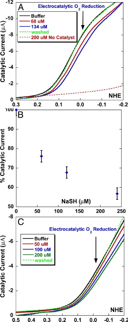
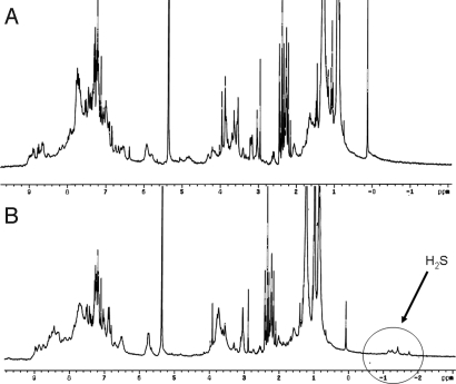
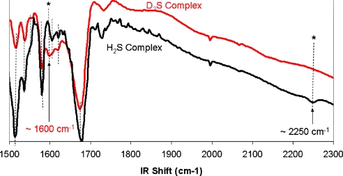
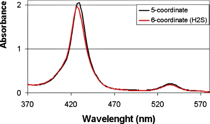
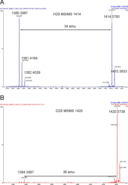
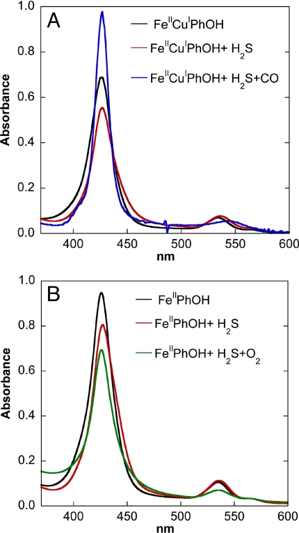
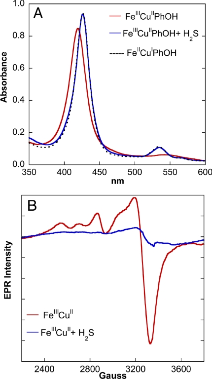
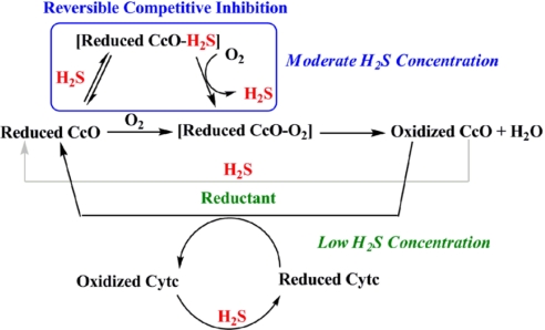
The toxic gas H(2)S is produced by enzymes in the body. At moderate concentrations, H(2)S elicits physiological effects similar to hibernation. Herein, we describe experiments that imply that the phenomenon probably results from reversible inhibition of the enzyme cytochrome c oxidase (CcO), which reduces oxygen during respiration. A functional model of the oxygen-reducing site in CcO was used to explore the effects of H(2)S during respiration. Spectroscopic analyses showed that the model binds two molecules of H2S. The electro-catalytic reduction of oxygen is reversibly inhibited by H(2)S concentrations similar to those that induce hibernation. This phenomenon derives from a weak, reversible binding of H(2)S to the Fe(II) porphyrin, which mimics heme a(3) in CcO's active site. No inhibition of CcO is detected at lower H(2)S concentrations. Nevertheless, at lower concentrations, H(2)S could have other biological effects on CcO. For example, H(2)S rapidly reduces Fe(III) and Cu(II) in both the oxidized form of this functional model and in CcO itself. H(2)S also reduces CcO's biological reductant, cytochrome c, which normally derives its reducing equivalents from food metabolism. Consequently, it is speculated that H(2)S might also serve as a source of electrons during periods of hibernation when food supplies are low.


Conflict of interest statement

The authors declare no conflict of interest.

Figures

Fig. 1.
Fig. 1.
Active site structures. CcO (A) and synthetic model (B) of CcO (Fe Cu and phenol analogue).
Fig. 2.
Fig. 2.
Schematic representation of the FeCuPhOH catalyst “clicked” onto an Au electrode (white) with Pt ring (gray) around it. “Cat” refers to the catalyst (Fig. 1B), excluding the terminal alkyne moiety.
Fig. 3.
Fig. 3.
The electrocatalytic reduction of oxygen is reversibly inhibited by H2S. (A) A linear sweep voltammogram of the FeCuPhOH catalyst modified electrode showing catalytic O2 reduction current under fast electron flux in the absence of NaSH (black), in the presence of 68 μM NaSH (red), 134 μM NaSH (blue), after removal of NaSH (dotted green), and in 200 μM NaSH in absence of any catalyst (dotted red). (B) Plot of percent catalytic current vs. NaSH concentration. (C) Linear sweep voltammogram of the FeCuPhOH catalyst modified electrode showing catalytic O2 reduction current under slow (rate determining) electron flux in the absence of NaSH (black), in the presence of 50 μM NaSH (red), 100 μM NaSH (blue), 200 μM NaSH (green, and afterremoval of NaSH (dotted green) .
Fig. 4.
Fig. 4.
1H-NMR spectrum (recorded in CD2Cl2 at −50 °C, 300 MHz) of FeII complexes (14 mM) before A and after B exposure to H2S.
Fig. 5.
Fig. 5.
Infrared spectrum of the H2S- and D2S-complexes (in black and red, respectively) on KBr pellets or in solution in dichloromethane in a KBr cell (10 mM).
Fig. 6.
Fig. 6.
UV/Vis spectrum of a 18 μM Fe(II) complex before reaction with H2S (dry, 5-coordinate, 430 nm in black) and after reaction (6-coordinate, 427 nm in red).
Fig. 7.
Fig. 7.
MSMS analysis on the most aboundant isotopic peak in the high-resolution nano-spray mass spectrum of the Fe(II) H2S (A, Calcd: 1414.3771, Found: 1414.3793) and D2S complexes (B, Calcd: 1420.4148, Found: 1420.3739), respectively. For this peak
Fig. 8.
Fig. 8.
The reactivity of Fe(II) H2S complexes with CO and O2 monitored by UV-Vis spectroscopy. (A) UV–Vis spectra of reduced 5 μM FeIICuIPhOH (black), reduced FeIICuIPhOH + 0.5 mM NaSH solution (red), and reduced FeIIPhOH + 0.5 mM NaSH solution + excess CO gas (blue). (B) Reduced 8 μM FeIIPhOH (black), reduced FeIIPhOH + 80 μM NaSH solution (red), and reduced FeIIPhOH + 80 μM NaSH solution + excess O2 gas (green).
Fig. 9.
Fig. 9.
The reactivity of fully oxidized complexes with H2S monitored by UV-Vis and EPR spectroscopies. (A) UV–Vis spectra (in a 50:50 volume ratio of pH 7 aqueous buffer and acetonitrile) of 10 μM oxidized FeIIICuIIPhOH (red), oxidized FeIIICuIIPhOH + 70 μM NaSH solution (blue) overlaid with a reduced FeIICuIPhOH spectrum (dotted black). (B) EPR spectra of 1 mM oxidized FeIIICuII (red) in THF and oxidized FeIIICuII + 1 mM (t-Bu4N)SH solution (blue) at 77 K.
Scheme 1.
Scheme 1.
Schematic representation of the possible roles of H2S affecting CcO activity. The blue box indicates that H2S binds to the reduced CcO active site, but can be subsequently replaced by O2. This inhibition should occur at moderate H2S concentrations. At lower concentrations, H2S does not compete with the substrate O2 but can still reduce CcO's active site and/or cytochrome c (Cytc) during catalytic O2 reduction.

Similar articles

Cited by

References

    1. Collman JP, Gagne RR, Reed CA. A paramagnetic dioxygen complex of iron II derived from a picket fence porphyrin further models for hemoproteins. J Am Chem Soc. 1974;96:2629–2631. - PubMed
    1. Collman JP, Gagne RR, Halbert TR, Marchon JC, Reed CA. Reversible oxygen adduct formation in ferrous complexes derived from a picket fence porphyrin model for oxy myoglobin. J Am Chem Soc. 1973;95:7868–7870. - PubMed
    1. Collman JP, Brauman JI, Rose E, Suslick KS. Cooperativity in O2 binding to iron porphyrins. Proc Natl Acad Sci USA. 1978;75:1052–1055. - PMC - PubMed
    1. Collman JP, et al. Picket-Fence Porphyrins. Synthetic Models for Oxygen Binding Hemoproteins. J Am Chem Soc. 1975;97:1427–1439. - PubMed
    1. Collman JP, Brauman JI, Suslick KS. Oxygen binding to iron porphyrins. J Am Chem Soc. 1975;97:7185–7186. - PubMed

Publication types

MeSH terms

LinkOut - more resources