. 2009 Dec 22;106(51):21608-13.
doi: 10.1073/pnas.0912101106. Epub 2009 Dec 9.

Structural analysis of the catalytically inactive kinase domain of the human EGF receptor 3

Affiliations

Structural analysis of the catalytically inactive kinase domain of the human EGF receptor 3

Natalia Jura et al. Proc Natl Acad Sci U S A. .

Abstract

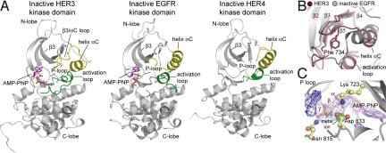
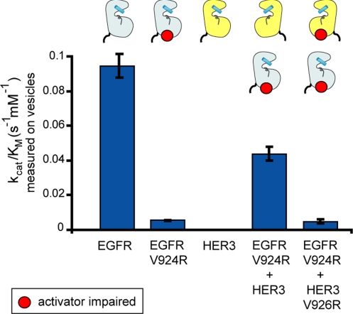
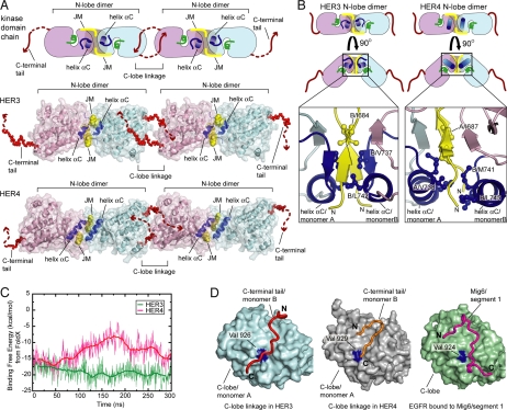
The kinase domain of human epidermal growth factor receptor (HER) 3/ErbB3, a member of the EGF receptor (EGFR) family, lacks several residues that are critical for catalysis. Because catalytic activity in EGFR family members is switched on by an allosteric interaction between kinase domains in an asymmetric kinase domain dimer, HER3 might be specialized to serve as an activator of other EGFR family members. We have determined the crystal structure of the HER3 kinase domain and show that it appears to be locked into an inactive conformation that resembles that of EGFR and HER4. Although the crystal structure shows that the HER3 kinase domain binds ATP, we confirm that it is catalytically inactive but can serve as an activator of the EGFR kinase domain. The HER3 kinase domain forms a dimer in the crystal, mediated by hydrophobic contacts between the N-terminal lobes of the kinase domains. This N-lobe dimer closely resembles a dimer formed by inactive HER4 kinase domains in crystal structures determined previously, and molecular dynamics simulations suggest that the HER3 and HER4 N-lobe dimers are stable. The kinase domains of HER3 and HER4 form similar chains in their respective crystal lattices, in which N-lobe dimers are linked together by reciprocal exchange of C-terminal tails. The conservation of this tiling pattern in HER3 and HER4, which is the closest evolutionary homolog of HER3, might represent a general mechanism by which this branch of the HER receptors restricts ligand-independent formation of active heterodimers with other members of the EGFR family.


Conflict of interest statement

The authors declare no conflict of interest.

Figures

Fig. 1.
Fig. 1.
(A) Schematic diagram of EGFR domain structure and the ligand-dependent activation process. (B) Multiple sequence alignment of the portions of kinase domains of EGFR family members. The residues that are identical in all four sequences are highlighted in gray, with sequence alterations unique to HER3 highlighted in orange. Residues that are part of the receiver interface are marked by dark blue stars, residues at the activator interface are marked by pink dots, and residues participating in the binding of the JM-B segment are marked by green triangles.
Fig. 2.
Fig. 2.
Crystal structure of the HER3 kinase domain. (A) Structure of the HER3 kinase domain in complex with AMP-PNP is compared with the crystal structure of the inactive EGFR kinase domain in complex with AMP-PNP (PDB ID code 2GS7) and the inactive HER4 kinase domain (PDB ID code 3BBW). (B) Detailed view of helix αC in the structure of HER3 (purple) and inactive EGFR (gray). (C) Detailed view of the ATP-binding site in the HER3 kinase domain, showing the electron density (at 3.5 σ above the mean value) around the AMP-PNP molecule and the metal ion. The difference electron density maps shown were calculated using a model of the protein at a stage before the inclusion of the nucleotide in the refinement.
Fig. 3.
Fig. 3.
HER3 is catalytically impaired as a receiver kinase but can function as an activator kinase. Catalytic efficiency (kcat/KM) of the kinase constructs (residues 672–998 in EGFR and residues 674–1,001 in HER3) on vesicles is measured using the continuous coupled-kinase assay. The values of kcat/KM were obtained from the linear dependence of reaction velocity on substrate concentration at a low substrate concentration, and the error bars are derived from the linear fit (7).
Fig. 4.
Fig. 4.
Analysis of the concatenated HER3 and HER4 kinase domains in the crystal lattices. (A) Pattern of kinase monomer interactions observed in the crystal lattices of the kinase domains of HER3 and inactive HER4 (PDB ID code 3BBW). The C-terminal tail exchanging dimers are propagated through the N-lobe dimer interface. JM refers to portion of the juxtamembrane segment. (B) Detailed view of the HER3 and HER4 N-lobe dimers. Hydrophobic residues are shown in stick representation. (C) Empirical estimates of binding free energy of HER3 and HER4 N-lobe homodimers using conformations obtained from all-atom molecular dynamics simulations. (D) Comparison between the C-terminal tail interaction with the C-lobe in the HER3 kinase domain dimer and the HER4 kinase domain dimer (PDB ID code 3BBW) and interaction of Mig6/segment 1 with the C-lobe of the EGFR kinase domain (PDB ID code 2RFE).
Fig. 5.
Fig. 5.
Model for oligomerization of the kinase domains of HER3 and inactive HER4 at the plasma membrane. (A) Flexible C-terminal tail/C-lobe interaction creates a possibility for the formation of branched HER3 or inactive HER4 oligomers, resulting in a 2D mesh. (B) Empirical estimates of binding free energy of the HER3/HER4 N-lobe homodimer, calculated using conformations obtained from an all-atom molecular dynamics simulation.

Similar articles

Cited by

References

    1. Yarden Y, Sliwkowski MX. Untangling the ErbB signalling network. Nat Rev Mol Cell Biol. 2001;2:127–137. - PubMed
    1. Guy PM, Platko JV, Cantley LC, Cerione RA, Carraway KL., 3rd Insect cell-expressed p180erbB3 possesses an impaired tyrosine kinase activity. Proc Natl Acad Sci USA. 1994;91:8132–8136. - PMC - PubMed
    1. Wieduwilt MJ, Moasser MM. The epidermal growth factor receptor family: Biology driving targeted therapeutics. Cell Mol Life Sci. 2008;65:1566–1584. - PMC - PubMed
    1. Baselga J, Swain SM. Novel anticancer targets: Revisiting ERBB2 and discovering ERBB3. Nat Rev Cancer. 2009;9:463–475. - PubMed
    1. Soltoff SP, Carraway KL, 3rd, Prigent SA, Gullick WG, Cantley LC. ErbB3 is involved in activation of phosphatidylinositol 3-kinase by epidermal growth factor. Mol Cell Biol. 1994;14:3550–3558. - PMC - PubMed

Publication types

Associated data