Comparative Study
. 2010 Mar;82(3):617-27.
doi: 10.1095/biolreprod.109.080408. Epub 2009 Dec 9.

Development and characterization of a three-dimensional organotypic human vaginal epithelial cell model

Affiliations
Comparative Study

Development and characterization of a three-dimensional organotypic human vaginal epithelial cell model

Brooke E Hjelm et al. Biol Reprod. 2010 Mar.

Abstract

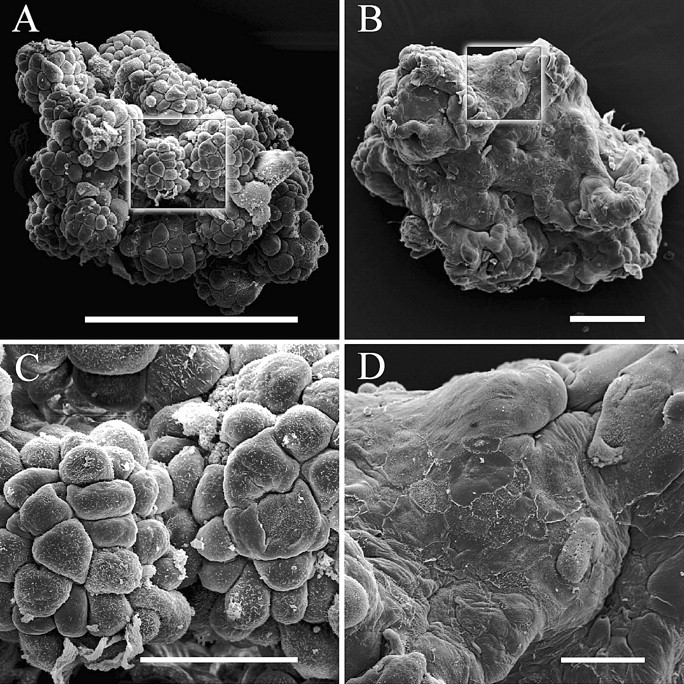
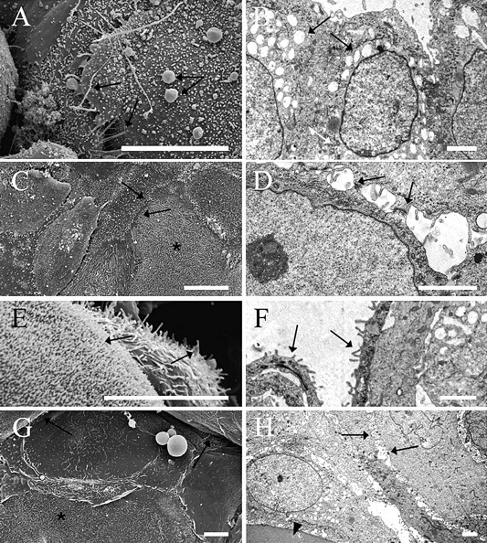
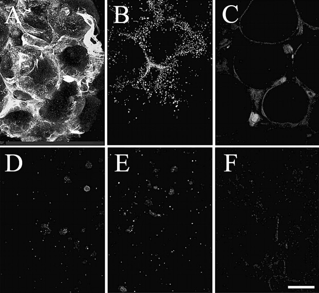
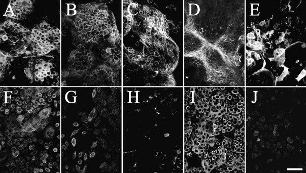
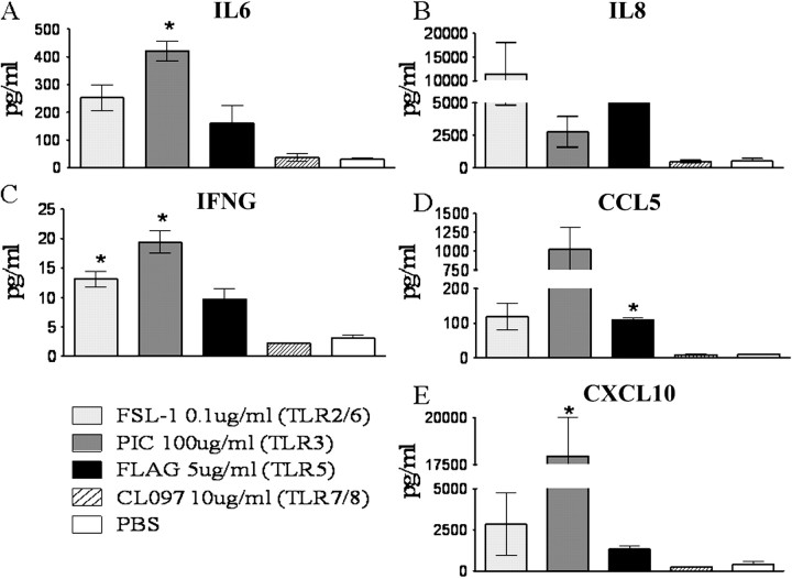
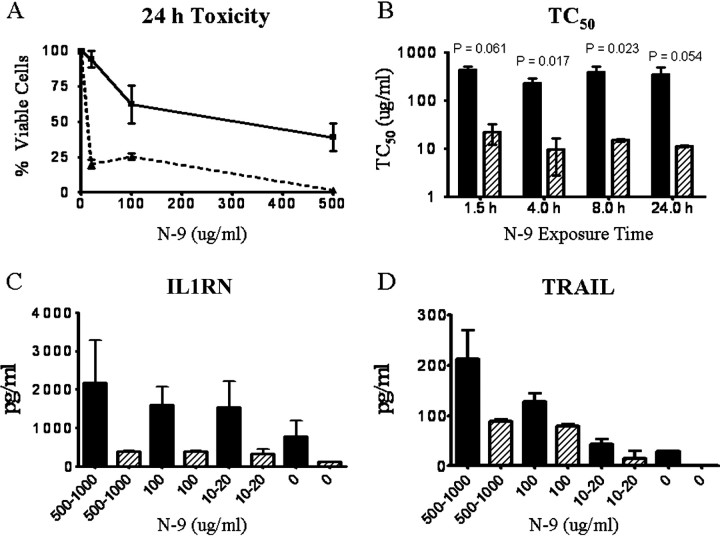
We have developed an in vitro human vaginal epithelial cell (EC) model using the innovative rotating wall vessel (RWV) bioreactor technology that recapitulates in vivo structural and functional properties, including a stratified squamous epithelium with microvilli, tight junctions, microfolds, and mucus. This three-dimensional (3-D) vaginal model provides a platform for high-throughput toxicity testing of candidate microbicides targeted to combat sexually transmitted infections, effectively complementing and extending existing testing systems such as surgical explants or animal models. Vaginal ECs were grown on porous, collagen-coated microcarrier beads in a rotating, low fluid-shear environment; use of RWV bioreactor technology generated 3-D vaginal EC aggregates. Immunofluorescence and scanning and transmission electron microscopy confirmed differentiation and polarization of the 3-D EC aggregates among multiple cell layers and identified ultrastructural features important for nutrient absorption, cell-cell interactions, and pathogen defense. After treatment with a variety of toll-like receptor (TLR) agonists, cytokine production was quantified by cytometric bead array, confirming that TLRs 2, 3, 5, and 6 were expressed and functional. The 3-D vaginal aggregates were more resistant to nonoxynol-9 (N-9), a contraceptive and previous microbicide candidate, when compared to two-dimensional monolayers of the same cell line. A dose-dependent production of tumor necrosis factor-related apoptosis-inducing ligand and interleukin-1 receptor antagonist, biomarkers of cervicovaginal inflammation, correlated to microbicide toxicity in the 3-D model following N-9 treatment. These results indicate that this 3-D vaginal model could be used as a complementary tool for screening microbicide compounds for safety and efficacy, thus improving success in clinical trials.


Figures

Fig. 1
Fig. 1
Scanning electron microscopy of early and late development of 3-D organotypic vaginal epithelial cell (EC) model. A) Low magnification SEM of early development (Days 19–21) displaying single layer of vaginal EC. B) Low magnification SEM of late development (Days 39–42) displaying multiple, flattened cell layers typical of stratified squamous epithelia. C) High magnification image from A (boxed area). D) High magnification image from B (boxed area). Bars = 200 μm (A and B) and 50 μm (C and D).
Fig. 2
Fig. 2
Scanning electron microscopy (left panel) and transmission electron microscopy (right panel) of physiologically relevant ultrastructural features observed in 3-D organotypic vaginal epithelial cell (EC) model. Arrows indicate extracellular mucus vesicles and string-like secretions (A), intracellular secretory vesicles (B), tightly adjoining cells on apical surface (C), internal cells with prominent hemidesmosomes (D), short, stubby, and long protruding microvilli on adjacent cells (E), microvilli on apical cell surface (F), microfolds (i.e., rugae) (G), and intercellular space with residual microvilli (or cytoplasmic processes) (H). Asterisks in C and G indicate cells densely covered with microridges. Arrowhead in H indicates microcarrier bead. Bars = 20 μm (A, C, E, G) and 2 μm (B, D, F, H).
Fig. 3
Fig. 3
Mucin production is highly localized and specific in the 3-D vaginal EC model. Immunofluorescence of mucin glycoproteins imaged by laser scanning confocal microscopy. Z-Stacks were reconstructed (optical sections: 1–2 μm, 50–100 sections) demonstrating protein expression levels and localization patterns in the 3-D organotypic vaginal EC model (top panel) compared to confluent monolayer cultures of the same cell line (bottom panel). MUC1 (A, D), MUC4 (B, E), and MUC5AC (C, F). Bar = 100 μm.
Fig. 4
Fig. 4
The 3-D vaginal aggregates demonstrate more specific staining of junctional differentiation and protein markers compared to monolayer counterparts. Immunofluorescence (optical sections: 1–2 μm, 50–100 sections) of markers were imaged by laser scanning confocal microscopy, demonstrating protein expression levels and localization patterns in the 3-D organotypic vaginal EC model (top panel) compared to confluent monolayer cultures of the same cell line (bottom panel). TJP1 (A, F), E-cadherin (B, G), ESA (C, H), CD1D (D, I), and involucrin (E, J). Bar = 100 μm.
Fig. 5
Fig. 5
Cytokine production in response to toll-like receptor (TLR) agonist stimulation in the 3-D vaginal EC model. Cytokines were quantified by cytometric bead array 24 h poststimulation of 3-D organotypic vaginal EC aggregates with FSL-1, PIC, FLAG, and CL097. IL6 (A), IL8 (B), IFNG (C), CCL5 (RANTES; D), and CXCL10 (IP-10; E). *P < 0.05; one-tailed t-test compared to the PBS group. Error bars show the standard error of the mean.
Fig. 6
Fig. 6
Validation of the 3-D vaginal EC model for microbicide evaluation. A) Nonoxynol-9 dose-dependent viability curve 24 h posttreatment of 3-D organotypic vaginal EC model compared to confluent monolayer cultures of the same cell line. B) Nonoxynol-9 TC50 levels of 3-D model and confluent monolayers at 1.5, 4, 8, and 24 h posttreatment. TC50 levels were calculated from two independent experiments. C) Nonoxynol-9 dose-dependant production of cytokine IL1RN 24 h posttreatment of 3-D model compared to confluent monolayers. D) Nonoxynol-9 dose-dependent production of cytokine TRAIL 24 h posttreatment of 3-D model compared to confluent monolayers. Solid lines and bars represent the 3-D vaginal EC model, whereas dashed lines and hatched bars represent confluent monolayers of vaginal EC. Error bars show the standard error of the means.

Similar articles

Cited by

References

    1. Herbst-Kralovetz MM, Quayle AJ, Ficarra M, Greene S, Rose WA II, Chesson R, Spagnuolo RA, Pyles RB. Quantification and comparison of toll-like receptor expression and responsiveness in primary and immortalized human female lower genital tract epithelia. Am J Reprod Immunol 2008; 59:212–224. - PubMed
    1. Quayle AJ. The innate and early immune response to pathogen challenge in the female genital tract and the pivotal role of epithelial cells. J Reprod Immunol 2002; 57:61–79. - PubMed
    1. Fichorova RN, Rheinwald JG, Anderson DJ. Generation of papillomavirus-immortalized cell lines from normal human ectocervical, endocervical, and vaginal epithelium that maintains expression of tissue-specific differentiation proteins. Biol Reprod 1997; 57:847–855. - PubMed
    1. DeSouza MM, Lagow E, Carson DD. Mucin functions and expression in mammalian reproductive tract tissues. Biochem Biophys Res Comm 1998; 247:1–6. - PubMed
    1. Fedele L, Bianchi S, Berlanda N, Fontana E, Raffaelli R, Bulfoni A, Braidotti P. Neovaginal mucosa after Vecchietti's laparoscopic operation for Rokitansky syndrome: structural and ultrastructural study. Am J Obstet Gynecol 2006; 195:56–61. - PubMed

Publication types

MeSH terms