. 2010 Feb;152(2):821-36.
doi: 10.1104/pp.109.146183. Epub 2009 Dec 9.

SEUSS and SEUSS-LIKE transcriptional adaptors regulate floral and embryonic development in Arabidopsis

Affiliations

SEUSS and SEUSS-LIKE transcriptional adaptors regulate floral and embryonic development in Arabidopsis

Fang Bao et al. Plant Physiol. 2010 Feb.

Abstract

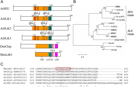
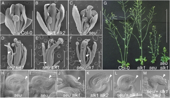
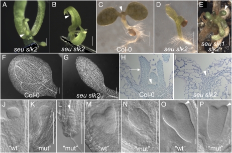
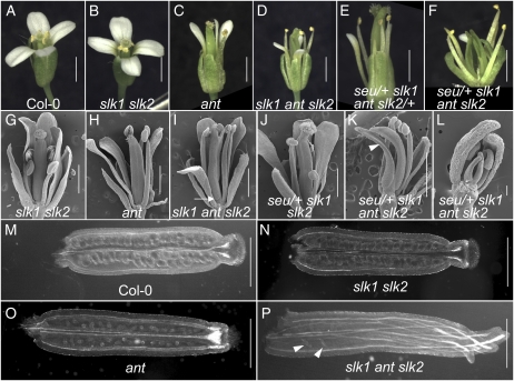
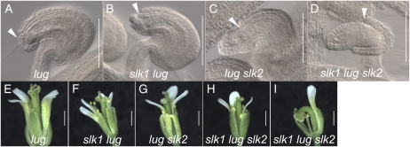
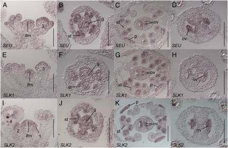
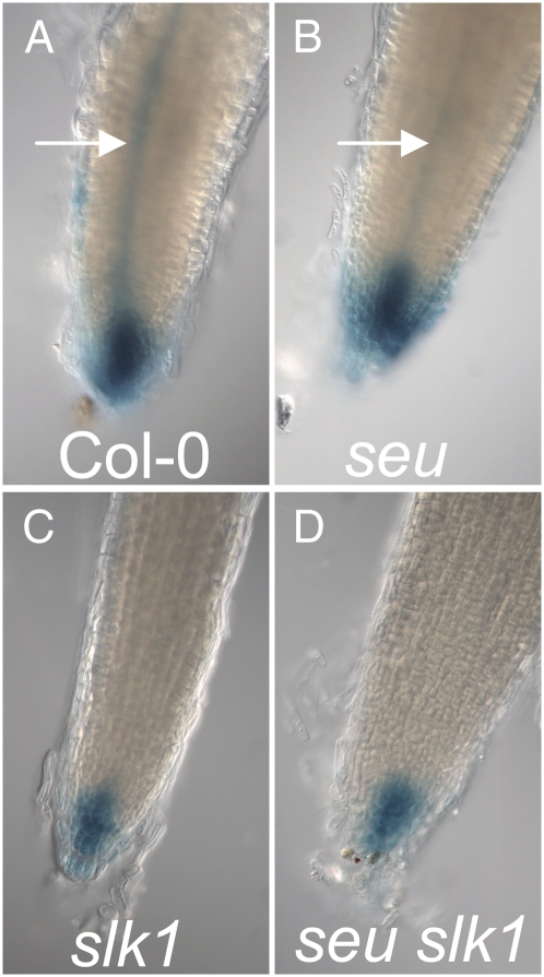
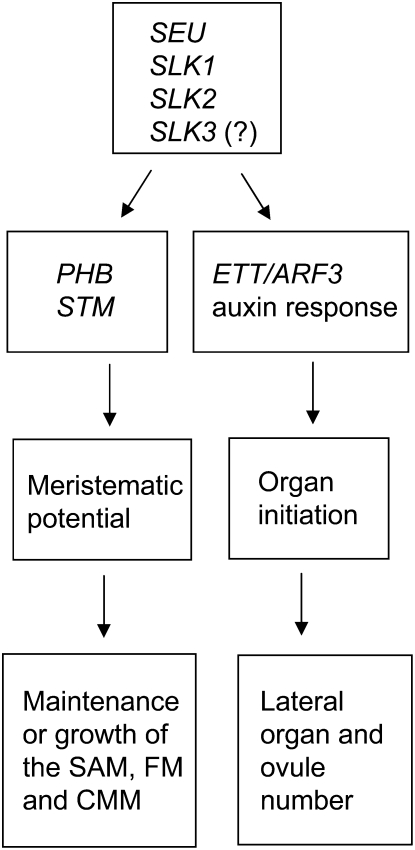
Multimeric protein complexes are required during development to regulate transcription and orchestrate cellular proliferation and differentiation. The Arabidopsis (Arabidopsis thaliana) SEUSS (SEU) gene encodes a transcriptional adaptor that shares sequence similarity with metazoan Lim domain-binding transcriptional adaptors. In Arabidopsis, SEU forms a physical complex with the LEUNIG transcriptional coregulator. This complex regulates a number of diverse developmental events, including proper specification of floral organ identity and number and the development of female reproductive tissues derived from the carpel margin meristem. In addition to SEU, there are three Arabidopsis SEUSS-LIKE (SLK) genes that encode putative transcriptional adaptors. To determine the functions of the SLK genes and to investigate the degree of functional redundancy between SEU and SLK genes, we characterized available slk mutant lines in Arabidopsis. Here, we show that mutations in any single SLK gene failed to condition an obvious morphological abnormality. However, by generating higher order mutant plants, we uncovered a degree of redundancy between the SLK genes and between SLK genes and SEU. We report a novel role for SEU and the SLK genes during embryonic development and show that the concomitant loss of both SEU and SLK2 activities conditions severe embryonic and seedling defects characterized by a loss of the shoot apical meristem. Furthermore, we demonstrate that SLK gene function is required for proper development of vital female reproductive tissues derived from the carpel margin. We propose a model that posits that SEU and SLK genes support organ development from meristematic regions through two different pathways: one that facilitates auxin response and thus organ initiation and a second that sustains meristematic potential through the maintenance of SHOOTMERISTEM-LESS and PHABULOSA expression.


Figures

Figure 1.
Figure 1.
Structural and evolutionary relationships of SEU, SLK, and Ldb proteins. A, Conserved protein domains in AtSEU, AtSLK, and select metazoan Ldb proteins. The Arabidopsis proteins share sequence similarity to the DD of Ldb1 (orange) and the LCCD of Chip (teal). The LID domain in metazoan Ldb proteins (purple) does not appear to be conserved in the AtSEU and AtSLK proteins. Numbers indicate amino acid positions, and black dividing lines indicate the locations of exon/exon boundaries that are conserved in the Arabidopsis proteins. Mutant allele insertion sites are indicated for SLK genes. B, The evolutionary history of full-length SLK proteins inferred using the neighbor-joining method (Saitou and Nei, 1987). The optimal tree (shown) suggests that the AtSLK proteins fall into a clade that is distinct from that containing AtSEU. The percentages of bootstrap support are shown next to the branches (Felsenstein, 1985). Evolutionary distances are in units of number of amino acid substitutions per site. C, ClustalW2 analysis of the LCCD region of metazoan Ldb and Arabidopsis SLK proteins. LCCD spans amino acids 201 to 249 in MmLdb1 and 387 to 435 in DmChip; the red boxed area indicates 10 amino acids deleted from Ldb1 that specifically disrupt activity of LCCD (van Meyel et al., 2003). At, Arabidopsis thaliana; Am, Antirrhinum majus; Dm, Drosophila melanogaster; Mm, Mus musculus; NLS, nuclear localization signal; Os, Oryza sativa. Protein identifiers not listed above are as follows: MmLdb2, NP034828; Os06g0126000, NP_001056655; Os11g10060, ABA91996; Os11g10070, ABA91997; AmSEU1, AJ620907; AMSEU2, AJ620908; AmSEU3A, AJ620909; AmSEU3B, AJ620910.
Figure 2.
Figure 2.
SLK1 shares redundant function with SEU during gynoecium and ovule development. A to F, Scanning electron micrograph images of flowers of the indicated genotypes. Bars = 1 mm. Some organs from the front of the flower have been removed to image internal whorls. D to F show three seu slk1 double mutant flowers that display enhanced splitting of the gynoecial apices and narrow or filamentous petals (arrowheads). Arrows indicate the basal extent of the carpel valves that is shifted toward the apex of the gynoecium. G, Whole plant phenotypes of slk1, seu, and seu slk1. Bar = 10 cm. H to M, Cleared ovules of the indicated genotypes. The extent of outer integument development is indicated with arrowheads. The majority (67%) of seu mutant ovules (H; Table III) display a wild-type extent of outer integument development (arrowhead). A minority (33%) of seu mutant ovules display a reduction in outer integument development (I; Table III). J shows enhanced ovule defects in the seu slk1 double mutant. In K, the slk1 slk2 double mutant appears morphologically wild type. In L and M, the disruption of ovule development is enhanced in seu/+ slk1 slk2 gynoecia (M) relative to seu/+ slk1 gynoecia (L). [See online article for color version of this figure.]
Figure 3.
Figure 3.
Floral, seedling, and embryonic phenotypes of seu slk2 double mutants. A, Early-arising flower from seu slk2 “escaper” plant. The arrowhead indicates a reduced gynoecial mound in whorl 4. B, Late-arising flower from seu slk2 escaper plant. The arrowhead indicates carpelloid whorl 1 sepals. C to H, Seedlings at 5 d post germination. C, Wild-type (Col-0) seedling. The arrowhead indicates leaves initiating from the SAM. D, The seu slk2 double mutant displays narrow cotyledons and a lack of true leaf development. E, A seu slk1 slk2 triple mutant seedling that has escaped embryonic lethality displays bulbous and very reduced rosette leaves (arrowhead). F, Cleared wild-type (Col-0) cotyledon shows vascular loops. G, The seu slk2 double mutant displays a very narrow cotyledon with a single unbranched central vascular element. H, Longitudinal section of a Col-0 seedling. SAM (arrowhead) and rosette leaf (arrow) are indicated. I, SAM and rosette leaves are not detected in the seu slk2 seedling. The arrowhead indicates the expected location of the SAM if wild type. J to P, Embryos segregating from a slk2/slk2 seu/+ parental self-cross. Embryos were classified as morphologically wild type (wt) or mutant (mut). See text for details. J to L, Globular-stage sibling embryos displayed wild-type (J), weakly disrupted (K), or severely disrupted (L) morphologies. The arrowhead in L indicates a globular domain with reduced cell number. M and N, Heart-stage embryos. While cotyledon primordia are apparent in morphologically wild-type embryos (M), morphologically mutant sibling embryos (N) lack obvious cotyledon primordia. O and P, Reduced cotyledon development (arrowhead) is apparent at the torpedo stage in those embryos displaying mutant morphologies (compare P with O). Bars = 100 μm in A, E, F to I, and J to N, 200 μm in B, 1 mm in C and D, and 10 μm in O and P. [See online article for color version of this figure.]
Figure 4.
Figure 4.
SLK1 and SLK2 are required with ANT and SEU for ovule initiation from the CMM. A, Col-0 flower. B, Morphology of a slk1 slk2 mutant flower is near that of Col-0. C, ant mutant. D, Gynoecial disruptions are enhanced in slk1 ant slk2 triple mutant relative to the ant and slk1 slk2 mutants. E and F, More severe reductions of CMM-derived tissues are observed in seu/+ slk1 ant slk2 (F) relative to the seu/+ slk1 ant slk2/+ (E) genotype. G to L, Scanning electron micrographs of flowers from the indicated genotypes. In I, the arrow indicates the basal valve boundary. In K, the seu/+ slk1 ant slk2 genotype conditions complete loss of ovule primordia and severe reduction of CMM-derived tissues (arrowhead). L shows a stage 10 flower. Disruption of the CMM is already evident at this stage. M to P, Optical cross sections from cleared gynoecia of the indicated genotypes. Arrowheads in P indicate the few ovules that have initiated. Bars = 1 mm in A to K, 100 μm in L, and 0.5 mm in M to P. [See online article for color version of this figure.]
Figure 5.
Figure 5.
Mutations in SLK1 and SLK2 enhance the lug ovule and floral phenotypes. A to D, Cleared ovules from gynoecia of the indicated genotypes. Arrowheads indicate the extent of outer integument development. Bars = 100 μm. E to I, Floral phenotypes of the indicated genotypes. H and I show the two-carpel and one-carpel phenotypes observed in the slk1 lug slk2 triple mutant flowers, respectively. The one-carpel gynoecia are often curved and bent over. Bars = 1 mm. [See online article for color version of this figure.]
Figure 6.
Figure 6.
SEU, SLK1, and SLK2 are expressed in young flower meristems, ovules, and the CMM. In situ hybridization on wild-type (Col-0) tissue with a SEU (A–D), SLK1 (E–H), or SLK2 (I–L) antisense probe (A–E and G–H). A, E, and I, Inflorescence longitudinal sections. Hybridization signal is detected in the inflorescence meristem (ifm) and throughout stage 1, 2, and early 3 floral primordia. Numbers indicate floral stages (Smyth et al., 1990). B, F, and J, Stage 6 floral cross section. Expression is detected throughout gynoecial mound (g) and stamen (st) primordia. C, G, and K, Stage 9 or early 10 floral cross sections. Expression is detected in ovules (ov) and petals (p). Within the stamens, expression is detected most strongly in the locules, pollen mother cells, and the tapetum. D, H, and L, Gynoecial cross sections show expression throughout the ovules at floral stages 10 or 11. Bars = 100 μm.
Figure 7.
Figure 7.
The seu, slk1, and seu slk1 mutants display reduced DR5-GUS activity. DR5:GUS reporter activity is shown in primary root apices of the indicated genotypes. Arrows in A and B indicate stele (root vasculature). [See online article for color version of this figure.]
Figure 8.
Figure 8.
Model of functional roles of SEU and SLK genes in the SAM and CMM. In this model, we propose that the SEU, SLK1, SLK2, and possibly SLK3 genes support the development of organs from the SAM, the floral meristem (FM), and the CMM through two gene regulatory events (right and left sides). SEU and the SLK genes support auxin responses that are required for organ initiation events in the CMM, floral meristem, and embryonic SAM. Additionally, SEU and SLK genes support the maintenance or growth of these meristematic regions by enabling the expression of PHB and STM.

Similar articles

Cited by

References

    1. Abel S, Oeller PW, Theologis A (1994) Early auxin-induced genes encode short-lived nuclear proteins. Proc Natl Acad Sci USA 91 326–330 - PMC - PubMed
    1. Agulnick AD, Taira M, Breen JJ, Tanaka T, Dawid IB, Westphal H (1996) Interactions of the LIM-domain-binding factor Ldb1 with LIM homeodomain proteins. Nature 384 270–272 - PubMed
    1. Alonso JM, Stepanova AN, Leisse TJ, Kim CJ, Chen H, Shinn P, Stevenson DK, Zimmerman J, Barajas P, Cheuk R, et al (2003) Genome-wide insertional mutagenesis of Arabidopsis thaliana. Science 301 653–657 - PubMed
    1. Azhakanandam S, Nole-Wilson S, Bao F, Franks RG (2008) SEUSS and AINTEGUMENTA mediate patterning and ovule initiation during gynoecium medial domain development. Plant Physiol 146 1165–1181 - PMC - PubMed
    1. Benkova E, Michniewicz M, Sauer M, Teichmann T, Seifertova D, Jurgens G, Friml J (2003) Local, efflux-dependent auxin gradients as a common module for plant organ formation. Cell 115 591–602 - PubMed

Publication types

MeSH terms