. 2010 Mar;298(3):C465-76.
doi: 10.1152/ajpcell.00449.2009. Epub 2009 Dec 9.

High concentrations of HGF inhibit skeletal muscle satellite cell proliferation in vitro by inducing expression of myostatin: a possible mechanism for reestablishing satellite cell quiescence in vivo

Affiliations

High concentrations of HGF inhibit skeletal muscle satellite cell proliferation in vitro by inducing expression of myostatin: a possible mechanism for reestablishing satellite cell quiescence in vivo

Michiko Yamada et al. Am J Physiol Cell Physiol. 2010 Mar.

Abstract

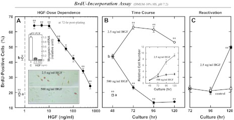
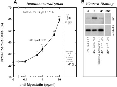
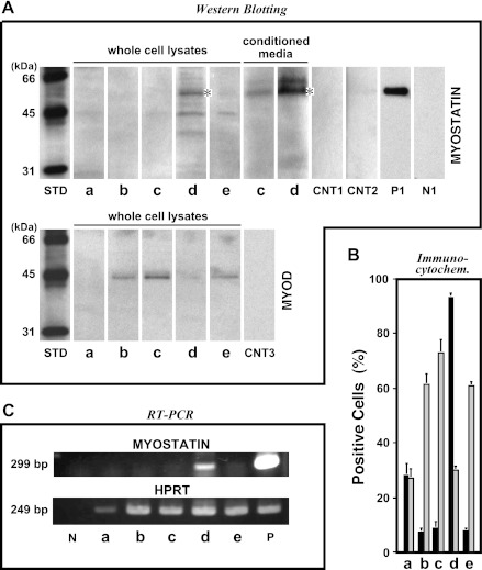
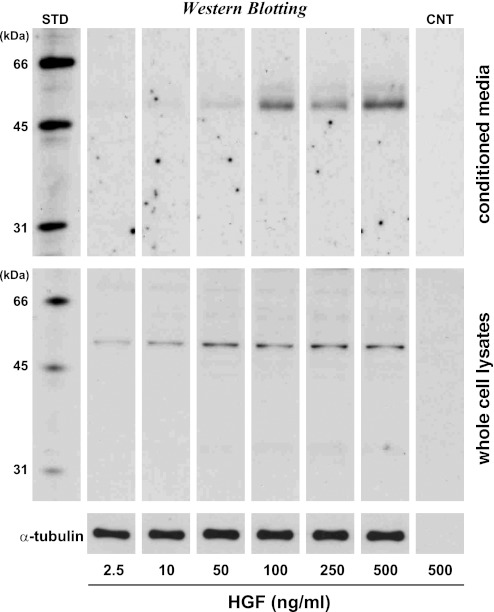
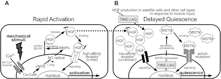
Skeletal muscle regeneration and work-induced hypertrophy rely on molecular events responsible for activation and quiescence of resident myogenic stem cells, satellite cells. Recent studies demonstrated that hepatocyte growth factor (HGF) triggers activation and entry into the cell cycle in response to mechanical perturbation, and that subsequent expression of myostatin may signal a return to cell quiescence. However, mechanisms responsible for coordinating expression of myostatin after an appropriate time lag following activation and proliferation are not clear. Here we address the possible role of HGF in quiescence through its concentration-dependent negative-feedback mechanism following satellite cell activation and proliferation. When activated/proliferating satellite cell cultures were treated for 24 h beginning 48-h postplating with 10-500 ng/ml HGF, the percentage of bromodeoxyuridine-incorporating cells decreased down to a baseline level comparable to 24-h control cultures in a HGF dose-dependent manner. The high level HGF treatment did not impair the cell viability and differentiation levels, and cells could be reactivated by lowering HGF concentrations to 2.5 ng/ml, a concentration that has been shown to optimally stimulate activation of satellite cells in culture. Coaddition of antimyostatin neutralizing antibody could prevent deactivation and abolish upregulation of cyclin-dependent kinase (Cdk) inhibitor p21. Myostatin mRNA expression was upregulated with high concentrations of HGF, as demonstrated by RT-PCR, and enhanced myostatin protein expression and secretion were revealed by Western blots of the cell lysates and conditioned media. These results indicate that HGF could induce satellite cell quiescence by stimulating myostatin expression. The HGF concentration required (over 10-50 ng/ml), however, is much higher than that for activation, which is initiated by rapid release of HGF from its extracellular association. Considering that HGF is produced by satellite cells and spleen and liver cells in response to muscle damage, local concentrations of HGF bathing satellite cells may reach a threshold sufficient to induce myostatin expression. This time lag may delay action of the quiescence signaling program in proliferating satellite cells during initial phases of muscle regeneration followed by induction of quiescence in a subset of cells during later phases.


Figures

Fig. 1.
Fig. 1.
High-concentration hepatocyte growth factor (HGF) treatments reduce the proliferation activity of satellite cells in cultures. Rat satellite cells were stimulated for activation for 24 h from 24- to 48-h postplating by 2.5 ng/ml human recombinant HGF in DMEM-10% normal horse serum (HS) (b), then incubated with higher concentrations of HGF for the next 72-h period followed by 5-bromo-2′-deoxyuridine (BrdU)-incorporation assay at 24-h intervals of time. A: HGF dose dependence monitored at 72-h postplating by the proliferation index decreasing down to a baseline level comparable to the 24-h control culture not receiving 2.5 ng/ml HGF (a). Typical immunomicrographs are shown in bottom inset with positive (brown) and negative cells. Cell lysates of companion cultures were analyzed for the mRNA expression of a differentiation marker myogenin at 72-h postplating by real-time quantitative (q)RT-PCR run under the TaqMan Probe assay standardized with hypoxanthine guanine phosphoribosyl transferase (HPRT; top inset); bar C, control without HGF; bars HGF, with 2.5 and 500 ng/ml HGF. B: time courses of the BrdU-incorporation activity and the relative cell density (inset) in cultures treated with 2.5 ng/ml (○) and 500 ng/ml HGF (●) from 48- to 120-h postplating. C: reactivation activity of cells that received 500 ng/ml HGF for 24-h period from 48- to 72-h postplating and then maintained in the presence (■) or absence (□) of 2.5 ng/ml HGF in DMEM-10% HS for the next 48 h. Data points represent means ± SE for four cultures per treatment; significant differences from starting culture means (data point b in A and B and the 72-h data point in C) are indicated (*P < 0.05; **P < 0.01).
Fig. 2.
Fig. 2.
Myostatin mediates high-level HGF-induced deactivation of satellite cells. Satellite cell activation was stimulated by 2.5 ng/ml HGF as shown in Fig. 1. Cultures were then treated with 500 ng/ml HGF for the next 24 h from 48- to 72-h postplating in the presence of various concentrations of polyclonal antimyostatin neutralizing antibody (0.1–10 μg/ml, plotted on a log scale as the x-axis) and evaluated for the BrdU-incorporation activity at 72-h postplating (A). Experiments included the following control cultures: c, with 2.5 ng/ml HGF; d″, with 500 ng/ml HGF plus 10 μg/ml control immunoglobulin (goat IgG). Data points depict the means ± SE for four cultures per treatment; significant differences from the control culture mean (leftmost data point without neutralizing antibody) are indicated (*P < 0.05; **P < 0.01). B: companion cultures were analyzed for p21 expression by enhanced chemiluminescence (ECL)-Western blotting of cell lysates; α-tubulin protein levels were included to standardize the cell loading. CNT, control blots of 500 ng/ml HGF-treated cells without primary antibody and with secondary reagents.
Fig. 3.
Fig. 3.
High-level HGF stimulates myostatin expression and secretion in proliferating satellite cell cultures. Satellite cell cultures were activated by 2.5 ng/ml HGF as shown in Fig. 1 and incubated with 500 ng/ml HGF for the next 24 h from 48- to 72-h postplating. Cultures were evaluated for myostatin expression by ECL-Western blotting of whole cell lysates and conditioned media from a constant number of cells (A), indirect immunocytochemistry (B, solid bars, representing means ± SE), and RT-PCR standardized with internal HPRT (C). Counterpart expression levels of MyoD protein are included in A (bottom row) and B (light-grayed bars). STD, biotinylated molecular weight standards; a, culture before activation treatment at 24 h; b, 2.5 ng/ml HGF culture at 48 h; c, 2.5 ng/ml HGF culture at 72 h; d, 500 ng/ml HGF culture at 72 h; e, 2.5 ng/ml HGF reactivation culture at 120 h. CNT1, CNT2, and CNT3, control blots of the cell lysate (d), conditioned medium (d), and cell lysate (c) without primary antibody and with secondary reagents, respectively; P1, positive control [conditioned medium from human embryonic kidney (HEK)293 cells transfected with His-tagged myostatin-expressing plasmid]; N1, negative control [conditioned medium from HEK293 cells transfected with enhanced green fluorescent protein (EGFP)-expressing plasmid]; P, rat skeletal muscle cDNA; N, no template. *52-kDa pro-myostatin form.
Fig. 4.
Fig. 4.
HGF dose dependence of myostatin expression and secretion. Activated satellite cell cultures were incubated with 2.5–500 ng/ml HGF for 24 h from 48- to 72-h postplating as shown in Fig. 1. Myostatin expression and secretion were evaluated by ECL-Western blotting of whole cell lysates (middle row) and conditioned media (top row) standardized relative to α-tubulin protein levels (bottom row). STD, biotinylated molecular weight standards; CNT, control blots of 500 ng/ml HGF culture without primary antibody and with secondary reagents.
Fig. 5.
Fig. 5.
Delayed action model for satellite cell quiescence. HGF may play dual roles in cell activation and quiescence in a dose-dependent manner as follows. A: possible mechanism for the activation: quiescent satellite cells are activated to reenter the cell cycle in response to mechanical perturbation of muscle tissue through a molecular cascade of events including calcium ion influx from extracellular compartment through a stretch-activated (SA)-Ca2+ channel, calcium-calmodulin (Ca-CaM) formation, nitric oxide (NO) radical production by activated constitutive NO synthase (cNOS; neuronal NOS and endothelial NOS), matrix metalloproteinase (MMP) activation, rapid release of HGF from its extracellular tethering (possibly with associated extracellular segment of proteoglycans) to give rise to ng/ml level of the growth factor, and the subsequent presentation to the high-affinity receptor c-met to generate a signal for satellite cell activation. [From Tatsumi and Allen (87) and Tatsumi (90), with modification.] B: satellite cell quiescence through a negative feedback mechanism following satellite cell activation and proliferation. HGF synthesis is initiated in satellite cells and spleen and liver cells in response to muscle damage. Local concentrations of HGF bathing proliferating satellite cells reach a threshold after an appropriate time lag for activation and proliferation of satellite cells. HGF can bind to an unknown low-affinity receptor responsible for signaling myostatin expression. The myostatin protein (MSTN) is secreted and processed to the active form that associates with activin type IIAB receptors that signal satellite cell quiescence. l-Arg, l-arginine.

Comment in

Similar articles

Cited by

References

    1. Allen RE, Boxhorn LK. Regulation of skeletal muscle satellite cell proliferation and differentiation by transforming growth factor-beta, insulin-like growth factor I, and fibroblast growth factor. J Cell Physiol 138: 311–315, 1989 - PubMed
    1. Allen RE, Sheehan SM, Taylor RG, Kendall TL, Rice GM. Hepatocyte growth factor activates quiescent skeletal muscle satellite cells in vitro. J Cell Physiol 165: 307–312, 1995 - PubMed
    1. Allen RE, Temm-Grove CJ, Sheehan SM, Rice GM. Skeletal muscle satellite cell cultures. Methods Cell Biol 52: 155–176, 1997 - PubMed
    1. Allen RE, Goll DE. Cellular and developmental biology of skeletal muscle as related to muscle growth. In: Biology of Growth of Domestic Animals, edited by Scanes CG. Ames, IA: Iowa State Press, 2003, p. 148–169
    1. Amthor H, Otto A, Macharia R, McKinnell I, Patel K. Myostatin imposes reversible quiescence on embryonic muscle precursors. Dev Dyn 235: 672–680, 2006 - PubMed

Publication types

MeSH terms

LinkOut - more resources