

AF4 is a critical regulator of the IGF-1 signaling pathway during Purkinje cell development
- PMID: 20007461
- PMCID: PMC6666107
- DOI: 10.1523/JNEUROSCI.5188-09.2009
AF4 is a critical regulator of the IGF-1 signaling pathway during Purkinje cell development
Abstract
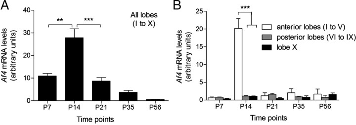
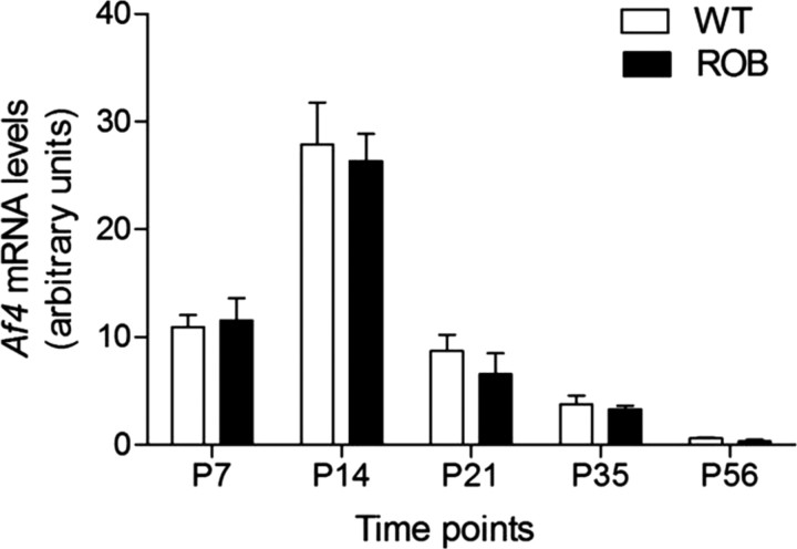
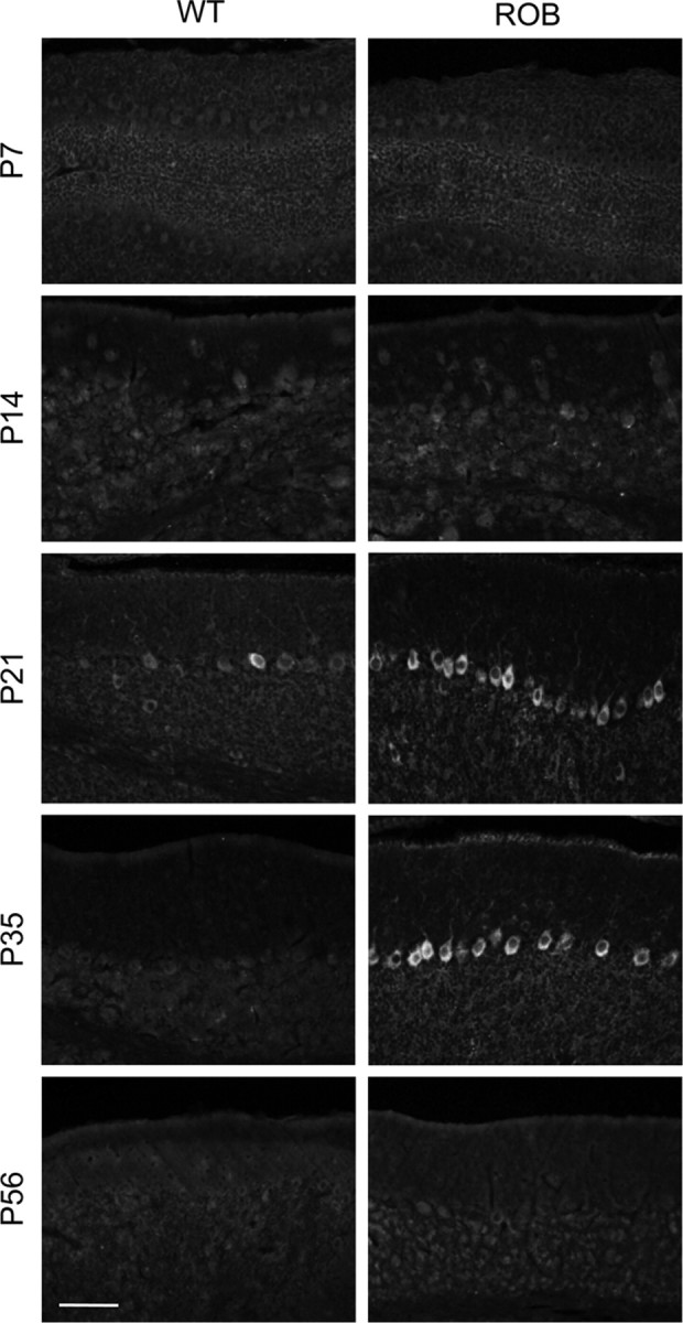
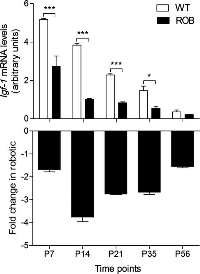
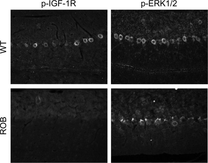
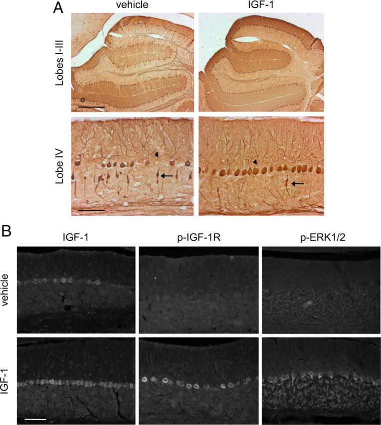
Deregulation of the insulin-like growth factor 1 (IGF-1) signaling pathway is a recurrent finding in mouse models and human patients with cerebellar ataxia and thus represents a common pathological cascade in neuronal cell death that may be targeted for therapy. We have previously identified a point mutation in AF4, a transcription cofactor of RNA polymerase II elongation and chromatin remodeling, that causes progressive and highly specific Purkinje cell (PC) death in the ataxic mouse mutant robotic, leading to the accumulation of AF4 in PCs. Here we confirm that the spatiotemporal pattern of PC degeneration in the robotic cerebellum correlates with the specific profile of AF4 upregulation. To identify the underlying molecular pathways, we performed microarray gene expression analysis of PCs obtained by laser capture microdissection (LCM) at the onset of degeneration. Igf-1 was significantly downregulated in robotic PCs compared with wild-type controls before and throughout the degenerative process. Consistently, we observed a decrease in the activation of downstream signaling molecules including type 1 IGF receptor (IGF-1R) and the extracellular signal-regulated kinase (ERK) 1 and ERK2. Chromatin immunoprecipitation confirmed that Igf-1 is a direct and the first validated target of the AF4 transcriptional regulatory complex, and treatment of presymptomatic robotic mice with IGF-1 indeed markedly delayed the progression of PC death. This study demonstrates that small changes in the levels of a single transcriptional cofactor can deleteriously affect normal cerebellum function and opens new avenues of research for the manipulation of the IGF-1 pathway in the treatment of cerebellar ataxia in humans.
Figures
Similar articles
-
The robotic mouse: unravelling the function of AF4 in the cerebellum.Cerebellum. 2005;4(4):250-60. doi: 10.1080/14734220500325897. Cerebellum. 2005. PMID: 16321881 Review.
-
The robotic mouse: understanding the role of AF4, a cofactor of transcriptional elongation and chromatin remodelling, in purkinje cell function.Cerebellum. 2009 Sep;8(3):175-83. doi: 10.1007/s12311-009-0101-0. Epub 2009 Apr 2. Cerebellum. 2009. PMID: 19340490 Review.
-
Fluoro-jade identification of cerebellar granule cell and purkinje cell death in the alpha1A calcium ion channel mutant mouse, leaner.Neuroscience. 2003;118(3):667-80. doi: 10.1016/s0306-4522(03)00019-8. Neuroscience. 2003. PMID: 12710975
-
Decreased IGF-I gene expression during the apoptosis of Purkinje cells in pcd mice.Brain Res Dev Brain Res. 1997 Feb 20;98(2):164-76. doi: 10.1016/s0165-3806(96)00168-x. Brain Res Dev Brain Res. 1997. PMID: 9051257
-
The mixed-lineage leukemia fusion partner AF4 stimulates RNA polymerase II transcriptional elongation and mediates coordinated chromatin remodeling.Hum Mol Genet. 2007 Jan 1;16(1):92-106. doi: 10.1093/hmg/ddl444. Epub 2006 Nov 29. Hum Mol Genet. 2007. PMID: 17135274
Cited by
-
Ischemia-induced autophagy contributes to neurodegeneration in cerebellar Purkinje cells in the developing rat brain and in primary cortical neurons in vitro.Biochim Biophys Acta. 2015 Sep;1852(9):1902-11. doi: 10.1016/j.bbadis.2015.06.007. Epub 2015 Jun 11. Biochim Biophys Acta. 2015. PMID: 26071643 Free PMC article.
-
Regulation of AMP-activated protein kinase signaling by AFF4 protein, member of AF4 (ALL1-fused gene from chromosome 4) family of transcription factors, in hypothalamic neurons.J Biol Chem. 2012 Jun 8;287(24):19985-96. doi: 10.1074/jbc.M112.367854. Epub 2012 Apr 23. J Biol Chem. 2012. PMID: 22528490 Free PMC article.
-
Insulin receptor in the brain: Mechanisms of activation and the role in the CNS pathology and treatment.CNS Neurosci Ther. 2018 Sep;24(9):763-774. doi: 10.1111/cns.12866. Epub 2018 Apr 24. CNS Neurosci Ther. 2018. PMID: 29691988 Free PMC article. Review.
-
Frataxin deficiency unveils cell-context dependent actions of insulin-like growth factor I on neurons.Mol Neurodegener. 2012 Oct 5;7:51. doi: 10.1186/1750-1326-7-51. Mol Neurodegener. 2012. PMID: 23039828 Free PMC article.
-
In Search of Molecular Markers for Cerebellar Neurons.Int J Mol Sci. 2021 Feb 12;22(4):1850. doi: 10.3390/ijms22041850. Int J Mol Sci. 2021. PMID: 33673348 Free PMC article. Review.
References
-
- Babu VR, Roberson JR, Van Dyke DL, Weiss L. Interstitial deletion of 4q35 in a familial satellited 4q in a child with developmental delay. Am J Hum Genet. 1987;41(Suppl):A113.
-
- Baker J, Liu JP, Robertson EJ, Efstratiadis A. Role of insulin-like growth factors in embryonic and postnatal growth. Cell. 1993;75:73–82. - PubMed
-
- Bale LK, Conover CA. Regulation of insulin-like growth factor binding protein-3 messenger ribonucleic acid expression by insulin-like growth factor I. Endocrinology. 1992;131:608–614. - PubMed
-
- Baskaran K, Erfurth F, Taborn G, Copeland NG, Gilbert DJ, Jenkins NA, Iannaccone PM, Domer PH. Cloning and developmental expression of the murine homolog of the acute leukemia proto-oncogene AF4. Oncogene. 1997;15:1967–1978. - PubMed
Publication types
MeSH terms
Substances
Grants and funding
LinkOut - more resources
Full Text Sources
Other Literature Sources
Miscellaneous