. 2009 Dec 9;29(49):15489-98.
doi: 10.1523/JNEUROSCI.3561-09.2009.

No Nogo66- and NgR-mediated inhibition of regenerating axons in the zebrafish optic nerve

Affiliations

No Nogo66- and NgR-mediated inhibition of regenerating axons in the zebrafish optic nerve

Houari Abdesselem et al. J Neurosci. .

Abstract

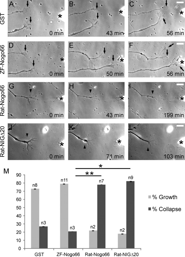
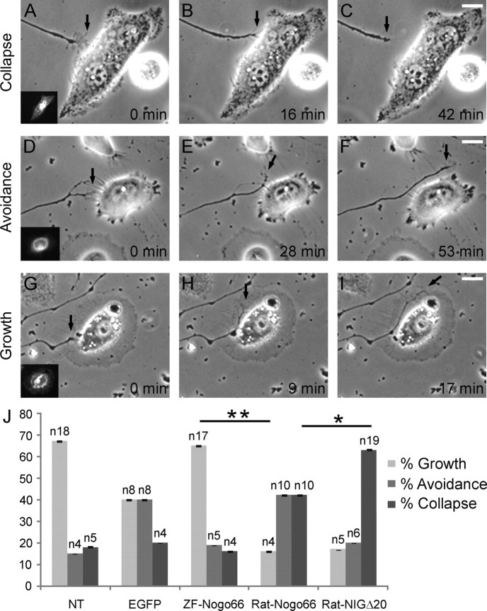
In contrast to mammals, lesioned axons in the zebrafish (ZF) optic nerve regenerate and restore vision. This correlates with the absence of the NogoA-specific N-terminal domains from the ZF nogo/rtn-4 (reticulon-4) gene that inhibits regeneration in mammals. However, mammalian nogo/rtn-4 carries a second inhibitory C-terminal domain, Nogo-66, being 70% identical with ZF-Nogo66. The present study examines, (1) whether ZF-Nogo66 is inhibitory and effecting similar signaling pathways upon Nogo66-binding to the Nogo66 receptor NgR and its coreceptors, and (2) whether Rat-Nogo66 on fish, and ZF-Nogo66 on mouse neurons, cause inhibition via NgR. Our results from "outgrowth, collapse and contact assays" suggest, surprisingly, that ZF-Nogo66 is growth-permissive for ZF and mouse neurons, quite in contrast to its Rat-Nogo66 homolog which inhibits growth. The opposite effects of ZF- and Rat-Nogo66 are, in both fish and mouse, transmitted by GPI (glycosylphosphatidylinositol)-anchored receptors, including NgR. The high degree of sequence homology in the predicted binding site is consistent with the ability of ZF- and mammalian-Nogo66 to bind to NgRs of both species. Yet, Rat-Nogo66 elicits phosphorylation of the downstream effector cofilin whereas ZF-Nogo66 has no influence on cofilin phosphorylation--probably because of significantly different Rat- versus ZF-Nogo66 sequences outside of the receptor-binding region effecting, by speculation, recruitment of a different set of coreceptors or microdomain association of NgR. Thus, not only was the NogoA-specific domain lost in fish, but Nogo66, the second inhibitory domain in mammals, and its signaling upon binding to NgR, was modified so that ZF-Nogo/RTN-4 does not impair axon regeneration.


Figures

Figure 1.
Figure 1.
ZF-Nogo66 expression in the ZF CNS. A, RT-PCR analysis on normal (NON) and regenerating zebrafish optic nerves (RON) shows expression of ZF-RTN4L mRNA. A reverse transcriptase negative control (Tr−) was performed with each sample. RT-PCR with GAPDH-specific primers (GAPDH) served as a loading control in each reaction. H2O, no template control. B, Western blot analysis with proteins from the ZF optic nerve (ON), brain (Br) and CNS myelin (M) and AB against ZF Nogo-66 which detects 36 kDa RTN4L in the optic nerve and brain, and 27 and 22 kDa bands in CNS myelin. C, ZF-Nogo66 AB on cross sections of the unlesioned zebrafish optic nerve showed staining (red) across the entire nerve and was enriched in fascicle boundaries (arrows) made by astrocytes. #, Territory of axons and glial cells in fascicles. The insert shows the entire nerve and the region from which the magnification was derived. D, Same section as in C, showing GFAP-staining (green) in addition to Nogo66-staining (red) which colocalize at fascicle boundaries and GFAP-positive processes (arrows). E, After optic nerve lesion, ZF-Nogo66 staining (red) was associated with patches and diffuse structures, probably myelin debris (*), some astrocyte processes (arrows), and with cells some of which may represent oligodendrocytes (GFPA-negative; arrowheads). The insert shows the entire nerve and the region from which the magnification was derived. F, Same section as in E, showing GFAP-staining (green) in addition to Nogo66-staining (red). Fascicle boundaries made up by astrocytes (arrows) and some astrocyte processes within fascicles are double-labeled. DAPI (blue) marks nuclei. Scale bars: 20 μm. G–I, Oligodendrocyte, isolated from the regenerating ZF optic nerve, is O4-positive (G, green) and Nogo66-negative (H) after staining on live cells but is stained by anti-ZF-Nogo66 AB (I, red) after fixation. Scale bars: 20 μm. J, Nogo-66 is associated with regenerating ZF RGC axons and growth cones after exposure to AB against ZF-Nogo66. Scale bars: 20 μm.
Figure 2.
Figure 2.
Axon outgrowth assay. Histogram showing the average number of ZF RGC axons per retina mini-explant after 24 h in culture. The quantification includes six separate experiments with a total number of ∼100 retina mini-explants per substrate. Substrates are as indicated below each column. GST was used as a positive control for axon outgrowth. Bars in each column represent SEM, and asterisks indicate significant difference (*p < 0.05; **p < 0.01; ***p < 0.001) by Student's t test. Number of axons per explant on ZF-Nogo66 is significantly higher compared to Rat-Nogo66 (and GST), and growth on Rat-Nogo66 is significantly higher compared to Rat-NIGΔ20.
Figure 3.
Figure 3.
Collapse assay. A–L, Time-lapse microscopy of ZF RGC growth cones responding to GST alone (A–C), to GST-ZF-Nogo66 (D–F), GST-Rat-Nogo66 (G–I), and GST-Rat-NIGΔ20 (J–L). Time of application in minutes is indicated. The position of the femtotip is marked by an asterisk (*). A–F, The growth cones (arrows) elongate after application of soluble GST (A–C) or GST-ZF-Nogo66 (D–F). G–L, Cessation of growth cone motility after application of Rat-GST-Nogo66 (G–I) or Rat-GST-NIGΔ20 (J–L). Arrowheads indicate retraction bulbs of collapsed growth cones. Note the contact between the lower of the two growth cones (F, arrow) and the tip of the needle (*). Scale bar: 10 μm. M, Quantification of growth cone responses: growth (gray bars), collapse (black bars) following application of GST-ZF-Nogo66, GST-Rat-Nogo66, GST-Rat-NIGΔ20 and GST. Bar in columns, SEM; n is the number of growth cones. A statistical difference exists between all experiments but only the two most important ones are indicated by asterisks (*p < 0.05; **p < 0.01 according to Student's t test). Note that ZF-Nogo66 allows a significantly higher percentage of growth as opposed to Rat-Nogo66 and Rat-NIGΔ20 which cause collapse in 79 and 82%, respectively, of the growth cones.
Figure 4.
Figure 4.
Contact assay. ZF RGC axons were cocultured with non- (NT) or transfected HeLa cells expressing ZF-Nogo66-EGFP-GPI, Rat-Nogo66-EGFP-GPI, Rat-NIGΔ20-EGFP-GPI, or EGFP-GPI (insert). A–I, Growth cone (arrows) contact with cells resulted in growth cone collapse (A–C), avoidance (D–F) and growth on the cell (G–I). Scale bar, 20 μm. J, Quantification of the growth cone responses in dependence of the expressed Nogo peptide. All differences between substrates are significant but only the two most important ones are labeled accordingly (*p < 0.05; **p < 0.01 according to Student's t test). Bar in columns, SEM; n, number of growth cones.
Figure 5.
Figure 5.
Axon/neurite growth assay under PI-PLC. A, ZF RGC axons were grown on GST (control), ZF- or Rat-Nogo66 in the presence of PI-PLC. The number of axons from ZF retinal explants growing on Rat-Nogo66 in the presence of PI-PLC increases significantly but decreases significantly on ZF-Nogo66 under these conditions whereas there is no change of axon number on GST. B, Mouse hippocampal neurons extend significantly longer neurites when ZF-Nogo66 is added to the culture but have significantly shorter neurites with Rat-Nogo66, both values in comparison to GST. In the presence of PI-PLC, the gain in neurite length on ZF-Nogo66 is lost (significant) and the reduction in length by Rat-Nogo66 is decreased (significant). Bar in columns, SEM *p < 0.05; **p < 0.01 according to Student's t test.
Figure 6.
Figure 6.
Nogo66-NgR binding assays and cofilin phosphorylation by ZF-Nogo66. A, Conditioned media from N2a cells expressing the soluble mouse- or ZF-NgR-Fc constructs (Mo-NgR1(310)-Fc and ZF-NgR(310)-Fc, respectively) were incubated with purified GST, GST-Rat-Nogo66 or GST-ZF-Nogo66 (Input). After precipitation, the mixtures were exposed to ABs against Fc and GST for detection of interacting proteins by Western blots (WB). Note that soluble mouse- and ZF-NgRs interact with both Rat- and ZF-Nogo66 but not with GST. B, N2a cells were treated with GST, GST-Rat-Nogo66 or GST-ZF-Nogo66 and the corresponding cell extracts were analyzed by WB with ABs against cofilin, phosphorylated-cofilin (P-cofilin), and phosphorylated-Erk1/2 (P-Erk1/2). Treatment with Rat-Nogo66 strongly increased the phosphorylation of cofilin (P-cofilin), whereas ZF-Nogo66 showed no effect. Activation of Erk1/2 was not affected by Rat- or ZF-Nogo66 treatment.

Similar articles

Cited by

References

    1. Ankerhold R, Stuermer CA. Fate of oligodendrocytes during retinal axon degeneration and regeneration in the goldfish visual pathway. J Neurobiol. 1999;41:572–584. - PubMed
    1. Atwal JA, Pinkston-Gosse, Syken J, Stawicki S, Wu Y, Shatz C, Tessier-Lavigne M. PirB is a functional receptor for myelin inhibitors of axonal regeneration. Science. 2008;323:967–970. - PubMed
    1. Barton WA, Liu BP, Tzvetkova D, Jeffrey PD, Fournier AE, Sah D, Cate R, Strittmatter SM, Nikolov DB. Structure and axon outgrowth inhibitor binding of the Nogo-66 receptor and related proteins. EMBO J. 2003;22:3291–3302. - PMC - PubMed
    1. Bastmeyer M, Bähr M, Stuermer CA. Fish optic nerve oligodendrocytes support axonal regeneration of fish and mammalian retinal ganglion cells. Glia. 1993;8:1–11. - PubMed
    1. Bastmeyer M, Beckmann M, Schwab ME, Stuermer CA. Growth of regenerating goldfish axons is inhibited by rat oligodendrocytes and CNS myelin but not by goldfish optic nerve tract oligodendrocytelike cells and fish CNS myelin. J Neurosci. 1991;11:626–640. - PMC - PubMed

Publication types

MeSH terms