. 2009 Dec 9;29(49):15520-30.
doi: 10.1523/JNEUROSCI.4630-09.2009.

Distinct dose-dependent cortical neuronal migration and neurite extension defects in Lis1 and Ndel1 mutant mice

Affiliations

Distinct dose-dependent cortical neuronal migration and neurite extension defects in Lis1 and Ndel1 mutant mice

Yong Ha Youn et al. J Neurosci. .

Abstract

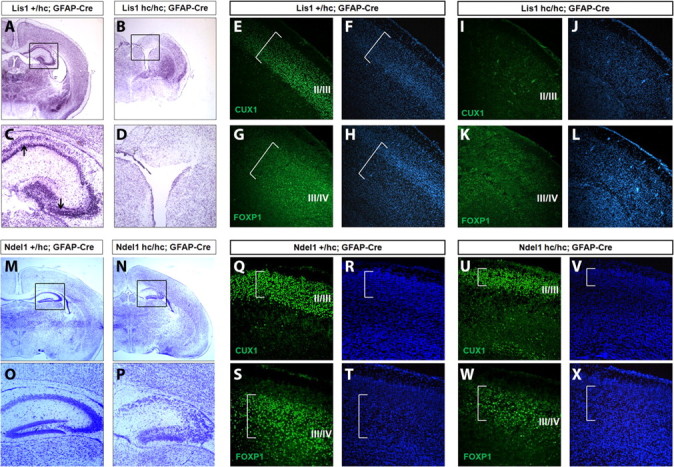
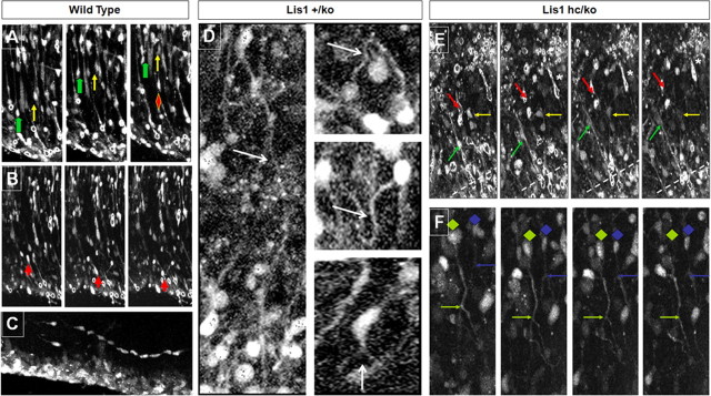
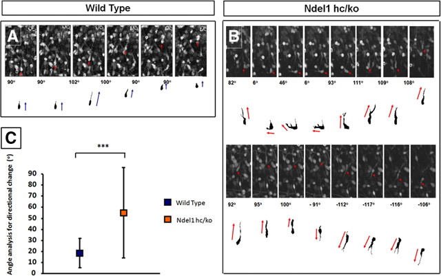
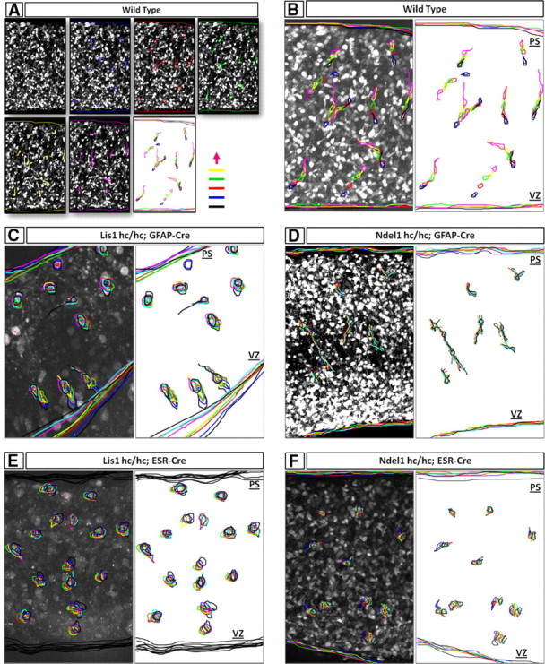
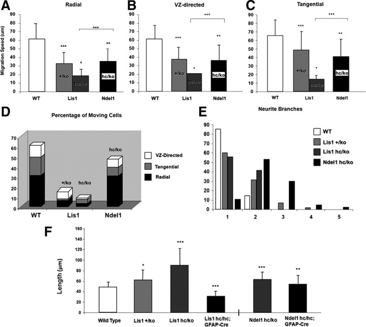
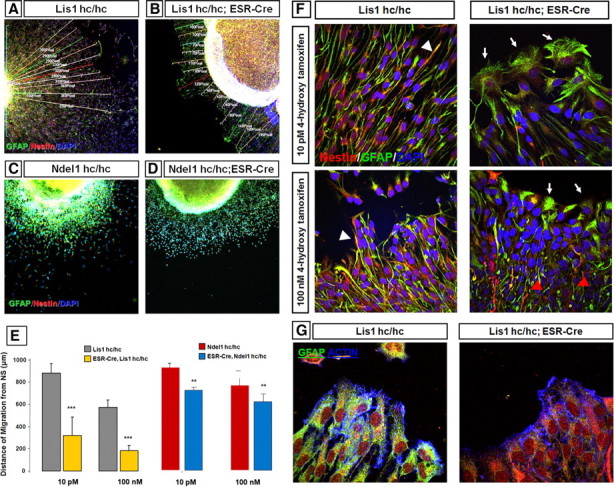
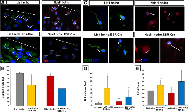
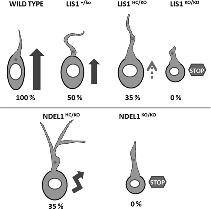
Haploinsufficiency of LIS1 results in lissencephaly, a human neuronal migration disorder. LIS1 is a microtubule- (MT) and centrosome- [microtubule organizing center (MTOC)] associated protein that regulates nucleokinesis via the regulation of dynein motor function and localization. NDEL1 (NudE isoform, NudE like) interacts with LIS1/dynein complex, and is phosphorylated by CDK5/P35. Previous reports using siRNA-mediated knock-down demonstrated similar critical roles for LIS1 and NDEL1 during neuronal migration, but neuronal migration has not been studied in genetic mutants for Lis1 and Ndel1 where protein levels are uniform in all cells. Brains from mice with complete loss of Lis1 and Ndel1 displayed severe cortical layering and hippocampal defects, but Lis1 mutants had more severe defects. Neuronal migration speed was reduced and neurite lengths were elongated in proportion to the reduction of LIS1 and NDEL1 protein levels in embryonic day 14.5 mutant cortical slices compared to wild type, using two-photon confocal time lapse videomicroscopy. Additionally, mice with 35% of wild-type NDEL1 levels displayed diverse branched migration modes with multiple leading processes, suggesting defects in adhesion and/or polarity. Complete loss of Lis1 or Ndel1 resulted in the total inhibition of nuclear movement in cortical slice assays, and in neurosphere assays, the percentage of migrating neurons with correctly polarized MTOC location was significantly reduced while nuclear-centrosomal distance was extended. Neurite lengths were increased after complete loss Ndel1 but reduced after complete loss of Lis1. Thus, Lis1 and Ndel1 are essential for normal cortical neuronal migration, neurite outgrowth, and function of the MTOC in a dose-dependent manner.


Figures

Figure 1.
Figure 1.
Cortical and hippocampal defects in Lis1 and Ndel1 genetic mutant mice after cresyl violet staining (A–D, M–P). Arrows indicate some splitting of the hippocampal pyramidal cell layer. E–L, Q–X, Laminar patterning is disrupted in Lis1 and Ndel1 mutant cortex. Identical immunostained (E, G, I, K, Q, S, U, W) and DAPI (F, H, J, L, R, T, V, X, respectively) counterstained sections were placed side-by-side. Cux1 (E, I, Q, U) and FoxP1 (G, K, S, W) were used as cortical markers specific for layer II/III and III/IV, respectively for Lis1hc/hc;hGFAPCre and mice (E–L) or Ndel1hc/hc;hGFAPCre mice (Q–X) at P5. In the matched immunostained and DAPI stained sections, the brackets indicate the extent of immunostaining to facilitate localization of the pattern of expression to the entire cortex.
Figure 2.
Figure 2.
Abnormal and slow neuronal migration of Lis1 mutant precursor cells in organotypic cortical slice cultures using time lapse confocal videomicroscopy. A–C, Three distinct types of movement patterns (radial, tangential and VZ-directed migration) were observed in cortical slice cultures from wild-type mice at E14.5. The yellow and green arrows indicate radially migrating cells (A), and the red arrow a VZ-directed migrating cell (B). For tangential migration (C), kymography denotes the serial movement patterns. D, Unique migration patterns with curled and long leading processes in cortical slices from Lis1+/ko mice. E, F, In cortical slices from Lis1hc/ko mice, most of the neuronal precursor cells in the IZ did not move and displayed a long leading process. The cell bodies were mostly stationary (triangles). Arrows indicate branch points in leading process.
Figure 3.
Figure 3.
Multidirectional movements, slow migration and increased lengths of leading processes in neural precursors from of Ndel1hc/ko precursor cells in organotypic cortical slice cultures using time lapse confocal videomicroscopy. A, B, Images of single moving cell in the IZ at different time points illustrated the boundaries of cell bodies and processes (silhouettes) from each cell in a time-dependent manner. Various branched and multidirectional movements as well as stationary state cells were observed in Ndel1hc/ko cortical slices. C, The angles of change in moving direction. Migrating cells from wild-type slices moved in a mostly straightforward direction with an average angle of 18.16°. However, the cells in Ndel1hc/ko slices frequently changed their direction of movement with an average angle of 59.55°, p < 0.00005 (Student's t test).
Figure 4.
Figure 4.
Impairment of migration in hGFAPCre- and CreER-induced knock-out of Lis1 and Ndel1. Merged images of cell boundaries of individual cells from the initial to final time are illustrated by different colors. Serial panels for time-dependent migration (each color indicates 90 min) (A) and merged images of those panels for migration of wild-type cells (B) are shown. Neuronal migration in GFAP-Cre;Lis1hc/hc (C) and GFAP-Cre;Ndel1hc/hc cortical slices (D) is shown. Migration was completely blocked, and there were fewer numbers of leading processes and branches. In both Cre-ER;Lis1hc/hc (E) and Cre-ER;Ndel1hc/hc (F) cortical slices, neuronal movements were severely inhibited after acute loss of LIS1 and NDEL1 through the administration of tamoxifen by intraperitoneal injection of pregnant dams. The last images (green) were not different in position compared to the first boundary images (black) in cells from Cre-ER;Lis1hc/hc and Cre-ER;Ndel1hc/hc cortical slices.
Figure 5.
Figure 5.
Quantitation of movement, branching and neurite length of neural precursor cells undergoing neuronal migration. Average migration speed and SD of cells from wild-type, Lis1 (+/ko with 50% and hc/ko with 35% of wild-type LIS1 levels) and Ndel1 (hc/ko with 35% of wild-type NDEL1 levels) mutants undergoing radial (A), VZ-directed (B), and tangential (C) migration. D, The total number of moving cells and the proportion of moving cells in each genotype that were moving in radial, VZ-directed or tangential directions is shown. E, The percentage of migrating neural precursor cells with secondary branches in each genotype is shown. Note that in Ndel1hc/ko cells, 90% of cells had more than one secondary branch and often had multiple branches, while in Lis1hc/ko cells, less than half had more than one branch and very few had more than two branches. F, Left of the hash mark: the length of the major leading process was extended in neural precursor cells from Lis1+/ko (light gray bar) and Lis1hc/ko (dark gray bar) cortical slices compared with wild-type (open bar), while the leading process was reduced in Lis1hc/hc;GFAP-Cre (black bar) cells. Right of the hash mark: the length of major leading process was extended in neural precursor cells from Ndel1hc/ko (dark gray bar) and in Ndel1hc/hc;GFAP-Cre (black bar) cortical slices compared to wild type. *p < 0.05; **p < 0.005, ***p < 0.0005 (Student's t test).
Figure 6.
Figure 6.
Migration defects of neurospheres of neuronal progenitors attached to the substratum from conditional knock-out mice for Lis1 or Ndel1 with or without Cre-ER after tamoxifen-induced gene deletion. AD, Low-magnification views of the radial migration patterns of neuronal precursor cells from Lis1hc/hc (A), Lis1hc/hc;Cre-ER (B), Ndel1hc/hc (C), and Ndel1hc/hc;Cre-ER (D) embryos treated with low (10 pm) doses of 4-hydroxy tamoxifen. The distance of radial movement of individual cells is indicated by individual white lines. Immunocytochemical staining is with GFAP (green), nestin (red) and DAPI. E, The average distance of radial migration of neuronal precursors from Lis1hc/hc, or Lis1hc/hc;Cre-ER neurospheres (left) and Ndel1hc/hc and Ndel1hc/hc;Cre-ER neurospheres (right) treated with low (10 pm) and high doses (100 nm) of 4-hydroxy tamoxifen, as indicated at the bottom of the panel. F, Early appearance of differentiated cells in Lis1hc/hc;Cre-ER neurosphere outgrowths. In cells from Lis1hc/hc embryos after 4-hydroxy tamoxifen treatment, nestin- (red) and GFAP- (green) positive cells were oriented radially, and the nuclei were elongated in the same direction. In contrast, in cells from Lis1hc/hc;Cre-ER embryos treated with 4-hydroxy tamoxifen, there were strong GFAP-positive cells localized in marginal area of outgrowth, with fewer nestin-positive cells that were disconnected and short glial fibers, and nuclei were more rounded in appearance. G, Actin protrusion was obvious in migrating cells from Lis1hc/hc embryos, but blunt ended actin bundles were observed in cells from Lishc/hc;Cre-ER embryos.
Figure 7.
Figure 7.
A, B, Polarization of the MTOC in migrating neural precursor cells from Lis1hc/hc, Lis1hc/hc;Cre-ER, Ndel1hc/hc and Ndel1hc/hc;Cre-ER embryos after 4-hydroxy tamoxifen treatment was measured. The orientation of the centrosome preceding the nucleus was measured based upon the frontier line of the outgrowth area, rather than the line of the wound in wound-healing studies of fibroblasts in culture as described previously (Etienne-Manneville and Hall, 2001) In controls, the MTOC was well polarized in >80% of cells. However, the polarization was significantly disrupted (<50%) both in neuronal progenitor cells from Lis1hc/hc;Cre-ER and Ndel1hc/hc;Cre-ER embryos. C, D, Distance between the nucleus and centrosome (N-C distance) of neural stem cells. The N-C distances were close to nucleus (<5 μm apart) in neuronal precursors from Lis1hc/hc and Ndel1hc/hc embryos, but in Lis1hc/hc;Cre-ER and Ndel1hc/hc;Cre-ER, the N-C distance was significantly extended (>20 μm). E, The length of major leading process was extended in neural precursor cells from Lis1hc/hc;Cre-ER and Ndel1hc/hc;Cre-ER embryos.
Figure 8.
Figure 8.
Distinct migration models of neurons in Lis1 and Ndel1 knock-out mice. Genotypes are above the cells, speeds of migration relative to wild-type are at the bottom of the cells, and the speed and direction of movement is indicated by the size and thickness of the arrow. “STOP” means that the cells did not move. Note the lengthened and curled processes of the Lis1 mutants, while the Ndel1 mutants displayed branching. Wild-type cells have an actively migrating elongated shape of the cell body, which is short, thin and moves straightforward into the leading process while maintaining a short N-C distance (centrosome; small dot and nucleus; circle). Lis1+/ko cells, which express 50% of wild-type LIS1 protein levels display a curved and thicker leading process with longer N-C distance, and Lis1 hc/ko (35% of Lis1 protein) cells are mostly inactive with a thick leading process and a longer N-C distance. Ndel1hc/ko cells display a multibranched leading process, a zig-zag moving pattern and longer N-C distance. In the absence of either Lis1 or Ndel1, the neuronal precursor cells did not substantially move. Thickness of the arrows indicates velocity of individual cells.

Similar articles

Cited by

References

    1. Ayala R, Shu T, Tsai LH. Trekking across the brain: the journey of neuronal migration. Cell. 2007;128:29–43. - PubMed
    1. Cahana A, Escamez T, Nowakowski RS, Hayes NL, Giacobini M, von Holst A, Shmueli O, Sapir T, McConnell SK, Wurst W, Martinez S, Reiner O. Targeted mutagenesis of Lis1 disrupts cortical development and LIS1 homodimerization. Proc Natl Acad Sci U S A. 2001;98:6429–6434. - PMC - PubMed
    1. Calaora V, Rogister B, Bismuth K, Murray K, Brandt H, Leprince P, Marchionni M, Dubois-Dalcq M. Neuregulin signaling regulates neural precursor growth and the generation of oligodendrocytes in vitro. J Neurosci. 2001;21:4740–4751. - PMC - PubMed
    1. Engele J, Bohn MC. Effects of acidic and basic fibroblast growth factors (aFGF, bFGF) on glial precursor proliferation: age dependency and brain region specificity. Dev Biol. 1992;152:363–372. - PubMed
    1. Etienne-Manneville S, Hall A. Integrin-mediated activation of Cdc42 controls cell polarity in migrating astrocytes through PKCzeta. Cell. 2001;106:489–498. - PubMed

Publication types

MeSH terms

Substances