. 2009 Dec 9;29(49):15531-41.
doi: 10.1523/JNEUROSCI.2961-09.2009.

Cellular mechanisms underlying burst firing in substantia nigra dopamine neurons

Affiliations

Cellular mechanisms underlying burst firing in substantia nigra dopamine neurons

Sarah N Blythe et al. J Neurosci. .

Abstract

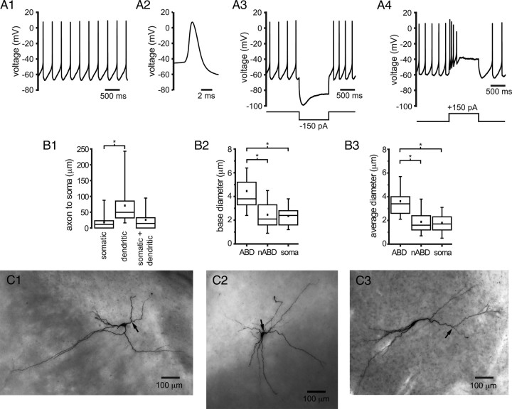
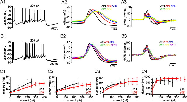
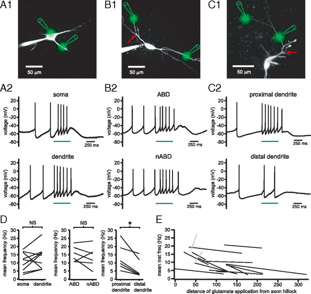
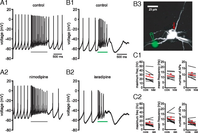
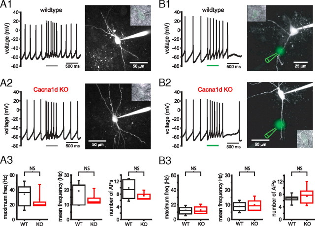
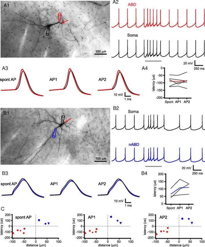
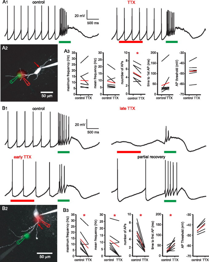
Burst firing of substantia nigra dopamine (SN DA) neurons is believed to represent an important teaching signal that instructs synaptic plasticity and associative learning. However, the mechanisms through which synaptic excitation overcomes the limiting effects of somatic Ca(2+)-dependent K(+) current to generate burst firing are controversial. Modeling studies suggest that synaptic excitation sufficiently amplifies oscillatory dendritic Ca(2+) and Na(+) channel currents to lead to the initiation of high-frequency firing in SN DA neuron dendrites. To test this model, visually guided compartment-specific patch-clamp recording and ion channel manipulation were applied to rodent SN DA neurons in vitro. As suggested previously, the axon of SN DA neurons was typically found to originate from a large-diameter dendrite that was proximal to the soma. However, in contrast to the predictions of the model, (1) somatic current injection generated firing that was similar in frequency and form to burst firing in vivo, (2) the efficacy of glutamatergic excitation was inversely related to the distance of excitation from the axon, (3) pharmacological blockade or genetic deletion of Ca(2+) channels did not prevent high-frequency firing, (4) action potential bursts were invariably detected first at sites that were proximal to the axon, and (5) pharmacological blockade of Na(+) channels in the vicinity of the axon/soma but not dendritic excitation impaired burst firing. Together, these data suggest that SN DA neurons integrate their synaptic input in a more conventional manner than was hypothesized previously.


Figures

Figure 1.
Figure 1.
Physiological and morphological properties of SN DA neurons. SN DA neurons were identified by their characteristic physiological properties including slow (<4 Hz), autonomous firing (A1), broad (>2 ms duration) action potentials (A2), sag in response to hyperpolarizing current injection (A3), and depolarization block (A4) in response to depolarizing current injection. B1, Selection of neurons for single somatic or dual somatic/dendritic recordings were based on the appearance of the soma or the width of proximal dendrites, respectively. The distance of the axon hillock from the soma was significantly greater for the latter selection strategy. B2, B3, The base and average diameters of the primary parts of ABDs were significantly greater than the equivalent sections of nABDs. The base and average diameters of ABDs were also significantly greater than the dendrites of neurons in which the axon emerged from the soma. C, Examples of neurons in which the axon emerged from a proximal dendrite close (32.9 μm) to the soma and the soma and a secondary dendrite far (243 μm) from the soma. The axon hillock is denoted by an arrow. Note that the cut end of each axon forms a characteristic retraction ball. *p < 0.05.
Figure 2.
Figure 2.
Somatic current generation generates a burst of action potentials. A–C, Action potential bursts evoked by somatic current injection in juvenile (A, C; P14 ± P1) and more mature (B, C; P52 ± P7) SN DA neurons. A1, B1, Response of representative SN DA neurons to depolarizing current injections (200 pA for 1 s). A2, B2, Overlay of selected action potentials (AP1, AP3, AP5, AP7, AP9, AP11) generated by current injection reveals a progressive decrease and increase in the amplitude and duration of action potentials, respectively. A3, B3, Overlay of the first derivative of action potentials illustrated in A2 and B2 reveals the progressive reduction in the peak of the first derivative and separation of early (axonal) and later (somatic) components of action potentials. C1, C2, Maximum (max) and mean frequency (freq) of burst activity versus current amplitude. C3, C4, Number of burst action potentials (APs) and duration of evoked burst firing versus current amplitude.
Figure 3.
Figure 3.
The intensity of glutamate-evoked activity is inversely related to the distance of glutamate application from the axon. Glutamate was pressure-pulse applied to specific compartments of SN DA neurons in three distinct combinations: soma and dendrite (A); ABD and nABD (B); proximal and distal sections of the same dendrite (C). A1, B1, C1, 2PLSM z-series of neurons together with sites of glutamate application (green). The axon hillock (red arrow) was identifiable in B1 and C1. A2, B2, C2, Glutamate-evoked activity for each combination. Green line denotes time over which glutamate was applied through pressure pulses. Applications of glutamate to the soma, ABDs and non-nABDs, and proximal and distal dendrites generated burst firing. D, E, Population data. Each line denotes an individual neuron. D, The frequencies of activity evoked by somatic versus dendritic or ABD versus nABD applications of glutamate were not significantly different. However, relatively proximal applications of glutamate generated significantly higher-frequency firing than more distal dendritic applications. E, Replotting of data with respect to distance of glutamate application from the axon reveals that, in the majority of neurons, glutamate applications relatively proximal to the axon generated higher-frequency activity than more distal applications (black lines). In only two neurons (gray lines) was an opposite trend observed. *p < 0.05.
Figure 4.
Figure 4.
Cav1 channel antagonists reduce the intensity of, but do not eliminate, burst firing in SN DA neurons. A, Generation of burst firing in an SN DA neuron by local synaptic stimulation (gray) under control conditions (A1) and in the presence of nimodipine (10 μm; A2). B, Generation of burst firing in a SN DA neuron by dendritic pressure-pulse application of glutamate (green) under control conditions (B1) and in the presence of isradipine (10 μm; B2). B3, 2PLSM z-series with site of glutamate application (green pipette) and axon hillock (red arrow) indicated. C, Population data. Individual neuron data indicated by black lines. Sample mean indicated by red line. Application of nimodipine (C1) or isradipine (C2) significantly reduced the intensity of evoked firing in most neurons tested and across the sample population compared with firing under control conditions. APs, Action potentials; CON, control; freq, frequency; ISR, isradipine; NIM, nimodipine. *p < 0.05.
Figure 5.
Figure 5.
Synaptic stimulation or dendritic application of glutamate generates burst firing in BAC D2 EGFP Cacna1d wild-type/knock-out SN DA neurons. Local synaptic stimulation (A, gray) or dendritic pressure-pulse application of glutamate (B, green) generated burst firing in SN DA neurons derived from wild type (WT; A1, B1) and Cacna1d knock-out (A2, B2) mice. 2PLSM z-series of each recorded neuron are adjacent to their respective electrophysiological records. The locations of glutamate application pipettes (green) are denoted. Insets, Overlays of Dodt contrast and EGFP fluorescent images confirm that each recorded neuron was BAC D2 EGFP positive. A3, B3, Population data. Burst firing was not significantly different for any parameter in WT and Cacna1d knock-out mice. APs, Action potentials; freq, frequency; KO, knock-out; NS, not significant.
Figure 6.
Figure 6.
The site of action potential initiation does not shift during synaptic stimulation-evoked burst firing. A–C, Simultaneous recordings from the soma (black) and an ABD (red) or the soma and a nABD (blue) were used to compare the relative timing of action potentials (time of dendritic action potential − time of somatic action potential) during spontaneous firing and synaptic stimulation-evoked action potential bursts. Spontaneous action potentials (spont AP) and the first (AP1) and second (AP2) action potentials of synaptic stimulation-evoked action potential bursts are indicated. A1, B1, Photomontages of recorded, biocytin-labeled neurons together with sites of somatic and dendritic recording locations relative to the axon hillock (red arrow). A2, B2, Local electrical stimulation at 50 Hz for 500 ms (gray bar) generated a burst of action potentials in each recorded neuron. A3, A4, Action potentials were first detected in the ABD and then the soma in a representative neuron (A3) and the sample population (A4; n = 5; black lines represent individual neuron data, red line represents sample mean). B3, B4, Action potentials were recorded in the soma and then the nABD in a representative neuron (B3) and the sample population (B4; n = 3; black lines represent individual neuron data, blue line represents sample mean). C, Timing of dendritic action potentials plotted with respect to the timing of somatic action potentials versus the relative distance of the dendritic and somatic recording electrodes from the axon hillock (distance = distance of dendritic electrode from axon hillock − distance of somatic electrode from axon hillock). Individual neuron data are represented by distinct symbols.
Figure 7.
Figure 7.
Initiation of action potential bursts is impaired by blockade of axo-somatic Nav channels but not Nav channels at the site of excitation. A, Application of TTX to the site of dendritic glutamate application had a minimal effect on spontaneous and glutamate-evoked activity. A1, A2, Representative example. A1, Application of TTX (red) 1 s before the dendritic pulsed application of glutamate (green) had little effect on evoked activity compared with that evoked under control conditions. A2, 2PLSM z-series with sites of TTX (red) and glutamate (green) application indicated for the example in A1. A3, Population data. Black lines represent individual cells, and red lines represent population mean. Dendritic TTX application significantly reduced the number of evoked action potentials (APs) but had no effect on any other parameter. B, Axo-somatic application of TTX reduced spontaneous activity and the intensity of burst firing that was evoked by dendritic application of glutamate. B1, B2, Representative example. B1, Proximal application of TTX (red) 1 s before the dendritic application of glutamate (green) reduced spontaneous and evoked activity compared with that observed under control conditions. The reduction in firing increased with successive applications of TTX; compare early (first) and late (ninth) trials. B2, 2PLSM z-series with sites of glutamate (green) and TTX (red) application indicated for the example in B2. B3, Population data. Black lines represent individual cells, and red lines represent population means. Axo-somatic TTX application significantly (red asterisk) reduced the intensity and increased the latency of burst firing and also increased the threshold of APs in each of five neurons tested. AP, Action potential. *p < 0.05.

Similar articles

Cited by

References

    1. Baufreton J, Atherton JF, Surmeier DJ, Bevan MD. Enhancement of excitatory synaptic integration by GABAergic inhibition in the subthalamic nucleus. J Neurosci. 2005;25:8505–8517. - PMC - PubMed
    1. Becker C, Jick SS, Meier CR. Use of antihypertensives and the risk of Parkinson disease. Neurology. 2008;70:1438–1444. - PubMed
    1. Beckstead MJ, Grandy DK, Wickman K, Williams JT. Vesicular dopamine release elicits an inhibitory postsynaptic current in midbrain dopamine neurons. Neuron. 2004;42:939–946. - PubMed
    1. Blythe SN, Atherton JF, Bevan MD. Synaptic activation of dendritic AMPA and NMDA receptors generates transient high-frequency firing in substantia nigra dopamine neurons in vitro. J Neurophysiol. 2007;97:2837–2850. - PubMed
    1. Centonze D, Picconi B, Gubellini P, Bernardi G, Calabresi P. Dopaminergic control of synaptic plasticity in the dorsal striatum. Eur J Neurosci. 2001;13:1071–1077. - PubMed

Publication types

MeSH terms