. 2009 Dec 9;29(49):15551-63.
doi: 10.1523/JNEUROSCI.3336-09.2009.

Activation of glycogen synthase kinase-3 beta is required for hyperdopamine and D2 receptor-mediated inhibition of synaptic NMDA receptor function in the rat prefrontal cortex

Affiliations

Activation of glycogen synthase kinase-3 beta is required for hyperdopamine and D2 receptor-mediated inhibition of synaptic NMDA receptor function in the rat prefrontal cortex

Yan-Chun Li et al. J Neurosci. .

Erratum in

  • J Neurosci. 2012 Feb 1;32(5):1914

Abstract

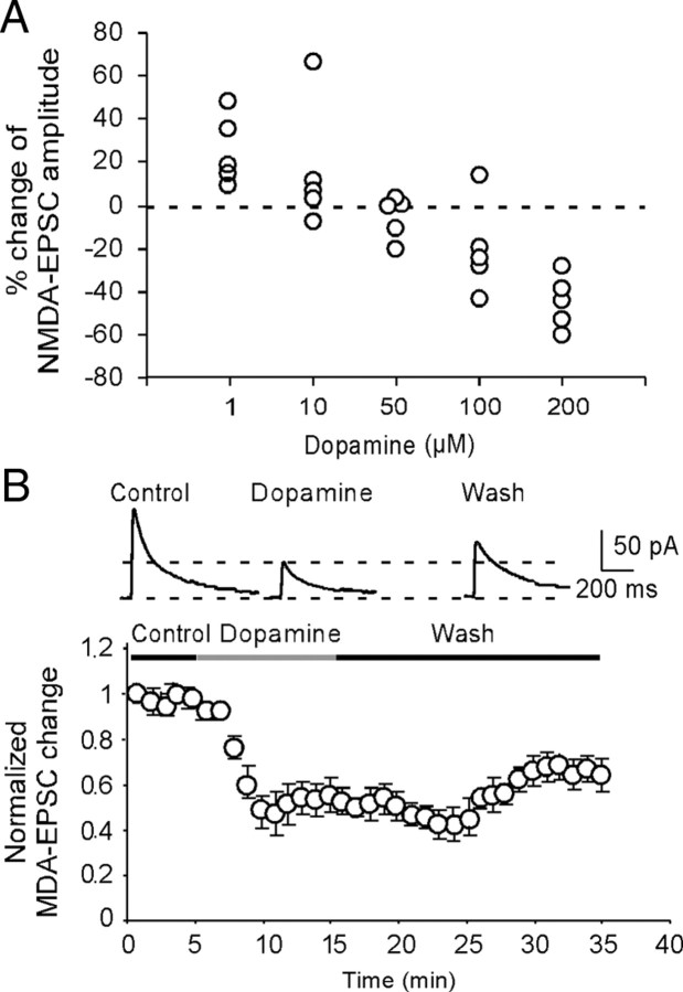
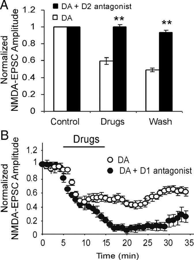
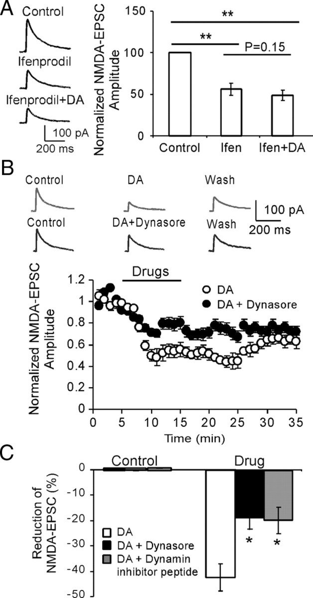
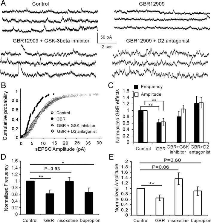
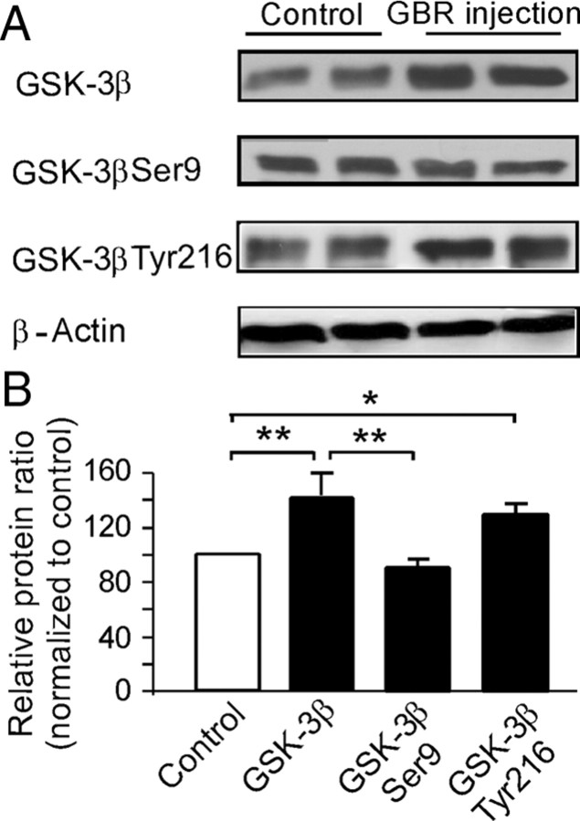
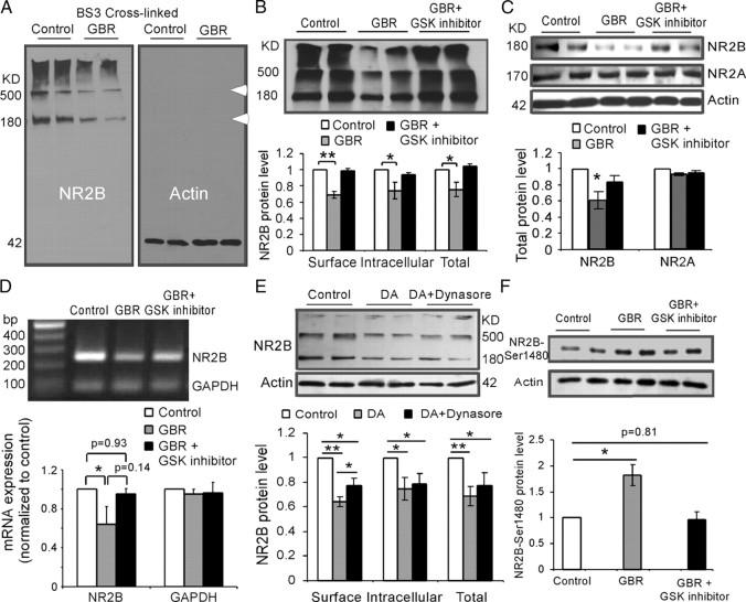
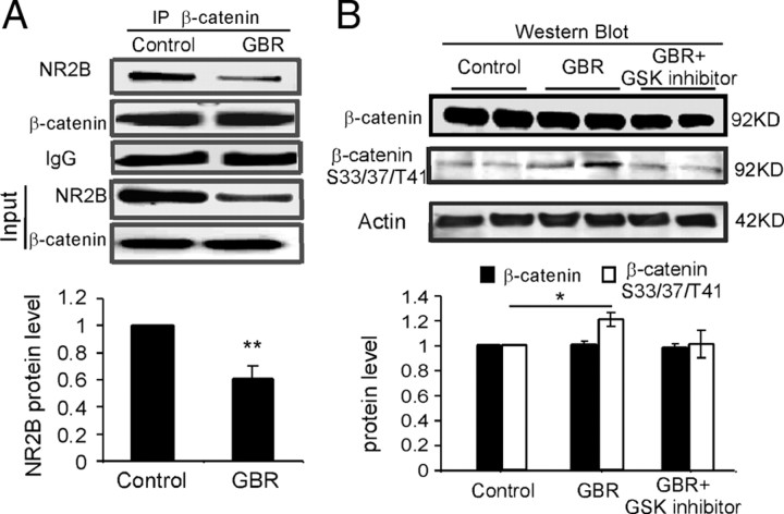
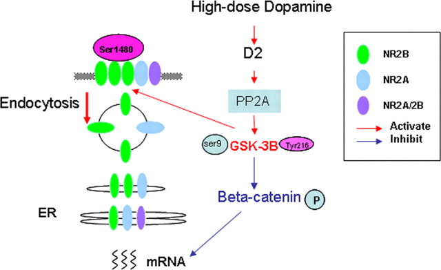
The interactions between dopamine and glutamate systems play an essential role in normal brain functions and neuropsychiatric disorders. The mechanism of NMDA receptor regulation through high concentrations of dopamine, however, remains unclear. Here, we show the signaling pathways involved in hyperdopaminergic regulation of NMDA receptor functions in the prefrontal cortex by incubating cortical slices with high concentration of dopamine or administering dopamine reuptake inhibitor 1-(2-[bis-(4-fluorophenyl)methoxy]ethyl)- 4-(3-phenylpropyl)piperazine (GBR12909) in vivo. We found that, under both conditions, the synaptic NMDA receptor-mediated currents were significantly attenuated by excessive dopamine stimulation through activation of D(2) receptors. Furthermore, high dose of dopamine failed to affect NMDA receptor-mediated currents after blockade of NR2B subunits but triggered a dynamin-dependent endocytosis of NMDA receptors. The high-dose dopamine/D(2) receptor-mediated suppression of NMDA receptors was involved in the increase of glycogen synthase kinase-3beta (GSK-3beta) activity, which in turn phosphorylates beta-catenin and disrupts beta-catenin-NR2B interaction, but was dependent on neither Gq11 nor PLC (phospholipase C). Moreover, the hyperdopamine induced by GBR12909 significantly decreased the expression of both surface and intracellular NR2B proteins, as well as NR2B mRNA levels, suggesting an inhibition of protein synthesis. These effects were, however, completely reversed by administration of either GSK-3beta inhibitor or D(2) receptor antagonist. These results therefore suggest that GSK-3beta is required for the hyperdopamine/D(2) receptor-mediated inhibition of NMDA receptors in the prefrontal neurons and these actions may underlie D(2) receptor-mediated psychostimulant effects and hyperdopamine-dependent behaviors in the brain.


Figures

Figure 1.
Figure 1.
DA induces bidirectional dose-dependent effects in synaptically evoked NMDA-EPSCs in layer 5 pyramidal neurons. A, Dose–response relationship for the effects of DA. The data were average effects of DA on NMDA-EPSCs determined at 10 min after DA application (n = 5 for each dose). B, Top panel, Examples of the NMDA-EPSCs recorded in presence of NBQX (20 μm) and picrotoxin (50 μm) in control, dopamine, and washout period. The representative traces indicated that high-dose DA (200 μm) significantly reduced the amplitudes of NMDA-EPSCs. Bottom panel, Summary graph showing the time course of high-dose (200 μm) DA effects on the amplitude of NMDA-EPSCs. This suppressing effect on NMDA currents was long-lasting with little recovery even after 15–20 min washout, suggesting the possibility of NMDA receptor internalization under this condition. Note that the data were normalized to the baseline EPSCs recorded in the first minute. Error bars indicate SEM.
Figure 2.
Figure 2.
High-dose DA attenuates NMDA-EPSCs through activation of D2, but not D1, receptors. A, Coapplication of selective D2 receptor antagonist L741,626 (10 μm) with DA (200 μm) significantly abolished DA-induced attenuation of NMDA-EPSCs (n = 6; p < 0.01). B, On the contrary, coapplication of selective D1 receptor antagonist SCH 23390 (10 μm) with DA (200 μm) potentiated DA-induced attenuation of NMDA-EPSCs (n = 6; p < 0.05). *p < 0.05; **p < 0.01 for all figures. Error bars indicate SEM.
Figure 3.
Figure 3.
High-dose DA-induced reduction of NMDA-EPSCs is partially dependent on dynamin-mediated internalization of NR2B subunits. A, Left panel, Sample traces showing the effects of NR2B antagonist ifenprodil (3 μm) and ifenprodil plus dopamine (200 μm) on NMDA-EPSCs. Right panel, Initial application of NR2B antagonist ifenprodil significantly decreased the amplitudes of NMDA-EPSCs (p < 0.01), whereas followed coapplication of ifenprodil with DA did not cause significant attenuation on the NMDA-EPSCs (p = 0.15), suggesting the involvement of NR2B subunits. B, Sample traces (top panel) and time course data (bottom panel) showing the effects of dynasore, a potent membrane-permeable inhibitor of endocytosis, on DA-induced inhibition of NMDA receptors (n = 6). C, The inhibitory effects of DA on NMDA-EPSCs were significantly blocked by dynamin inhibitory peptide (50 μm in pipette) and dynasore (100 μm in bath), which prevent receptor endocytosis (n = 6; p < 0.05 for both). Error bars indicate SEM.
Figure 4.
Figure 4.
PP2A-GSK-3β, but not Gq11, signaling pathway is involved in the high-dose DA/D2-induced depression of NMDA-EPSCs. A, Coapplication of GSK-3β inhibitors TDZD (10 μm) or SB216,763 (10 μm), the two structurally different GSK-3β inhibitors, completely abolished the DA-induced suppression in NMDA receptor mediated-EPSCs (n = 6; p < 0.01 for both). Both inhibitors themselves, however, did not show clear effects on the basal NMDA receptor-mediated transmissions (data not shown). B, Coapplication of Gq11 inhibitor (10 μm) with DA (100 or 200 μm), however, did not affect the effects of DA (n = 6; p > 0.05). C, D, PP2A inhibitor okadaic acid (100 nm) significantly blocked the DA effects on NMDA receptor-mediated EPSCs (n = 6; p < 0.05). Error bars indicate SEM.
Figure 5.
Figure 5.
Hyperdopamine induced by systemic administration of GBR12909 significantly decreases the spontaneous NMDA-EPSCs, and the effects were completely blocked by systemic coadministration of GBR12909 and either D2 antagonist or GSK-3β inhibitor TDZD. A, Representative traces of the spontaneous NMDA-EPSCs. B, Cumulative probability showing the obvious shift of the spontaneous NMDA-EPSC in GBR12909 administration. C, The decreases of both spontaneous EPSC frequency (p < 0.01) and amplitude (p < 0.01) induced by GBR12909 were completely reversed by coadministration of GBR12909 with either D2 antagonist or GSK-3β inhibitor TDZD in vivo. D, E, Effects of selective NET inhibitior nisoxetine (10 mg/kg, i.p.) and a nonselective DA and NE reuptake blocker bupropion (10 mg/kg, i.p.) on NMDA currents. Nisoxetine exhibited no effect on EPSC frequency but increased the amplitude of NMDA current, although not statistically significantly (n = 6; p = 0.93 for frequency and p = 0.06 for amplitude). In contrast, bupropion significantly decreased EPSC frequency (n = 6; p < 0.05), similar to GBR12909, but had no significant effects on the EPSC amplitude (n = 6; p = 0.60). Error bars indicate SEM.
Figure 6.
Figure 6.
Hyperdopamine induced by systemic administration of GBR12909 significantly increases GSK-3β expression in the PFC. A, Analysis by Western blots showed that a single dose of GBR12909 (10 mg/kg, i.p.) caused significant increase of total protein levels of GSK-3β as well as the levels of the phosphorylated form of GSK-3β Tyr216 but not GSK-3β Ser9 at 1 h after injection. B, Quantitative analysis showed that the total protein levels of GSK-3β were significantly increased by ∼40% after GBR12909 administrations relative to saline vehicle control (p < 0.01). A similar increase of ∼30% was observed in the levels of phosphor-GSK-3β Tyr216 relative to saline vehicle control (p < 0.05). In contrast, the levels of phosphor-GSK-3β Ser9 remained unchanged compared with control levels (p = 0.67) and but were significantly lower compared with total protein levels of GSK-3β (p < 0.01). Error bars indicate SEM.
Figure 7.
Figure 7.
Systemic administration of GBR12909 significantly decreases both surface and intracellular NR2B proteins, total NR2B, NR2B mRNA expression, as well as NR2B pS1480, in the PFC. A, Representative images of Western blot analysis from the PFC slices incubated with the BS3 cross-linking reagent. BS3-linked surface NR2B was presented as high-molecular-weight aggregates (>500 kDa), whereas the intracellular NR2B remained in a monomeric form represented by a single molecular weight band at 180 kDa. In contrast, β-actin was located exclusively within cytoplasm as a monomer band, confirming effectiveness of the cross-linking. B, Top panel, Representative blots showing the protein levels of NR2B subunits after systemic administration of GBR12909 and coapplication of GBR12909 and GSK-3β inhibitor TDZD. Bottom panel, Quantitative analysis showed that relative protein levels of the presumptive surface, intracellular, and total NR2B subunits relative to the control levels (*p < 0.05; **p < 0.01). C, GBR12909 (10 mg/kg, i.p.; 1 h) significantly decreased the total protein expression of NR2B (p < 0.05) but not NR2A (p > 0.05). D, The mRNA expression of NR2B subunits was significantly decreased by systemic injection of GBR12909 (p < 0.05) and this effect was reversed by coapplication of GBR12909 and GSK-3β inhibitor TDZD (p = 0.93). In contrast, the GAPDH expressions were stable, without clear change. E, The DA-induced endocytosis of NR2B subunit was further confirmed by incubating the prefrontal cortical slices with membrane-impermeable cross-linking reagent BS3. Both surface and intracellular protein levels of NR2B subunits were significantly decreased by DA treatment (200 μm; 10 min; n = 4; p < 0.01). When DA was coapplied with dynasore (100 μm), the effect of DA on surface, but not intracellular, expression of NR2B, was partially but significantly blocked (n = 4; p < 0.05). F, GBR12909 injection (10 mg/kg, i.p.; 1 h) significantly increased the expression of NR2B pS1480 (p < 0.05), and this increase was blocked by preinjection of GSK-3β inhibitor TDZD 30 min in advance (1 mg/kg, i.p.). Error bars indicate SEM.
Figure 8.
Figure 8.
Hyperdopamine induces phosphorylation of β-catenin and β-catenin–NR2B interaction. A, β-Catenin was immunoprecipitated with anti-β-catenin antibody and was then probed for the coimmunoprecipitated NR2B with specific anti-NR2B antibody. Under control conditions, NR2B coprecipitated with β-catenin, whereas β-tubulin was not detected in β-catenin immunoprecipitates (data not shown), suggesting a potential protein interaction between β-catenin and NR2B. Consistently, NR2B binding, as well as input NR2B expression, was significantly decreased by GBR12909 administration (10 mg/kg, i.p.) (p < 0.01). B, The total protein level of β-catenin was not changed by GBR12909 treatment in Western blot detection (p > 0.05). In contrast, the expression of phosphor-β-catenin detected with antibody against Ser33/37/Tyr41 sites was significantly increased by GBR12909 treatment (p < 0.05). These results indicate that β-catenin may mediate the action of GSK-3β in NMDA receptor trafficking and protein synthesis. Error bars indicate SEM.
Figure 9.
Figure 9.
Schematic graph showing the signaling pathway involved in hyperdopamine/D2-mediated inhibition of synaptic NMDA receptors. High concentration of DA/activation of D2 receptor activates GSK-3β via its upstream regulator PP2A. GSK-3β in turn phosphorylates β-catenin (Ser33/37/Tyr41), which interacts with NR2B, controls the NR2B gene transcription, and thus affects the protein synthesis of NR2B subunits. Phosphorylation of NR2B-Ser1480 and dynamin play an important role for NR2B endocytosis in the DA-induced inhibition of NMDA current.

Similar articles

Cited by

References

    1. Aberle H, Bauer A, Stappert J, Kispert A, Kemler R. beta-Catenin is a target for the ubiquitin-proteasome pathway. EMBO J. 1997;16:3797–3804. - PMC - PubMed
    1. Al-Hallaq RA, Conrads TP, Veenstra TD, Wenthold RJ. NMDA di-heteromeric receptor populations and associated proteins in rat hippocampus. J Neurosci. 2007;27:8334–8343. - PMC - PubMed
    1. Andersen PH. The dopamine uptake inhibitor GBR 12909: selectivity and molecular mechanism of action. Eur J Pharmacol. 1989;166:493–504. - PubMed
    1. Beaulieu JM, Sotnikova TD, Marion S, Lefkowitz RJ, Gainetdinov RR, Caron MG. An Akt/beta-arrestin 2/PP2A signaling complex mediates dopaminergic neurotransmission and behavior. Cell. 2005;122:261–273. - PubMed
    1. Beaulieu J-M, Tirotta E, Sotnikova TD, Masri B, Salahpour A, Gainetdinov RR, Borrelli E, Caron MG. Regulation of Akt signaling by D2 and D3 dopamine receptors in vivo. J Neurosci. 2007a;27:881–885. - PMC - PubMed

Publication types

MeSH terms

LinkOut - more resources