. 2010 Jan-Feb;3(1-2):57-72.
doi: 10.1242/dmm.003228. Epub 2009 Dec 9.

Lack of host SPARC enhances vascular function and tumor spread in an orthotopic murine model of pancreatic carcinoma

Affiliations

Lack of host SPARC enhances vascular function and tumor spread in an orthotopic murine model of pancreatic carcinoma

Shanna A Arnold et al. Dis Model Mech. 2010 Jan-Feb.

Abstract

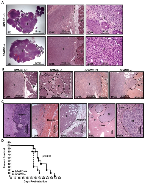
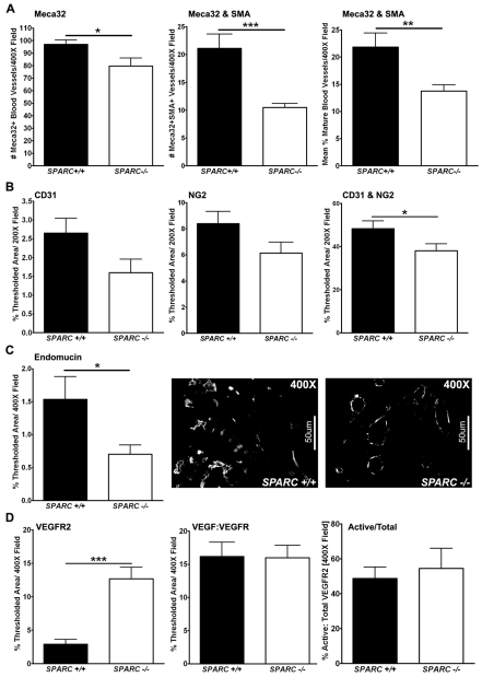
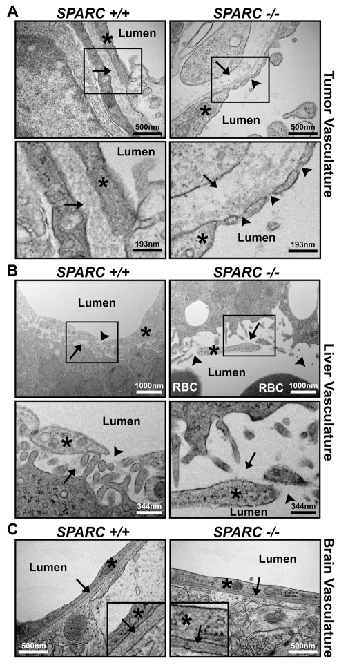
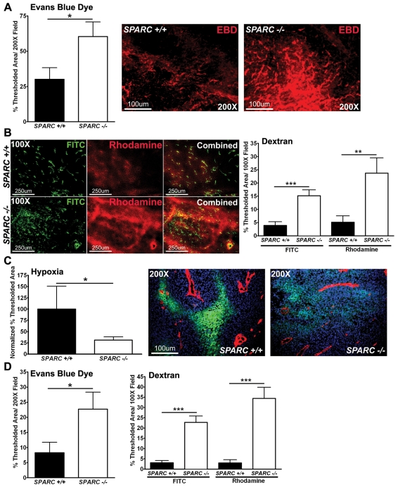
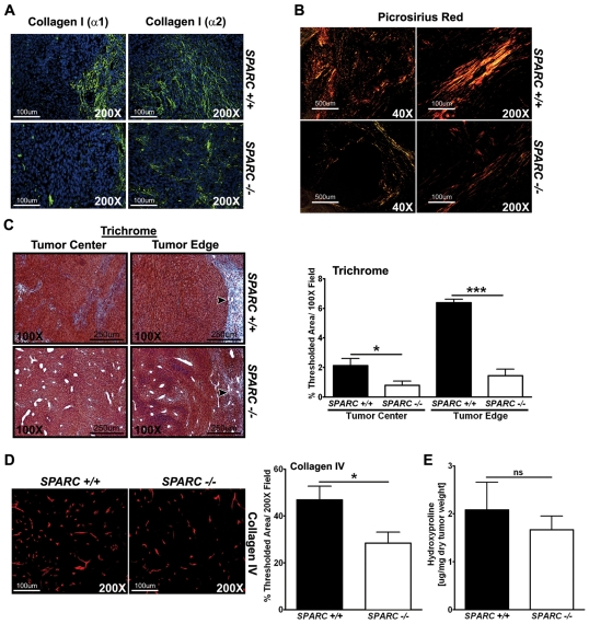
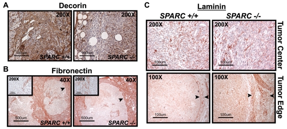
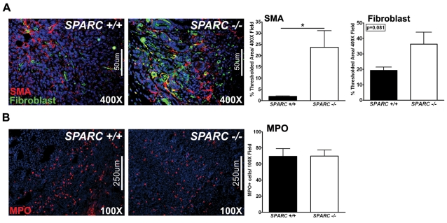
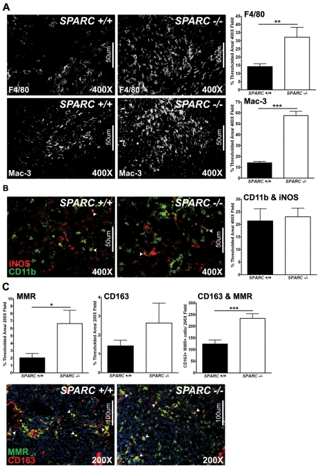
Utilizing subcutaneous tumor models, we previously validated SPARC (secreted protein acidic and rich in cysteine) as a key component of the stromal response, where it regulated tumor size, angiogenesis and extracellular matrix deposition. In the present study, we demonstrate that pancreatic tumors grown orthotopically in Sparc-null (Sparc(-/-)) mice are more metastatic than tumors grown in wild-type (Sparc(+/+)) littermates. Tumors grown in Sparc(-/-) mice display reduced deposition of fibrillar collagens I and III, basement membrane collagen IV and the collagen-associated proteoglycan decorin. In addition, microvessel density and pericyte recruitment are reduced in tumors grown in the absence of host SPARC. However, tumors from Sparc(-/-) mice display increased permeability and perfusion, and a subsequent decrease in hypoxia. Finally, we found that tumors grown in the absence of host SPARC exhibit an increase in alternatively activated macrophages. These results suggest that increased tumor burden in the absence of host SPARC is a consequence of reduced collagen deposition, a disrupted vascular basement membrane, enhanced vascular function and an immune-tolerant, pro-metastatic microenvironment.


Figures

Fig. 1.
Fig. 1.
Orthotopic tumor local invasion and metastasis. A total of 1×10⁶ PAN02 cells were injected into the tail of the pancreas of Sparc+/+ and Sparc−/− mice. Tumors were allowed to grow for 6–8 weeks. (A-C) Hematoxylin and eosin (H&E) stained tissue sections reveal the invasive nature of tumor growth in Sparc−/− mice. Dotted lines demarcate the normal pancreas (P) and adjacent tissue from the primary tumor (T), invasive lesions (I) or metastatic lesions (M). (A) Low (5×) magnification images of the primary tumor sections show the residual pancreas, medium (100×) magnification images show the primary tumor border, and high (400×) magnification images show the tumor cell morphology in Sparc+/+ mice compared with Sparc−/− mice. (B) These images reveal the finger-like invasion of tumors grown in the absence of host SPARC at both the primary pancreatic site (P) and into the adjacent tissue (muscle), in direct contrast to the well-defined borders of the tumors grown in Sparc+/+ controls. (C) These images display PAN02 local invasion into the spleen, abdominal muscle and visceral fat (adipose), as well as metastasis (M) to the liver. (D) Survival curve of Sparc+/+ and Sparc−/− mice challenged with orthotopic PAN02 tumors. n=10/group; P=0.018, Gehan-Breslow-Wilcoxon test. Bars, 5 mm (A, left panels); 250 μm (A, center panels; B; C, four panels on the left); 50 μm (A, right panels; C, right panel).
Fig. 2.
Fig. 2.
Tumor MVD and maturity. Fluorescence immunohistochemistry was utilized to quantify MVD, pericyte association, and blood vessel activation in PAN02 orthotopic tumors grown in Sparc+/+ and Sparc−/− mice (A-D). (A) The number of Meca-32-positive vessels, and the number of Meca-32 and SMA colocalized vessels, was quantified in methyl Carnoy’s-fixed tumor sections stained with rat anti-mouse endothelial cell Meca-32 (Hallmann et al., 1995) and rabbit anti-SMA. The percentage of mature vessels was calculated as the number of Meca-32 and SMA colocalized vessels divided by the total number of Meca-32-positive vessels in each image. (B) The percentage of thresholded area was quantified for CD31, NG2, and CD31 and NG2 colocalization in frozen tumor sections stained with rat anti-mouse CD31 and rabbit anti-NG2. (C) Quantification of rat anti-endomucin, another blood vessel marker, was used to confirm MVD in methyl Carnoy’s-fixed tumor sections. Representative images of endomucin staining in tumors from Sparc+/+ and Sparc−/− mice are shown. (D) The levels of VEGFR2 and VEGF:VEGFR complex were quantified in frozen tumor sections stained with rat anti-VEGFR2 (RAFL-2) (Ran et al., 2003) and biotinylated mouse anti-VEGF:VEGFR complex (Gv39M) (Brekken et al., 1998). The total VEGFR2 and activated VEGFR (VEGF:VEGFR) staining was quantified as the percentage of thresholded area in an entire field. The amount of activated vasculature (Active/Total) was calculated by dividing the colocalized area of RAFL-2 and Gv39M by the total RAFL-2 area and was recorded as a percentage. *P<0.05, **P<0.01, ***P<0.001; Student’s t-test. Bars, 50 μm.
Fig. 3.
Fig. 3.
Microvessel structure in organs from tumor-bearing Sparc+/+ and Sparc−/− mice. TEM images of blood vessels within the tumor (A), liver (B) and brain (C) of Sparc+/+ and Sparc−/− animals bearing orthotopic PAN02 tumors. Red blood cells (RBC) and blood vessel lumens are labeled. (A) Note the reduction of ECM deposition under (arrows), and gaps between (arrowheads), endothelial cells (*) in tumors from Sparc−/− mice compared with Sparc+/+ animals. The lower panels are magnified regions taken from the upper panels, as indicated. (B) Note the differences in how the sinusoidal endothelial cells (*) associate with the underlying hepatocytes (arrows) and each other (arrowheads) in the liver of tumor-bearing Sparc−/− mice compared with their Sparc+/+ counterparts. The lower panels are magnified regions taken from the upper panels, as indicated. (C) Tumor-bearing Sparc−/− mice present a normal blood-brain barrier and normal endothelial cell (*) attachment to the vascular basement membrane (arrows). Insets are regions enlarged by approximately fourfold to aid in the visualization of the vascular basement membrane within the brain. Bars, 500 nm (A, top panels; C); 193 nm (A, bottom panels); 1000 nm (B, top panels); 344 nm (B, bottom panels).
Fig. 4.
Fig. 4.
Permeability, perfusion and vascular function. Mice bearing orthotopic PAN02 tumors were injected intravenously with EBD (A), fluorescent dextrans (B) or a hypoxia marker (Hypoxyprobe-1) (C). Tissue was snap-frozen, sectioned (10μm) and analyzed by fluorescence microscopy. (A) Quantification of EBD permeability within tumors was recorded as the percentage of thresholded area. Representative images of EBD fluorescence (red) in tumors grown in Sparc+/+ and Sparc−/− mice are shown. (B) FITC-dextran (2 million MW) (green) and rhodamine-dextran (10,000 MW) (red) permeability in tumors. The percentage of thresholded area was quantified. (C) The amount of hypoxia within tumors was quantified using an antibody, FITC-conjugated mouse anti-pimonidazole, directed against an adduct that forms when Hypoxyprobe-1 enters hypoxic tissue. The percentage of thresholded area was quantified and two separate experiments were combined by normalization to data from Sparc+/+ mice. Images display hypoxia (green) near the vasculature (red) stained with the rat anti-mouse endothelial cell (Meca-32) antibody within tumors. Nuclei are marked with DAPI (blue). (D) Liver permeability was also affected by the lack of host SPARC, as measured by both EBD and the fluorescent dextrans. *P<0.05, **P<0.01, ***P<0.001; Student’s t-test. Bars, 100 μm (A,C); 250 μm (B).
Fig. 5.
Fig. 5.
Collagen deposition and maturation in PAN02 orthotopic tumors. The amount of collagen production, deposition and maturation was assessed in orthotopic tumors from Sparc−/− and Sparc+/+ mice. (A) Immunohistochemistry was performed on tumors with the antibodies rabbit anti-collagen I (α1) (LF-67, green) (Bernstein et al., 1996; Bernstein et al., 1995) and rabbit anti-collagen I (α2) (46425, green). Nuclei are stained with DAPI (blue). (B) Picrosirius Red staining reveals the maturity of the fibrillar collagen within tumors. Sparc−/− mice show less mature and/or smaller collagen fibrils (yellow and orange) compared with tumors grown in Sparc+/+ mice, which display more mature and/or larger collagen fibrils (orange and red) under polarized light. (C) Masson’s trichrome staining shows the amount of fibrillar collagen (blue) deposited in and around tumors. Arrowheads point to the collagen capsule at the tumor border. Collagen deposition at the tumor edge and center was quantified on trichrome-stained tissue and was recorded as the percentage of thresholded area. (D) Immunohistochemistry with a rabbit anti-collagen type IV antibody (red), and quantification of the percentage of thresholded area. (E) The amount of collagen produced and secreted within tumors was quantified by hydroxyproline analysis. ns, not significant; *P<0.05, ***P<0.001; Student’s t-test. Bars, 100 μm (A; B, right panels; D); 500 μm (B, left panels); 250 μm (C).
Fig. 6.
Fig. 6.
ECM deposition and composition in PAN02 orthotopic tumors. Immunohistochemistry for decorin (A), fibronectin (B) and laminin (C) was performed on methyl Carnoy’s-fixed, paraffin-embedded tumor sections. (A) Immunohistochemistry with a goat anti-mouse decorin antibody (brown). (B) Immunohistochemistry with a rabbit anti-fibronectin antibody (red). Insets are higher magnification images of the tumor center. (C) Immunohistochemistry with a rabbit anti-laminin antibody (red). Images show the tumor center and edge. Arrowheads in B and C specify the tumor capsule. Bars, 100 μm (A,C); 500 μm (B).
Fig. 7.
Fig. 7.
Host-cell infiltration in PAN02 orthotopic tumors. Fluorescence immunohistochemistry was performed to evaluate the stromal response in tumors grown in Sparc+/+ and Sparc−/− mice (A,B). (A) Methyl Carnoy’s-fixed tumors were stained with rabbit anti-SMA (red) and rat anti-pan-reticular fibroblast marker (fibroblasts, green) antibodies, and the percentage of thresholded area was quantified. (B) Snap-frozen tumors were stained with a rabbit anti-MPO antibody (red) and the number of MPO-positive cells per field was quantified. Nuclei are stained with DAPI (blue). *P<0.05; Student’s t-test. Bars, 50 μm (A); 250 μm (B).
Fig. 8.
Fig. 8.
Macrophage recruitment and activation in PAN02 orthotopic tumors. Fluorescence immunohistochemistry was utilized to assess macrophage recruitment and activation in tumors grown in Sparc+/+ and Sparc−/− mice (A-C). (A) Methyl Carnoy’s-fixed tumors were stained with a rat anti-F4/80 or a rat anti-Mac-3 antibody and the percentage of thresholded area was quantified. (B) Snap-frozen tumors were stained with rat anti-CD11b (green) and rabbit anti-iNOS (red) antibodies. The percentage of thresholded area showing CD11b colocalized with iNOS was quantified. (C) Snap-frozen tumors were stained with rat anti-mannose receptor (MMR, green) and rabbit anti-CD163 (red) antibodies. The percentage of thresholded area showing MMR alone, CD163 alone, and MMR and CD163 colocalization was quantified. Nuclei are stained with DAPI (blue). *P<0.05, **P<0.01, ***P<0.001; Student’s t-test. Bars, 50 μm (A,B); 100 μm (C).

Similar articles

Cited by

References

    1. Arnold S, Mira E, Muneer S, Korpanty G, Beck AW, Holloway SE, Manes S, Brekken RA. (2008). Forced expression of MMP9 rescues the loss of angiogenesis and abrogates metastasis of pancreatic tumors triggered by the absence of host SPARC. Exp Biol Med. 233, 860–873 - PMC - PubMed
    1. Baluk P, Hashizume H, McDonald DM. (2005). Cellular abnormalities of blood vessels as targets in cancer. Curr Opin Genet Dev. 15, 102–111 - PubMed
    1. Bernstein EF, Fisher LW, Li K, LeBaron RG, Tan EM, Uitto J. (1995). Differential expression of the versican and decorin genes in photoaged and sun-protected skin. Comparison by immunohistochemical and northern analyses. Lab Invest. 72, 662–669 - PubMed
    1. Bernstein EF, Chen YQ, Kopp JB, Fisher L, Brown DB, Hahn PJ, Robey FA, Lakkakorpi J, Uitto J. (1996). Long-term sun exposure alters the collagen of the papillary dermis. Comparison of sun-protected and photoaged skin by northern analysis, immunohistochemical staining, and confocal laser scanning microscopy. J Am Acad Dermatol. 34, 209–218 - PubMed
    1. Bingle L, Brown NJ, Lewis CE. (2002). The role of tumour-associated macrophages in tumour progression: implications for new anticancer therapies. J Pathol. 196, 254–265 - PubMed

Publication types

MeSH terms