. 2010 Apr;24(4):1229-38.
doi: 10.1096/fj.09-145508. Epub 2009 Dec 9.

Acid ceramidase improves the quality of oocytes and embryos and the outcome of in vitro fertilization

Affiliations

Acid ceramidase improves the quality of oocytes and embryos and the outcome of in vitro fertilization

Efrat Eliyahu et al. FASEB J. 2010 Apr.

Abstract

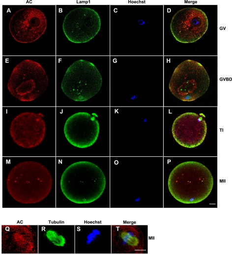
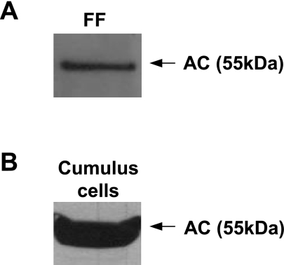
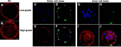
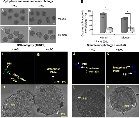
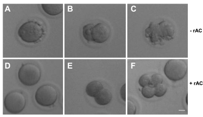
A major challenge of assisted reproduction technologies (ARTs) is to mimic the natural environment required to sustain oocyte and embryo survival. Herein, we show that the ceramide-metabolizing enzyme, acid ceramidase (AC), is expressed in human cumulus cells and follicular fluid, essential components of this environment, and that the levels of this enzyme are positively correlated with the quality of human embryos formed in vitro. These observations led us to develop a new approach for oocyte and embryo culture that markedly improved the outcome of in vitro fertilization (IVF). The addition of recombinant AC (rAC) to human and mouse oocyte culture medium maintained their healthy morphology in vitro. Following fertilization, the number of mouse embryos formed in the presence of rAC also was improved (from approximately 40 to 88%), leading to approximately 5-fold more healthy births. To confirm these observations, immature bovine oocytes were matured in vitro and subjected to IVF in the presence of rAC. Significantly more high-grade blastocysts were formed, and the number of morphologically intact, hatched embryos was increased from approximately 24 to 70%. Overall, these data identify AC as an important component of the in vivo oocyte and embryo environment, and provide a novel technology for enhancing the outcome of assisted fertilization. Eliyahu, E., Shtraizent, N., Martinuzzi, K., Barritt, J., He, X., Wei, H., Chaubal, S., Copperman, A. B., Schuchman, E. H. Acid ceramidase improves the quality of oocytes and embryos and the outcome of in vitro fertilization.


Figures

Figure 1
Figure 1
AC is highly expressed in human oocytes at different maturation stages. GV (A–D), GVBD (E–H), TI (I–L), and MII (M–T) oocytes. Oocytes were labeled using specific antibodies for AC (red; A, E, I, M, Q), Lamp1 (green; B, F, J, N), and tubulin (green; R), and for DNA using Hoechst stain (C, G, K, O, S). Localization of the primary antibodies was visualized using a fluorescent second antibody, Cy-3/2, and laser-scanning confocal microscopy. Merged images indicate colocalization of labeled proteins and/or DNA (D, H, L, P, T). As a control, oocytes were labeled with secondary antibodies only (data not shown). Scale bars = 10 μm. Images are representative of 3 independent experiments.
Figure 2
Figure 2
AC is present in human follicular fluid (FF) and cumulus cells. AC expression was determined by Western blotting in 10 μl of human FF (diluted to 1 μg/μl total protein concentration) (A), and 24 μg of total protein from cumulus cell extracts (B). Western blot analysis was performed using mouse anti-human AC IgG, revealing the human AC precursor (at 55 kDa). Western blot images are representative of ≥3 independent experiments.
Figure 3
Figure 3
AC expression in human embryos. A, B) Low AC expression in the blastocele fluid and outer cell mass of a representative low-grade human embryo (A) compared with high AC expression in a high-grade embryo (B), detected using goat IgG against human AC. C–J), Lower AC expression and higher apoptosis (TUNEL) in the outer cell mass of a low-grade embryo compared to the inner cell mass of the same embryo, which had higher AC expression and less apoptosis. AC was detected using goat IgG against human AC (E, I) and TUNEL assay (D, H); DNA labeling by Hoechst (C, G); merged images indicate colocalization (F, J). Scale bars = 10 μm. Data are representative of 3 independent experiments.
Figure 4
Figure 4
rAC supplementation improves the quality of oocytes cultured in vitro. Mature MII mouse oocytes (A, B) or immature and mature human oocytes (C, D, F–M), retrieved following superovulation, were incubated in vitro for 24 h in the absence or presence of rAC. Oocyte quality and apoptosis were assessed on the basis of membrane, cytoplasm, and DNA morphology using light imaging, TUNEL assay, and Hoechst DNA labeling. Fluorescence was visualized using confocal microscopy. AD) Representative images show the apoptotic appearance of mouse and human oocytes cultured in the absence of rAC (B, D) compared to those cultured with rAC (A, C). E) Percentages of apoptotic mouse and human oocytes cultured with and without rAC. Data represent analysis of 100 oocytes from each group (P<0.001 between groups with and without rAC). Black bars, without rAC; gray bars, with rAC. F–I) DNA fragmentation (apoptosis) assessed by TUNEL staining in a representative human oocyte incubated in the absence of rAC (F) compared to an oocyte cultured in the presence of rAC (G). PBI, first polar body (positive control for apoptosis). JM) Condensed appearance of DNA in a human oocyte incubated in the absence of rAC (J), compared to an intact metaphase plate in an oocyte cultured in the presence of rAC (K). Scale bars = 10 μm. Images represent analysis of 3 different oocytes with and without rAC treatment.
Figure 5
Figure 5
rAC improves the quality and number of mouse embryos derived by IVF and the number of mouse pups born following reimplantation. MII mouse oocytes were retrieved following superovulation and incubated in vitro for 6 h (aged oocytes) in order to mimic the conditions of human IVF, followed by fertilization in the absence (A–C) or presence (D–F) of rAC-supplemented fertilization medium. Representative images were recorded, and percentages of oocytes developing to the 2- to 4-cell stage were determined. Images illustrate the apoptotic morphology of aged oocytes (A) and embryos (B, C) derived from IVF in the absence of rAC, compared to the normal appearance of oocytes (D) and embryos (E, F) derived by IVF in the presence of rAC, assessed by light microscopy. Scale bar = 10 μm. Images are representative of ≥3 independent experiments.

Similar articles

Cited by

References

    1. Kolialexi A., Mavrou A., Spyrou G., Tsangaris G. T. Mass spectrometry-based proteomics in reproductive medicine. Mass Spectrom Rev. 2008;27:624–634. - PubMed
    1. Revelli A., Delle Piane L., Casano S., Molinari E., Massobrio M., Rinaudo P. Follicular fluid content and oocyte quality: from single biochemical markers to metabolomics. Reprod Biol Endocrinol. 2009;7:40–53. - PMC - PubMed
    1. Perez G. I., Tao X. J., Tilly J. L. Fragmentation and death (aka apoptosis) of ovulated oocytes. Mol Hum Reprod. 1999;5:414–420. - PubMed
    1. Galli C., Lazzari G. The manipulation of gametes and embryos in farm animals. Reprod Domest Anim. 2008;43(Suppl. 2):1–7. - PubMed
    1. Pukazhenthi B. S., Wildt D. E. Which reproductive technologies are most relevant to studying, managing and conserving wildlife? Reprod Fertil Dev. 2004;16:33–46. - PubMed

Publication types

LinkOut - more resources