. 2010 Mar;298(3):R584-98.
doi: 10.1152/ajpregu.00452.2009. Epub 2009 Dec 9.

A peripheral neuroimmune link: glutamate agonists upregulate NMDA NR1 receptor mRNA and protein, vimentin, TNF-alpha, and RANTES in cultured human synoviocytes

Affiliations

A peripheral neuroimmune link: glutamate agonists upregulate NMDA NR1 receptor mRNA and protein, vimentin, TNF-alpha, and RANTES in cultured human synoviocytes

Terry A McNearney et al. Am J Physiol Regul Integr Comp Physiol. 2010 Mar.

Abstract

Human primary and clonal synovial cells were incubated with glutamate receptor agonists to assess their modulating influence on glutamate receptors N-methyl-d-aspartate (NMDA) NR1 and NR2 and inflammatory cytokines to determine potential for paracrine or autocrine (neurocrine) upregulation of glutamate receptors, as has been shown for bone and chondrocytes. Clonal SW982 synoviocytes constitutively express vimentin, smooth muscle actin (SMA), and NMDA NR1 and NR2. Coincubation (6 h) with glutamate agonists NMDA (5 microM), and the NMDA NR1 glycine site activator (+/-)1-aminocyclopentane-cis-1,3-dicarboxylic acid (5 muM), significantly increases cellular mRNA and protein levels of glutamate receptors, as well as increasing vimentin, SMA, tumor necrosis factor-alpha, and RANTES (regulated on activation, normal T-cell expressed and secreted), assessed qualitatively and quantitatively with nucleotide amplification, image analysis of immunocytochemical staining, fluorescein-activated cell sorting, Western blotting, and immunoassays. Human primary synovial cells harvested from patients with arthritic conditions also constitutively expressed NMDA NR1 with increases after agonist treatment. Glutamate receptor agonist-induced increases were blocked by the noncompetitive glutamate antagonist MK-801 (8 microg/ml) and NR1 blocking antibody. Coincubation with glutamate agonists and phorbol 12-myristate 13-acetate, a protein kinase C activator, significantly enhanced mean levels of TNF-alpha and RANTES in SW982 cell supernatants compared with incubation with either agent alone. Increases were diminished with protein kinase inhibitor and NR1 blocking antibody. The functional activation of glutamate receptors on human synoviocytes establishes a neurogenic cell signaling link between neurotransmitter glutamate released from nerve terminals and target cells in the joint capsule. The influence of glutamate on subsequent release of cellular proinflammatory mediators in non-neural tissue for activation of downstream immune events supports a peripheral neuroimmune link in arthritis.


Figures
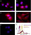
Fig. 1.
Fig. 1.
Glutamate N-methyl-d-aspartate (NMDA) NR1 receptors on human SW892 cultured synoviocytes increase after treatment with NMDA and (±)1-aminocyclopentane-cis-1,3-dicarboxylic acid (ACPD). A: glutamate NMDA NR1 receptor staining (red) is imaged in a high-power photomicrograph of control untreated synovial cells. B–D: increases in immunostaining for glutamate NMDA NR1 are evident in cells incubated concurrently with 5 μM NMDA and 5 μM ACPD for 2 (B), 6 (C), and 24 h (D). Cells shown were stained with rabbit anti-NMDA NR1 antibody (1:100). E: cells were stained with normal rabbit serum (NRS) as a nonspecific control (1:100). Dual nuclear counterstaining with 4,6-diamidino-2-phenylindole (DAPI) is shown in blue. Bar, 20 μm. F: 1 of 3 fluorescence-activated cell sorting (FACS) analyses used to assess expression of NMDA NR1 subunit protein in SW892 cultured synoviocytes treated for 6 h with 5 μM NMDA and 5 μM ACPD (except peak 3). Peak 1, with secondary antibody only; peak 2, nonspecific rabbit serum control; peak 3, untreated SW892 cultured synoviocytes with rabbit anti-NMDA NR1 antibody; peak 4, rabbit anti-NR1 antibody; peak 5, mouse monoclonal anti-vimentin antibody. Nonspecific antibody control peaks (not shown) were identical to peak 2 in both untreated and NMDA/ACPD-treated cultures. Fluorescence intensity was read at 530 nm.
Fig. 2.
Fig. 2.
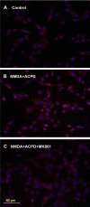
Preincubation with MK-801 blocks NMDA NR1 upregulation in synoviocytes. A: low-power photomicrograph of glutamate NMDA NR1 receptors (red) stained with rabbit anti-NMDA NR1 antibody on control untreated synovial cells. B: cells incubated concurrently with 5 μM NMDA and 5 μM ACPD for 6 h. C: cells preincubated for 1 h with 10 μM MK-801, a selective, noncompetitive NMDA receptor antagonist, and then incubated for 6 h with 5 μM NMDA and 5 μM ACPD. Dual nuclear counterstaining with DAPI is shown in blue. Bar, 80 μm.
Fig. 3.
Fig. 3.
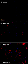
Immunocytochemical localization of glutamate NMDA NR1 on primary synovial cell cultures. Photomicrographs of cells derived from synovial fluid of a patient with rheumatoid arthritis are stained (red) immunocytochemically for glutamate NMDA NR1 receptor subunit. A: untreated synovial cells were stained with NRS as a nonspecific matched antibody control. B: immunostaining for NMDA NR1 receptor subunit is evident in synoviocyte cell cultures stained with NMDA NR1 subunit antibody. C: increases in the immunostaining for NMDA NR1 receptor subunit are evident in synoviocyte cultures stained with NMDA NR1 subunit antibody after coincubation with 5 μM NMDA and 5 μM ACPD for 6 h. Dual nuclear counterstaining with DAPI is shown in blue. Bar, 80 μm.
Fig. 4.
Fig. 4.
Glutamate NMDA NR2 receptor subunit expression on synoviocytes identified by immunostaining and RT-PCR. A: high-power photomicrograph of glutamate NMDA NR2 receptors (red) on control untreated clonal SW892 human synoviocytes synovial cells. B: immunostaining was increased in cells incubated concurrently with 5 μM NMDA and 5 μM ACPD for 6 h. Cells shown were stained with rabbit anti NMDA NR2 antibody. Bar, 20 μm. C: amplification of specific NMDA NR2 subunits was identified by RT-PCR in human synoviocytes. Cytosolic lysates of untreated SW982 cells (synoviocytes) and human brain (hippocampus) as control were applied to SDS-PAGE and transferred to a nitrocellulose filter. Lanes were primed for NMDA NR2 subunits A–D as shown. All 4 subunits were expressed in the lysates from human brain. NMDA NR2 subunits A, B, and D were expressed in lysates from untreated SW982 synoviocytes.
Fig. 5.
Fig. 5.
Vimentin expression on clonal SW982 synoviocytes. A and B: high-power photomicrographs of immunocytochemical staining for vimentin in control untreated synovial cells (A) and in cells coincubated with 5 μM NMDA and 5 μM ACPD for 6 h (B). Matched nonspecific antibody staining yielded no detectable fluorescence. Bar, 20 μm. C: bar graph showing the mean relative fluorescence densities of cellular proteins after glutamate receptor activation in SW982 cultured synoviocytes. Mean cellular protein densities were calculated from unbiased sampled fields using Bioquant software. The untreated cell culture groups are illustrated with open bars, and filled bars represent cell cultures that were coincubated for 6 h with 5 μM NMDA and 5 μM ACPD. Significant increases (*P < 0.05) in mean fluorescence densities were noted for vimentin, NMDA NR1, and NMDA NR2 staining in the NMDA- and ACPD-treated cells compared with untreated cells for each of these antibody pairs. No increases in fluorescence intensity were noted comparing control and treated cells with smooth muscle actin (SMA) staining. Negligible fluorescence densities were noted with nonspecific, anti-CD11b, or anti-CD21 antibody staining in untreated and treated synoviocyte cultures.
Fig. 6.
Fig. 6.
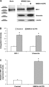
NMDA NR1 and vimentin expression by Western blot and real-time PCR analysis of clonal SW982 synoviocytes. A: cytosolic lysates were applied to SDS-PAGE and transferred to a nitrocellulose filter. Top row: human brain (hippocampus) was used as a positive control for NMDA NR1 subunit protein, at 116 kDa (left lane). Untreated SW982 human synovial cell lysate was stained with mouse monoclonal anti-NMDA NR1 antibody (middle lane). SW982 human synovial cell lysate was coincubated with 5 μM NMDA and 5 μM ACPD for 6 h and stained with mouse monoclonal anti-NMDA NR1 antibody(right lane). Middle row: the blot was stripped, and the lanes were then stained with mouse monoclonal anti-vimentin antibody. Middle lane, untreated cells. Right lane: cells were coincubated with 5 μM NMDA and 5 μM ACPD for 6 h. Bottom row: All lanes were also stained for β-actin as an internal control. Left lane was exposed for 10 s, and all other lanes were exposed for 30 s. Similar results were obtained on Western blot studies using a mouse monoclonal antibody specific for NMDA NR1 (shown in A) or a rabbit polyclonal antibody. B: corrected (normalized) densitometry determinations of Western blot staining for NMDA- and ACPD-treated cells showed a 3-fold increase in NMDA NR1 subunit protein and a 3.1-fold increase in vimentin protein compared with untreated cells (control, *P < 0.01). C: the mean relative NMDA NR1 gene expression level determined by real-time PCR was increased in NMDA- and ACPD-stimulated synoviocytes (5.8-fold) compared with untreated cells (control, *P < 0.01).
Fig. 7.
Fig. 7.
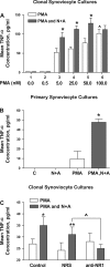
Mean tumor necrosis factor-α (TNF-α) levels increase in clonal and primary synovial cell cultures after treatment with glutamate receptor agonists measured by immunoassay. A: dose response for increasing doses of PMA with a fixed dose of 5 μM NMDA and 5 μM ACPD (N+A). Coincubation of SW982 human synoviocytes with PMA and N+A resulted in a significant increase in mean TNF-α concentrations compared with cell cultures incubated with PMA alone, demonstrating an enhancement of PMA activation by glutamate receptor agonists. *P < 0.05, PMA alone vs. PMA and N+A. ^P < 0.05, 100 nM PMA vs. 25 nM PMA. Each condition represents the mean of 9 samples. B: coincubation of primary synovial cell cultures derived from the synovial fluid of a patient with active psoriatic arthritis with 25 nM PMA, 5 μM NMDA, and 5 μM ACPD resulted in a significant increase (4.3-fold) in mean TNF-α concentrations compared with cell cultures incubated with PMA alone. *P < 0.05, PMA alone vs. PMA and N+A. Untreated cells and cells treated with 5 μM NMDA and 5 μM ACPD did not demonstrate increases in TNF-α levels in the culture supernatant. C: coincubation of SW892 human synoviocytes with 25 nM PMA, 5 μM NMDA, and 5 μM ACPD resulted in a significant increase in mean TNF-α concentrations compared with cell cultures incubated with PMA alone (*P < 0.05 compared with PMA alone). Preincubation of the cells with NRS did not diminish the enhancement seen with PMA, NMDA, and ACPD incubations (**P < 0.01 compared with PMA alone). However, preincubation with specific NMDA NR1 antibody blocked the increase in mean TNF-α levels seen with coincubation of SW892 synoviocytes with PMA and NMDA and ACPD (^P < 0.05 comparing cells preincubated with NRS to cells preincubated with NMDA NR1 antibody).
Fig. 8.
Fig. 8.
Preincubation of synoviocytes with bisindolylmaleimide-1 (Bis), a potent protein kinase C (PKC) inhibitor, markedly decreases mean TNF-α and RANTES concentrations. A: SW982 cells preincubated with Bis had significant reductions in supernatant TNF-α levels compared with levels generated with PMA alone or PMA and N+A incubations. *P < 0.05, Bis-treated cells compared with cells not treated with Bis. **P < 0.01 compared with cells preincubated with NMDA NRS. ^P < 0.05, cells coincubated with PMA and N+A compared with cells treated with PMA alone. The degree of inhibition was dose dependent. The control lanes (C) depict baseline levels obtained in the absence of PMA or in the absence of PMA but coincubated with NMDA and ACPD. B: in independent experiments, preincubation of SW982 synoviocyte cultures with Bis markedly decreased the mean RANTES levels generated with PMA alone or with PMA and NMDA+ACPD incubations. **P < 0.01, Bis-treated cells compared with cells not treated with Bis. ^P < 0.05, cells coincubated with PMA and NMDA+ACPD compared with cells treated with PMA alone.
Fig. 9.
Fig. 9.
Normal and inflamed rat knee joints: histological and immunocytochemical staining for NMDA NR1. Microphotographs of knee joint capsules from naive rats (A and B) and from rats 1 wk after induction of arthritis with complete Freund's adjuvant (CFA) (C and D). Note the thin synovial cell layer at the edge of the tissue section in the naive animal with no apparent NMDA N1 staining (arrows). The proliferation of synoviocytes in animals with inflamed knee joints is evident histologically (C) and with NMDA NR1 staining throughout most of the section in D (arrows). A and C, hematoxylin and eosin stain; B and D, NMDA NR1 immunohistochemical stain.

Similar articles

Cited by

References

    1. Behr TM, Spes CH, Pongratz DE, Weiss M, Meiser B, Uberfuhr P, Theisen K, Angermann CE. Adult human cardiomyocytes coexpress vimentin and Ki67 in heart transplant rejection and in dilated cardiomyopathy. J Heart Lung Transplant 17: 795–800, 1998 - PubMed
    1. Chenu C, Serre CM, Raynal C, Burt-Pichat B, Delmas PD. Glutamate receptors are expressed by bone cells and are involved in bone resorption. Bone 22: 295–299, 1998 - PubMed
    1. Dougherty PM, Sluka KA, Sorkin LS, Westlund KN, Willis WD. Neural changes in acute arthritis in monkeys. I. Parallel enhancement of responses of spinothalamic tract neurons to mechanical stimulation and excitatory amino acids. Brain Res Brain Res Rev 17: 1–13, 1992 - PubMed
    1. Ehlers MD, Fung ET, O'Brien RJ, Huganir RL. Splice variant-specific interaction of the NMDA receptor subunit NR1 with neuronal intermediate filaments. J Neurosci 18: 720–730, 1998 - PMC - PubMed
    1. Flood S, Parri R, Williams A, Duance V, Mason D. Modulation of interleukin-6 and matrix metalloproteinase 2 expression in human fibroblast-like synoviocytes by functional ionotropic glutamate receptors. Arthritis Rheum 56: 2523–2534, 2007 - PubMed

Publication types

MeSH terms