

Ectopic activation of Mycobacterium tuberculosis-specific CD4+ T cells in lungs of CCR7-/- mice
- PMID: 20007536
- PMCID: PMC2879893
- DOI: 10.4049/jimmunol.0901230
Ectopic activation of Mycobacterium tuberculosis-specific CD4+ T cells in lungs of CCR7-/- mice
Abstract
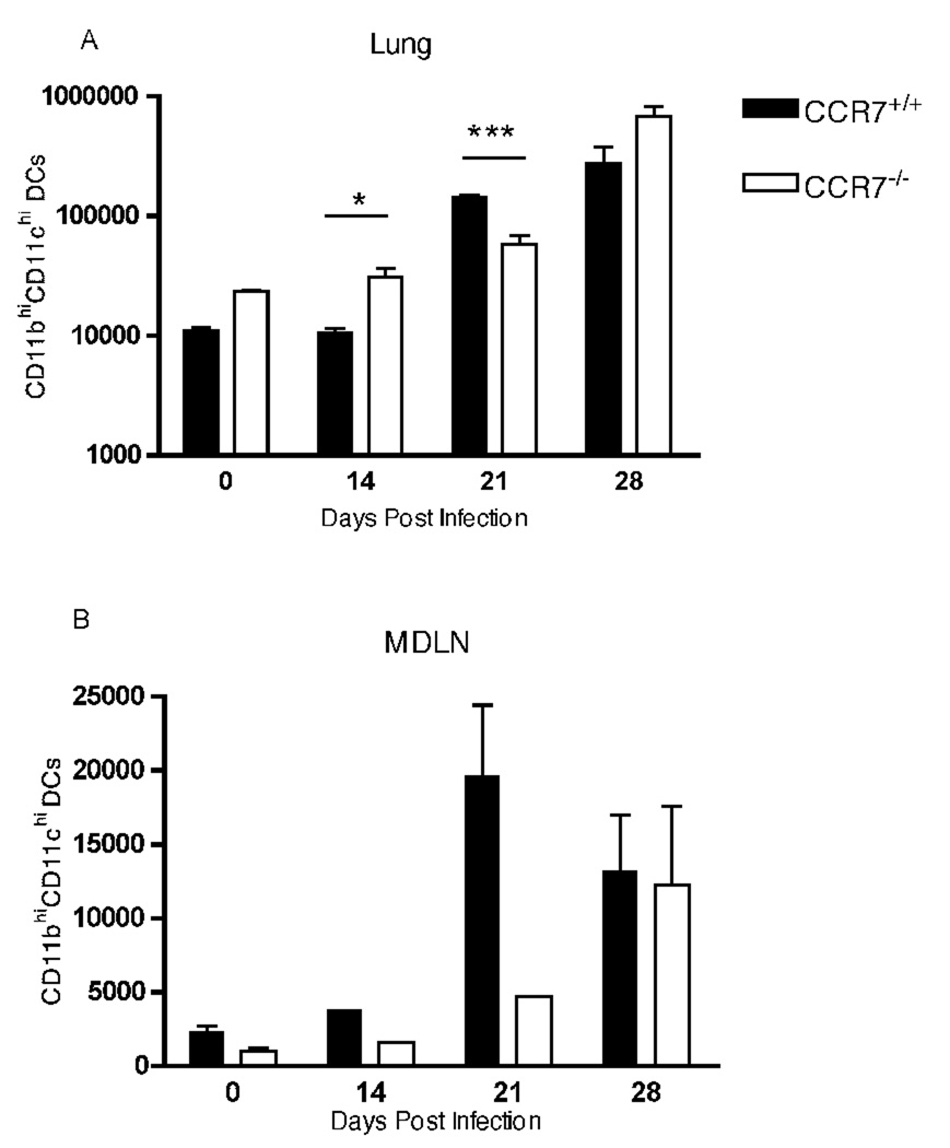
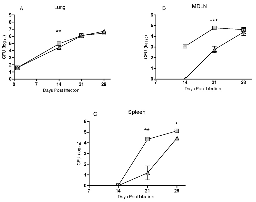
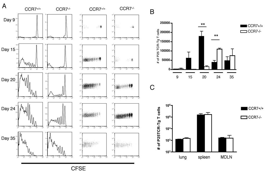
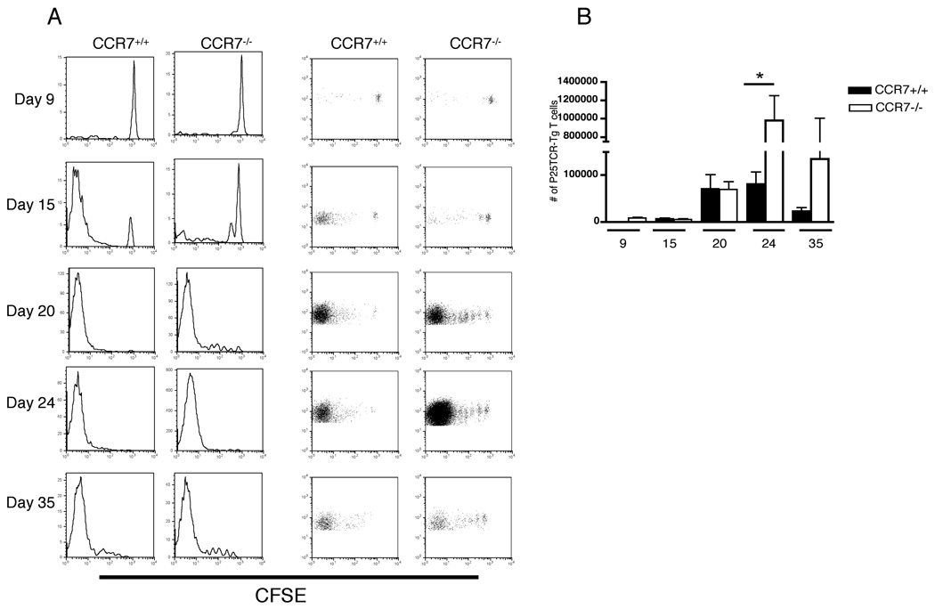
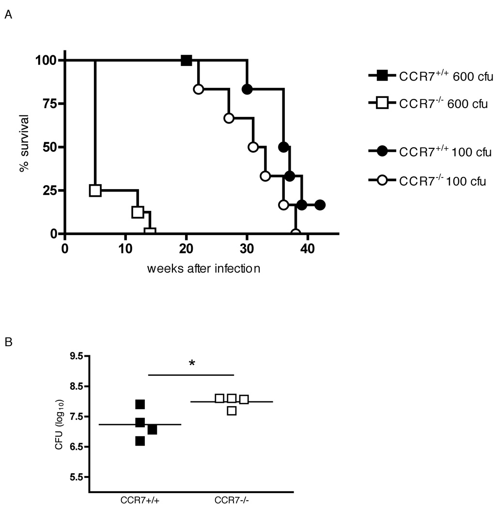
Initiation of an adaptive cellular immune response depends on intimate interactions with APCs and naive T lymphocytes. We previously reported that activation of naive Mycobacterium tuberculosis-specific CD4+ T cells depends on dendritic cell (DC) transport of live bacteria from the lungs to the mediastinal lymph node (MDLN). Because the migratory paths of DCs are largely governed by the chemokine receptor CCR7, which is expressed on DCs upon maturation by proinflammatory stimuli, we examined the quantitative contribution of CCR7-dependent DC migration in the context of tuberculosis. We found that early trafficking of DCs from the lungs to the MDLN depended on CCR7-mediated signaling, but alternative mechanism(s) are used later in infection. Impaired migration of DCs in CCR7(-/-) mice resulted in delayed dissemination of bacteria to MDLN and spleen and in delayed kinetics of activation of adoptively transferred Ag85B-specific CD4+ T cells. Furthermore, in contrast to control mice, we found that naive Ag85B-specific CD4+ T cells are activated to proliferate in the lungs of CCR7(-/-) mice and, when infected with higher doses of bacteria, resistance to M. tuberculosis infection in CCR7(-/-) mice is compromised compared with wild-type mice.
Figures
Similar articles
-
Lung neutrophils facilitate activation of naive antigen-specific CD4+ T cells during Mycobacterium tuberculosis infection.J Immunol. 2011 Jun 15;186(12):7110-9. doi: 10.4049/jimmunol.1100001. Epub 2011 May 9. J Immunol. 2011. PMID: 21555529 Free PMC article.
-
C-C Motif Chemokine Receptor 7 Exacerbates Hypertension Through Effects on T Lymphocyte Trafficking.Hypertension. 2020 Mar;75(3):869-876. doi: 10.1161/HYPERTENSIONAHA.119.14148. Epub 2020 Jan 27. Hypertension. 2020. PMID: 31983306 Free PMC article.
-
Mycobacterium tuberculosis infects dendritic cells with high frequency and impairs their function in vivo.J Immunol. 2007 Aug 15;179(4):2509-19. doi: 10.4049/jimmunol.179.4.2509. J Immunol. 2007. PMID: 17675513
-
Initiation of the adaptive immune response to Mycobacterium tuberculosis depends on antigen production in the local lymph node, not the lungs.J Exp Med. 2008 Jan 21;205(1):105-15. doi: 10.1084/jem.20071367. Epub 2007 Dec 24. J Exp Med. 2008. PMID: 18158321 Free PMC article.
-
Modulation of pulmonary dendritic cell function during mycobacterial infection.Infect Immun. 2008 Feb;76(2):671-7. doi: 10.1128/IAI.01079-07. Epub 2007 Nov 26. Infect Immun. 2008. PMID: 18039834 Free PMC article.
Cited by
-
Cytokines and Chemokines in Mycobacterium tuberculosis Infection.Microbiol Spectr. 2016 Oct;4(5):10.1128/microbiolspec.TBTB2-0018-2016. doi: 10.1128/microbiolspec.TBTB2-0018-2016. Microbiol Spectr. 2016. PMID: 27763255 Free PMC article. Review.
-
Initial immune response after exposure to Mycobacterium tuberculosis or to SARS-COV-2: similarities and differences.Front Immunol. 2023 Aug 17;14:1244556. doi: 10.3389/fimmu.2023.1244556. eCollection 2023. Front Immunol. 2023. PMID: 37662901 Free PMC article. Review.
-
Lung neutrophils facilitate activation of naive antigen-specific CD4+ T cells during Mycobacterium tuberculosis infection.J Immunol. 2011 Jun 15;186(12):7110-9. doi: 10.4049/jimmunol.1100001. Epub 2011 May 9. J Immunol. 2011. PMID: 21555529 Free PMC article.
-
Cell-to-cell transfer of M. tuberculosis antigens optimizes CD4 T cell priming.Cell Host Microbe. 2014 Jun 11;15(6):741-52. doi: 10.1016/j.chom.2014.05.007. Cell Host Microbe. 2014. PMID: 24922576 Free PMC article.
-
Biosafety Level 3 setup for multiphoton microscopy in vivo.Sci Rep. 2017 Apr 3;7(1):571. doi: 10.1038/s41598-017-00702-x. Sci Rep. 2017. PMID: 28373723 Free PMC article.
References
-
- Winslow GM, Roberts AD, Blackman MA, Woodland DL. Persistence and turnover of antigen-specific CD4 T cells during chronic tuberculosis infection in the mouse. J Immunol. 2003;170:2046–2052. - PubMed
Publication types
MeSH terms
Substances
Grants and funding
LinkOut - more resources
Full Text Sources
Medical
Molecular Biology Databases
Research Materials
Miscellaneous
