Comparative Study
. 2009 Dec 15;183(12):8244-57.
doi: 10.4049/jimmunol.0901958.

Differential role of the Fas/Fas ligand apoptotic pathway in inflammation and lung fibrosis associated with reovirus 1/L-induced bronchiolitis obliterans organizing pneumonia and acute respiratory distress syndrome

Affiliations
Comparative Study

Differential role of the Fas/Fas ligand apoptotic pathway in inflammation and lung fibrosis associated with reovirus 1/L-induced bronchiolitis obliterans organizing pneumonia and acute respiratory distress syndrome

Andrea D Lopez et al. J Immunol. .

Abstract

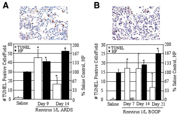
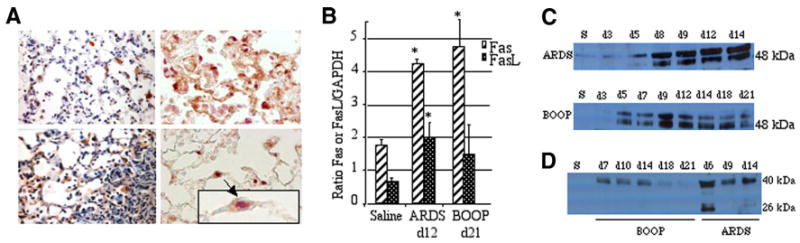
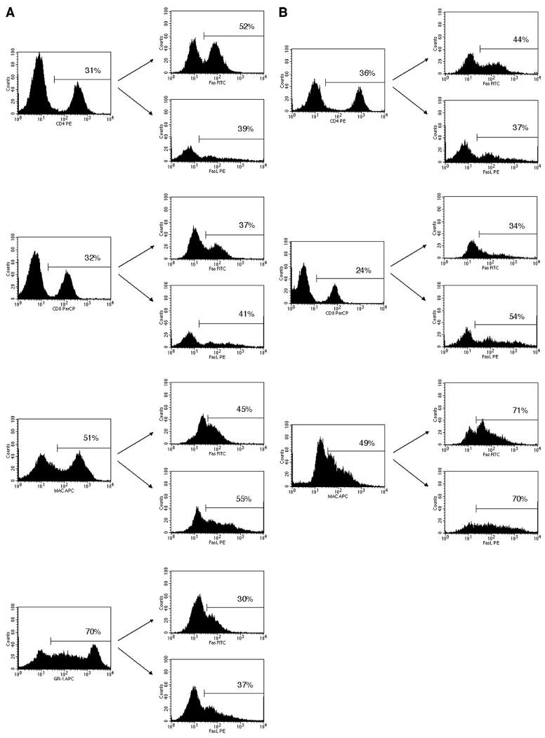
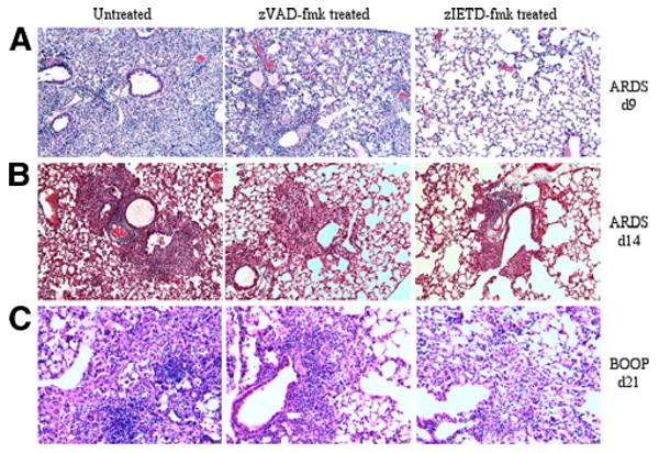
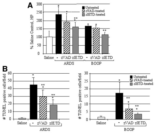
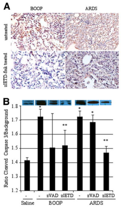
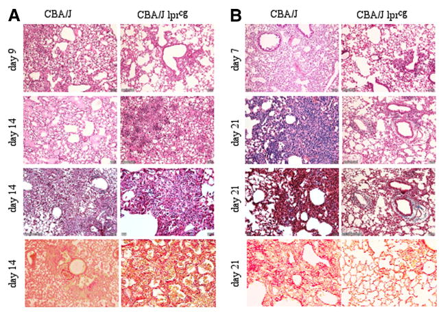
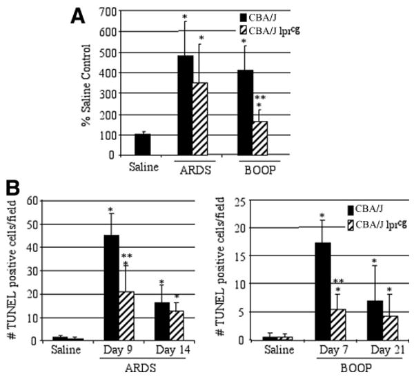
Bronchiolitis obliterans organizing pneumonia (BOOP) and acute respiratory distress syndrome (ARDS) are two clinically and histologically distinct syndromes sharing the presence of an inflammatory and fibrotic component. Apoptosis via the Fas/Fas ligand (FasL) pathway plays an important role in the development of acute lung injury and fibrosis characteristic of these and other pulmonary inflammatory and fibrotic syndromes. We evaluated the role of apoptosis via the Fas/FasL pathway in the development of pulmonary inflammation and fibrosis in reovirus 1/L-induced BOOP and ARDS. CBA/J mice were intranasally inoculated with saline, 1 x 10(6) (BOOP), or 1 x 10(7) (ARDS) PFU reovirus 1/L, and evaluated at various days postinoculation for in situ apoptosis by TUNEL analysis and Fas/FasL expression. Our results demonstrate the presence of apoptotic cells and up-regulation of Fas/FasL expression in alveolar epithelium and in infiltrating cells during the inflammatory and fibrotic stages of both reovirus 1/L-induced ARDS and BOOP. Treatment of mice with the caspase 8 inhibitor, zIETD-fmk, inhibited apoptosis, inflammation, and fibrotic lesion development in reovirus 1/L-induced BOOP and ARDS. However, CBA/KlJms-Fas(lpr-cg)/J mice, which carry a point mutation in the Fas cytoplasmic region that abolishes the ability of Fas to transduce an apoptotic signal, do not develop pulmonary inflammation and fibrotic lesions associated with reovirus 1/L-induced BOOP, but still develop inflammation and fibrotic lesions associated with reovirus 1/L-induced ARDS. These results suggest a differential role for the Fas/FasL apoptotic pathway in the development of inflammation and fibrotic lesions associated with BOOP and ARDS.


Conflict of interest statement

Disclosures: The authors have no financial conflicts of interest.

Figures

FIGURE 1
FIGURE 1
Apoptosis is evident in reovirus 1/L-induced ARDS and BOOP. CBA/J mice were i.n. inoculated with either 1 × 10⁶ PFU (BOOP) or 1 × 10⁷ PFU (ARDS) reovirus 1/L and sacrificed at the indicated time points. Apoptosis in situ was assessed using the TUNEL technique on paraffin-embedded lung sections. The extent of pulmonary fibrosis was also determined by estimating total lung collagen as reflected by HP content of the lung. A, Reovirus 1/L-induced ARDS; representative in situ TUNEL staining on day 9 postreovirus 1/L-induced ARDS (top). Histograms are TUNEL-positive cells (open) and HP content (filled) in saline-inoculated control (day 9) and on days 9 and 14 after reovirus 1/L-induced ARDS (bottom). B, Reovirus 1/L-induced BOOP; representative in situ TUNEL staining on day 14 after reovirus 1/L-induced BOOP (top). Histograms are TUNEL-positive cells (open) and HP content (filled) in saline-inoculated control (day 7) and on days 7, 14, and 21 after reovirus 1/L-induced BOOP (bottom). TUNEL data represent mean ± SD of eight 225-mm2 fields from two independent experiments with three mice per time point. HP data represent the mean ± SD of four mice per time point. Results are expressed as a percentage of HP content in saline-inoculated, control mice. ND, not determined. *, p < 0.05 compared with saline-inoculated, control mice.
FIGURE 2
FIGURE 2
Both Fas and FasL are expressed in reovirus 1/L-induced ARDS and BOOP. CBA/J mice were i.n. inoculated with either 1 × 10⁶ PFU (BOOP) or 1 × 10⁷ PFU (ARDS) reovirus 1/L and sacrificed at the indicated time points. A, Lungs of reovirus 1/L-induced ARDS were paraffin embedded and stained with an Ab to either Fas (top left) or FasL (bottom left) on day 9. IHC for Fas and FasL is representative of four experiments with two mice per time point. Lung sections from reovirus 1/L-induced ARDS were also dual stained with an Ab to Fas (brown-precipitate) and TTF-1, a marker for alveolar epithelial type II cells (purple precipitate) (top right and bottom right) on day 9. IHC for Fas and TTF-1 is representative of four individual mice. An enlarged image of an alveolar epithelial cell (arrow) stained positively for both TTF-1 (predominant purple staining nucleus) and Fas (predominant brown staining cytoplasm) is shown in the indicated boxed region. B, RNA was prepared from whole lung tissue of either saline or reovirus 1/L-inoculated mice. Relative expression of Fas or FasL on day 12 reovirus 1/L-induced ARDS or on day 21 reovirus 1/L-induced BOOP was determined by comparing the ratio of Fas or FasL mRNA with the housekeeping gene, GAPDH. Histograms represent densitometric data from the mean ± SD autoradiogram signals from three mice per time point. *, p < 0.05 compared with saline-inoculated, control mice. C, Western analysis from whole lung lysates for protein expression of Fas overtime in reovirus 1/L-induced ARDS and BOOP; representative of two independent experiments (S, saline day 9). D, Western analysis from BAL fluid for protein expression of FasL overtime in reovirus 1/L-induced ARDS and BOOP. Representative of two independent experiments (S, saline day 9).
FIGURE 3
FIGURE 3
Fas and FasL are expressed on the surface of infiltrating cells in reovirus 1/L-induced ARDS and BOOP. CBA/J mice were i.n. inoculated with either 1 × 10⁶ PFU (BOOP) or 1 × 10⁷ PFU (ARDS) reovirus 1/L and cells recovered by BAL were analyzed for the coexpression of cell surface phenotype markers with surface expression of Fas and FasL. Infiltrating cells were first stained with either anti-CD4, anti-CD8, anti-Mac1, or anti-GR1. The gated positive cells were then analyzed for the expression of either Fas or FasL. A, Cells recovered from ARDS-induced mice on day 9 postinoculation. B, Cells recovered from BOOP-induced mice on day 7 postinoculation. The percentage of the total cells expressing either anti-CD4, anti-CD8, anti-Mac1, or anti-GR1 is shown in the histograms on the left. The percentage of the gated population expressing either Fas or FasL is shown in the histograms on the right. The results shown represent one of the three independent experiments demonstrating similar results.
FIGURE 4
FIGURE 4
Both inflammation and fibrosis are diminished in zIETD-fmk-treated reovirus 1/L-induced ARDS or BOOP. CBA/J mice were i.n. inoculated with either 1 × 10⁶ PFU (BOOP) or 1 × 10⁷ PFU (ARDS) reovirus 1/L and were left untreated or treated with 5 mg/kg of either zVAD-fmk or zIETD-fmk daily beginning on day 3 postinoculation. A, Reovirus 1/L-induced ARDS at day 9 (H&E). B, Reovirus 1/L-induced ARDS at day 14 (Masson's trichrome). C, Reovirus 1/L-induced BOOP at day 21 (H&E). Objective magnification, ×20. Results are representative of four independent experiments evaluating four mice per time point.
FIGURE 5
FIGURE 5
Collagen deposition and apoptosis are diminished in zIETD-fmk treated reovirus 1/L-induced ARDS or BOOP. CBA/J mice were i.n. inoculated with either 1 × 10⁶ PFU (BOOP) or 1 × 10⁷ PFU (ARDS) reovirus 1/L and were left untreated or treated with 5 mg/kg of either zVAD-fmk or zIETD-fmk daily beginning on day 3 postinoculation until sacrifice. A, The extent of pulmonary fibrosis was determined by estimating total lung collagen as reflected by the measurement of the HP content of the lung. Results are expressed as a percentage of HP content in saline-inoculated, control mice. Results shown are for day 14 ARDS and day 21 BOOP. Each data point represents the mean ± SD of six mice. *, p < 0.05 compared with saline-inoculated, control mice (day 14); **, p < 0.05 compared with reovirus 1/L-inoculated mice. B, Apoptosis in situ was assessed using TUNEL labeling on paraffin-embedded lung sections in saline (day 9 ARDS, day 14 BOOP), reovirus 1/L-induced ARDS (day 9) and reovirus 1/L-induced BOOP (day 14). TUNEL data represent mean ± SD of eight 225-mm2 fields from two independent experiments with three mice per time point. *, p < 0.05 compared with saline-inoculated, control mice; **, p < 0.05 compared with reovirus 1/L-inoculated mice.
FIGURE 6
FIGURE 6
The presence of both caspase-8 and caspase-3 are diminished in zIETD-fmk reovirus 1/L-induced ARDS or BOOP. CBA/J mice were i.n. inoculated with either 1 × 10⁶ PFU (BOOP) or 1 × 10⁷ PFU (ARDS) reovirus 1/L and were left untreated or treated with 5 mg/kg of either zVAD-fmk or zIETD-fmk daily beginning on day 3 postinoculation until sacrifice. A, Lungs of reovirus 1/L-induced BOOP and ARDS on day 14 postinoculation were paraffin embedded and stained with a rabbit polyclonal Ab for active/cleaved caspase-8 in either untreated (top) or zIETD-fmk-treated (bottom) reovirus 1/L-iduced BOOP or ARDS. IHC for active/cleaved caspase-8 is representative of two experiments with two mice per time point. B, Western analysis from whole lung lysates for protein expression of cleaved caspase-3 was determined in reovirus 1/L-induced ARDS on day 9 postinoculation and in reovirus 1/L-induced BOOP on day 14 postinoculation. Relative expression of cleaved caspase-3 was determined by comparing the ratio of the cleaved caspase-3 band to background. Histograms represent densitometric data from the mean ± SD autoradiogram signals from three mice for the saline, ARDS-induced, and BOOP-induced mice and from at least four mice in the zVAD-fmk- and zIETD-fmk-treated groups. *, p < 0.05 compared with saline-inoculated, control mice; **, p < 0.05 compared with reovirus 1/L-inoculated mice.
FIGURE 7
FIGURE 7
Both inflammation and fibrosis are diminished in reovirus 1/L-induced BOOP in CBA/KlJms-Faslpr-cg/J mice but not in reovirus 1/L-induced ARDS in CBA/KlJms-Faslpr-cg/J mice. CBA/J or CBA/KlJms-Faslpr-cg/J mice were i.n. inoculated with either 1 × 10⁶ PFU (BOOP) or 1 × 10⁷ PFU (ARDS) reovirus 1/L. Paraffin-embedded lung sections were stained with H&E (first and second panels), Masson's trichrome (third panel), or Sirius red (fourth or bottom panels). A, Reovirus 1/L-induced ARDS in CBA/J or CBA/KlJms-Faslpr-cg/J mice; B, Reovirus 1/L-induced BOOP in CBA/J or CBA/KlJms-Faslpr-cg/J mice; Objective magnification, ×20. Representative of two experiments with two mice per time point.
FIGURE 8
FIGURE 8
Apoptosis is reduced in reovirus 1/L-inoculated CBA/K1Jms-Faslpr-cg/J mice. CBA/J or CBA/KlJms-Faslpr-cg/J mice were i.n. inoculated with either 1 × 10⁶ PFU (BOOP) or 1 × 10⁷ PFU (ARDS) reovirus 1/L and sacrificed at the indicated time points. A, Sirius red staining in both CBA/J or CBA/KlJms-Faslpr-cg/J mice was quantitated in reovirus 1/L-induced ARDS (day 14) and in reovirus 1/L-induced BOOP (day 21) using ImageJ software. Results are expressed as the percentage of Sirius red content in saline-inoculated, control mice. Sirius red data represent mean ± SD of two experiments with two mice per time point. *, p < 0.05 compared with saline-inoculated, control mice. **, p < 0.05 compared with reovirus 1/L-inoculated mice. B, Apoptosis in situ was assessed using TUNEL labeling on paraffin-embedded lung sections. TUNEL-positive cells in saline (day 9 ARDS; day 7 BOOP) and in reovirus 1/L-induced ARDS on days 9 and 14 or in reovirus 1/L-induced BOOP on days 7 and 21 are shown. TUNEL data represent mean ± SD of eight 225-mm2 fields from two experiments with two mice per time point. *, p < 0.05 compared with saline-inoculated, control mice; **, p < 0.05 compared with reovirus 1/L-inoculated mice.

Similar articles

Cited by

References

    1. Cooper JA., Jr Pulmonary fibrosis: pathways are slowly coming into light. Am J Resp Cell Mol Biol. 2000;22:520–523. - PubMed
    1. Kuwano K, Hagimoto N, Hara N. Molecular mechanisms of pulmonary fibrosis and current treatment. Curr Mol Med. 2001;1:551–573. - PubMed
    1. Katzenstein AL, Mukhopadhyay S, Myers JL. Diagnosis of usual interstitial pneumonia and distinction from other fibrosing interstitial lung diseases. Hum Pathol. 2008;39:1275–1294. - PubMed
    1. Afshar K, Sharma OP. Interstitial lung disease: trials and tribulations. Curr Opin Pulm Med. 2008;14:427–433. - PubMed
    1. Drakopanagiotakis F, Polychronopoulos V, Judson MA. Organizing pneumonia. Am J Med Sci. 2008;335:34–39. - PubMed

Publication types

MeSH terms