

The structure of the KlcA and ArdB proteins reveals a novel fold and antirestriction activity against Type I DNA restriction systems in vivo but not in vitro
- PMID: 20007596
- PMCID: PMC2836571
- DOI: 10.1093/nar/gkp1144
The structure of the KlcA and ArdB proteins reveals a novel fold and antirestriction activity against Type I DNA restriction systems in vivo but not in vitro
Abstract
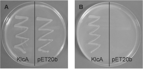
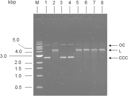
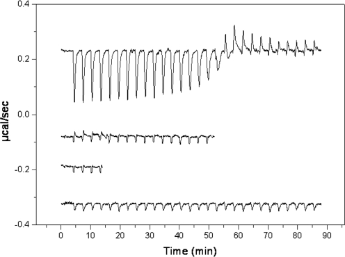
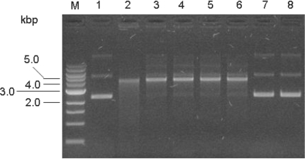
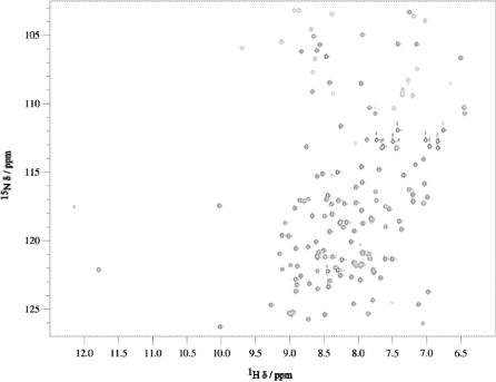
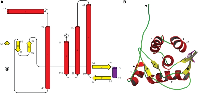
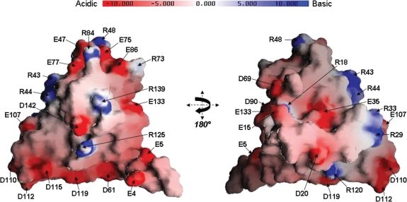
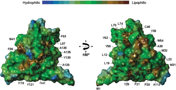
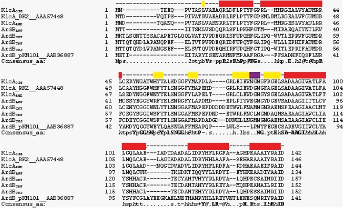
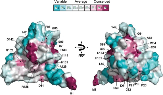
Plasmids, conjugative transposons and phage frequently encode anti-restriction proteins to enhance their chances of entering a new bacterial host that is highly likely to contain a Type I DNA restriction and modification (RM) system. The RM system usually destroys the invading DNA. Some of the anti-restriction proteins are DNA mimics and bind to the RM enzyme to prevent it binding to DNA. In this article, we characterize ArdB anti-restriction proteins and their close homologues, the KlcA proteins from a range of mobile genetic elements; including an ArdB encoded on a pathogenicity island from uropathogenic Escherichia coli and a KlcA from an IncP-1b plasmid, pBP136 isolated from Bordetella pertussis. We show that all the ArdB and KlcA act as anti-restriction proteins and inhibit the four main families of Type I RM systems in vivo, but fail to block the restriction endonuclease activity of the archetypal Type I RM enzyme, EcoKI, in vitro indicating that the action of ArdB is indirect and very different from that of the DNA mimics. We also present the structure determined by NMR spectroscopy of the pBP136 KlcA protein. The structure shows a novel protein fold and it is clearly not a DNA structural mimic.
Figures

Similar articles
-
Antirestriction activities of KlcA (RP4) and ArdB (R64) proteins.FEMS Microbiol Lett. 2018 Dec 1;365(23). doi: 10.1093/femsle/fny227. FEMS Microbiol Lett. 2018. PMID: 30239714
-
ArdB Protective Activity for Unmodified λ Phage Against EcoKI Restriction Decreases in UV-Treated Escherichia coli.Curr Microbiol. 2019 Nov;76(11):1374-1378. doi: 10.1007/s00284-019-01755-z. Epub 2019 Aug 12. Curr Microbiol. 2019. PMID: 31407052
-
[Anti-Restriction Activity of ArdB Protein against EcoAI Endonuclease].Mol Biol (Mosk). 2023 Jan-Feb;57(1):101-105. doi: 10.31857/S0026898423010056. Mol Biol (Mosk). 2023. PMID: 36976744 Russian.
-
[Antirestriction proteins ardA and Ocr as effective inhibitors of the type I restriction-modification enzymes].Mol Biol (Mosk). 2009 Mar-Apr;43(2):264-73. Mol Biol (Mosk). 2009. PMID: 19425495 Review. Russian.
-
Defining domains in type-I restriction and modification enzymes.Gene. 1988 Dec 25;74(1):239-41. doi: 10.1016/0378-1119(88)90295-8. Gene. 1988. PMID: 3074012 Review. No abstract available.
Cited by
-
A novel gene, ardD, determines antirestriction activity of the non-conjugative transposon Tn5053 and is located antisense within the tniA gene.FEMS Microbiol Lett. 2012 Dec;337(1):55-60. doi: 10.1111/1574-6968.12005. Epub 2012 Oct 3. FEMS Microbiol Lett. 2012. PMID: 22967207 Free PMC article.
-
Interrogating two extensively self-targeting Type I CRISPR-Cas systems in Xanthomonas albilineans reveals distinct anti-CRISPR proteins that block DNA degradation.Nucleic Acids Res. 2024 Jan 25;52(2):769-783. doi: 10.1093/nar/gkad1097. Nucleic Acids Res. 2024. PMID: 38015466 Free PMC article.
-
Combined comparative genomics and clinical modeling reveals plasmid-encoded genes are independently associated with Klebsiella infection.Nat Commun. 2022 Aug 1;13(1):4459. doi: 10.1038/s41467-022-31990-1. Nat Commun. 2022. PMID: 35915063 Free PMC article.
-
Type I restriction enzymes and their relatives.Nucleic Acids Res. 2014 Jan;42(1):20-44. doi: 10.1093/nar/gkt847. Epub 2013 Sep 24. Nucleic Acids Res. 2014. PMID: 24068554 Free PMC article. Review.
-
Comparative Genomics Suggests Mechanisms of Genetic Adaptation toward the Catabolism of the Phenylurea Herbicide Linuron in Variovorax.Genome Biol Evol. 2020 Jun 1;12(6):827-841. doi: 10.1093/gbe/evaa085. Genome Biol Evol. 2020. PMID: 32359160 Free PMC article.
References
-
- Thomas CM, Nielsen KM. Mechanisms of, and barriers to, horizontal gene transfer between bacteria. Nat. Rev. 2005;3:711–721. - PubMed
-
- Tock MR, Dryden DTF. The biology of restriction and anti-restriction. Curr. Opin. Microbiol. 2005;8:466–472. - PubMed
-
- Wilkins BM. Plasmid promiscuity: meeting the challenge of DNA immigration control. Env. Microbiol. 2002;4:495–500. - PubMed
Publication types
MeSH terms
Substances
Grants and funding
- BB/C511599/1/BB_/Biotechnology and Biological Sciences Research Council/United Kingdom
- BB/D522589/1/BB_/Biotechnology and Biological Sciences Research Council/United Kingdom
- G078780/Z/05/Z/WT_/Wellcome Trust/United Kingdom
- BB/D001870/1/BB_/Biotechnology and Biological Sciences Research Council/United Kingdom
- GR080463MA/WT_/Wellcome Trust/United Kingdom
LinkOut - more resources
Full Text Sources
Molecular Biology Databases
