. 2010 Mar;38(5):1461-77.
doi: 10.1093/nar/gkp1147. Epub 2009 Dec 9.

Rad26p, a transcription-coupled repair factor, is recruited to the site of DNA lesion in an elongating RNA polymerase II-dependent manner in vivo

Affiliations

Rad26p, a transcription-coupled repair factor, is recruited to the site of DNA lesion in an elongating RNA polymerase II-dependent manner in vivo

Shivani Malik et al. Nucleic Acids Res. 2010 Mar.

Abstract

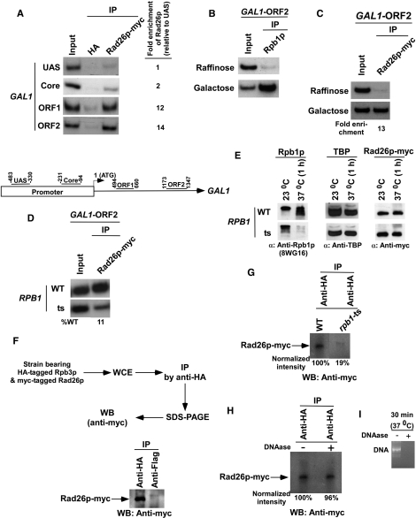
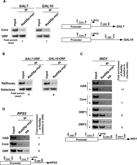
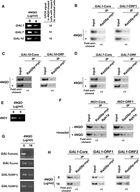
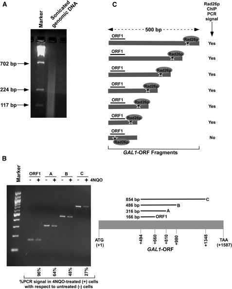
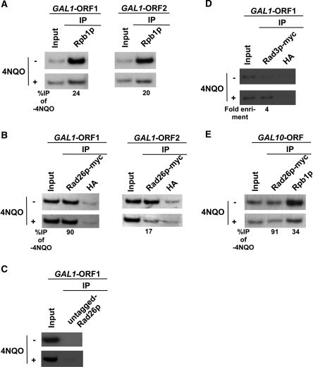
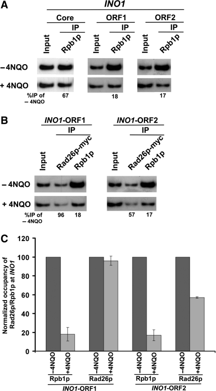
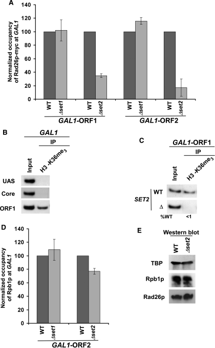
Rad26p, a yeast homologue of human Cockayne syndrome B with an ATPase activity, plays a pivotal role in stimulating DNA repair at the coding sequences of active genes. On the other hand, DNA repair at inactive genes or silent areas of the genome is not regulated by Rad26p. However, how Rad26p recognizes DNA lesions at the actively transcribing genes to facilitate DNA repair is not clearly understood in vivo. Here, we show that Rad26p associates with the coding sequences of genes in a transcription-dependent manner, but independently of DNA lesions induced by 4-nitroquinoline-1-oxide in Saccharomyces cerevisiae. Further, histone H3 lysine 36 methylation that occurs at the active coding sequence stimulates the recruitment of Rad26p. Intriguingly, we find that Rad26p is recruited to the site of DNA lesion in an elongating RNA polymerase II-dependent manner. However, Rad26p does not recognize DNA lesions in the absence of active transcription. Together, these results provide an important insight as to how Rad26p is delivered to the damage sites at the active, but not inactive, genes to stimulate repair in vivo, shedding much light on the early steps of transcription-coupled repair in living eukaryotic cells.


Figures

Figure 1.
Figure 1.
Rad26p is associated with the coding sequence but not promoter of the GAL1 gene in a transcription-dependent manner. (A) Top panel: analysis of the recruitment of Rad26p at the UAS, core promoter, ORF of the GAL1 gene. The yeast strain expressing myc-tagged Rad26p was grown in YPG up to an OD600 of 1.0 prior to cross-linking. The ChIP assay was performed as described in the Materials and Methods section. Immunoprecipitation was performed using a mouse monoclonal antibody against the c-myc epitope-tag (9E10; Santa Cruz Biotechnology, Inc.). An anti-HA (Santa Cruz Biotechnology, Inc.) was used as a non-specific antibody. Both the input and immunoprecipitated DNA samples were analyzed by PCR. The dilution factors for input and immunoprecipitated DNA samples are mentioned in the modified ChIP protocol (see ‘Materials and Methods’ section). The immunoprecipitated DNA was quantitated as the ratio of immunoprecipitate over the input in the autoradiogram. The fold enrichment of Rad26p at the GAL1 coding sequence with respect to UAS is presented. IP, immunoprecipitate. Bottom panel: the PCR primer pairs located at the UAS, core promoter, and towards the 5′-(ORF1) and 3′-(ORF2) ends of the ORF of the GAL1 gene. (B) RNA polymerase II is associated with the coding sequence of the active GAL1 gene. The yeast strain was grown in raffinose (YPR)- or galactose (YPG)-containing growth medium up to an OD600 of 1.0 prior to crosslinking. Immunoprecipitations were performed using a mouse monoclonal antibody 8WG16 (Covance) against the carboxy terminal domain of the largest subunit (Rpb1p) of RNA polymerase II. (C) Rad26p is associated with the ORF of the active GAL1 gene. The fold enrichment of Rad26p at the GAL1 coding sequence in galactose-containing growth medium in comparison to raffinose-containing growth medium is presented. (D) Rpb1p is essential for recruitment of Rad26p to the GAL1 coding sequence. (E) Analysis of the global levels of Rad26p in the rpb1-ts and wild-type strains at the non-permissive temperature. The yeast strain expressing myc epitope-tagged Rad26p were grown in YPD medium at permissive (23°C) and non-permissive (37°C) temperatures as discussed in the ‘Materials and Methods’ section. The WCE was run on SDS–polyacrylamide gel, and then analyzed by western blot assay. (F) Rad26p interacts with RNA polymerase II as revealed by the co-immunoprecipitation assay. (G) Analysis of Rad26p association with RNA polymerase II in the rpb1-ts mutant using a co-immunoprecipitation assay. (H) The co-immunoprecipitation assay as in panel G in the presence of DNAase. (I) Analysis of DNA digestion as a control for the experiments presented in (H). PCR-amplified DNA was treated with DNAase for 30 min at 37°C, and then was analyzed by agarose gel electrophoresis.
Figure 2.
Figure 2.
Rad26p is associated with the coding sequences but not promoters of the genes in a transcription-dependent manner. (A) Rad26p is associated with the coding sequences of the active GAL7 and GAL10 genes. The ChIP assay was performed as in Figure 1A. The fold enrichment of Rad26p at the coding sequence in comparison to the core promoter is presented. (B) Rad26p is associated with the ORFs of GAL7 and GAL10 in a transcription-dependent manner. The fold enrichment of Rad26p at the coding sequence in galactose-containing growth medium in comparison to raffinose-containing growth medium is presented. (C) Rad26p is associated with the coding sequence of the INO1 gene in a transcription-dependent manner. The yeast strain expressing myc-tagged Rad26p was induced for INO1 as described in the ‘Materials and Methods’ section. Primer pairs located at the UAS, core promoter, two different locations (ORF1 and ORF2) of ORF of INO1 (bottom panel) were used for the PCR analysis of the immunoprecipitated DNA samples. +, induced stated; and −, repressed state. The ChIP assay was performed as in Figure 1A. The fold enrichment of Rad26p at different locations of INO1 under transcriptionally active conditions in comparison to inactive conditions is presented. (D) Rad26p is associated with the coding sequence, but not promoter, of a constitutively active gene, RPS5. The yeast strain carrying myc epitope-tagged Rad26p was grown in YPD up to an OD600 of 1.0 prior to cross-linking. The ChIP assay was performed as in Figure 1A. The fold enrichment of Rad26p at the RPS5 coding sequence with respect to UAS is presented.
Figure 3.
Figure 3.
The DNA lesion alone does not target the recruitment of Rad26p in the absence of active transcription. (A) Analysis of DNA damage at the GAL1, GAL7 and GAL10 loci within the first 20 min of 4NQO treatment. The yeast cells were grown in YPR, and then treated with 4NQO for 20 min. The genomic DNA was prepared and was analyzed by PCR. (B) The DNA lesions at the GAL1 gene do not target the recruitment of Rad26p in the absence of active transcription. The yeast strain carrying myc-tagged Rad26p was grown in YPR up to an OD600 of 1.0, and then treated with 4NQO for 20 min prior to cross-linking. The ChIP assay was carried out as in Figure 1A. The fold change of Rad26p ChIP signal at GAL1 in 4NQO-treated cells in comparison to untreated cells under transcriptionally inactive conditions. The DNA lesions at the GAL10 (C) and GAL7 (D) genes also do not target the recruitment of Rad26p in the absence of active transcription. (E) Analysis of DNA damage at the INO1 gene within the first 20 min of 4NQO treatment. Yeast cells were grown in synthetic complete medium containing 100 µM inositol at 30°C up to an OD600 of 1.0, and then treated with 4NQO for 20 min. The whole INO1 gene was amplified by PCR, using the specific primer pairs as mentioned in Table 1. (F) The DNA lesions at the INO1 gene do not target the recruitment of Rad26p in the absence of active transcription. The yeast strain expressing myc-tagged Rad26p was grown in the synthetic complete medium containing 100 µM inositol at 30°C up to an OD600 of 1.0, and then treated with 4NQO for 20 min prior to cross-linking. (G) Analysis of DNA damage at the GAL1 locus within the first 10 min of 4NQO treatment at a concentration of 16 µg/ml. The yeast cells were grown in YPR and treated with 4NQO as in (A). The whole GAL1 locus, core promoter, ORF1 and ORF2 regions were amplified by PCR, using the specific primer pairs as mentioned in Table 1. (H) Analysis of the association of Rad26p with GAL1 following 4NQO treatment at the concentration of 16 µg/ml in the absence of transcription. Yeast cells were grown in YPR. The ChIP assay was performed as in Figure 1A. The fold change of Rad26p ChIP signal at GAL1 in 4NQO-treated cells in comparison to untreated cells under transcriptionally inactive conditions.
Figure 4.
Figure 4.
Analysis of DNA lesion in the GAL1 coding sequence. (A) Analysis of the size of DNA fragments in the ChIP assay. Yeast cells were grown in YPR prior to cross-linking as in Figure 3B. The genomic DNA was isolated following sonication (7 times, 10 s each) of WCE and was analyzed by agarose gel electrophoresis. (B) Analysis of DNA damage at GAL1 with varying lengths of coding sequence. Yeast cells were grown, cross-linked and sonicated as in (A). Cells were treated with 4NQO at a final concentration of 4 µg/ml as described in Figure 3A. DNA was isolated and analyzed by PCR using specific primer pairs. PCR products with different sizes were analyzed by 2% agarose gel electrophoresis. (C) The schematic diagram for the analysis of Rad26p recruitment at the site of DNA lesion in the ChIP assay. The ‘star’ represents the site of DNA lesion.
Figure 5.
Figure 5.
RNA polymerase II promotes the recruitment of Rad26p to the DNA lesions at the coding sequences of the active GAL1 and GAL10 genes. (A) Analysis of the association of RNA polymerase II with the GAL1 coding sequence in the presence and absence of 4NQO-induced DNA damage. The yeast strain was grown in YPR up to an OD600 of 0.8 at 30°C, and then transferred to YPG for 90 min to induce GAL1 prior to 4NQO treatment for 20 min. As a control, yeast cells were also grown under similar growth conditions without 4NQO treatment. These cells were used for the ChIP assay to analyze the level of Rpb1p at the GAL1 ORF in 4NQO-treated (+) and untreated (−) cells. The percentage of DNA immunoprecipitated in the 4NQO-treated cells relative to that of the untreated cells is represented as ‘%IP of -4NQO’. (B) Analysis of Rad26p recruitment towards the 5′- and 3′-ends of the GAL1 ORF in the presence and absence of 4NQO-induced DNA damage. The yeast strain carrying myc-tagged Rad26p was grown and crosslinked as in (A). Immunoprecipitation was performed as in Figure 1A. (C) The ChIP experiments at the GAL1 coding sequence using the strain that bears untagged Rad26p. An anti-myc antibody was used in the ChIP assay. (D) Analysis of Rad3p recruitment at the GAL1 coding sequence following 4 µg/ml 4NQO treatment. Immunoprecipitation was performed using the modified ChIP protocol as described in the ‘Materials and Methods’ section. (E) Analysis of the association of Rad26p and RNA polymerase II with the GAL10 ORF.
Figure 6.
Figure 6.
RNA polymerase II promotes the recruitment of Rad26p to the site of DNA lesion at the coding sequence of the active INO1 gene. (A) Analysis of association of RNA polymerase II with the INO1 coding sequence in the presence and absence of 4NQO-induced DNA damage. The INO1 gene was induced prior to 4NQO treatment (4 µg/ml) for 20 min as described in the ‘Materials and Methods’ section. As a control, yeast cells were also grown under similar growth conditions without 4NQO treatment. These cells were used for the ChIP assay to analyze the level of Rpb1p at the INO1 ORF in 4NQO-treated (+) and untreated (−) cells. (B) Analysis of the recruitment of Rad26p towards the 5′- and 3′-ends (ORF1 and ORF2, respectively) of the INO1 ORF in the presence and absence of the 4NQO-induced DNA damage. The yeast strain carrying myc-tagged Rad26p was grown and cross-linked as in (A). Immunoprecipitation was performed as in Figure 1A. (C) The data of the (A and B) were plotted in the form of a histogram.
Figure 7.
Figure 7.
Methylation of K36 but not K4 on histone H3 stimulates the association of Rad26p with the coding sequence of the active gene. (A) Histone H3K36 methylation stimulates the recruitment of Rad26p to the coding sequence of the active GAL1 gene. The deletion mutants of SET1 and SET2, and wild-type strains were grown in YPR up to an OD600 of 0.8 at 30°C, and then transferred to YPG for 90 min to induce GAL1 prior to cross-linking. The immunoprecipitations were performed as described in Figure 1A. The normalized occupancies of Rad26p at the GAL1 ORF in the wild-type and deletion mutant strains were plotted in the form of a histogram. (B) Histone H3 associated with the coding sequence of the active GAL1 gene is methylated on K36. Yeast cells were grown and cross-linked as in (A). Immunoprecipitation was performed using an anti-H3K36me3 antibody (Abcam-ab9050) as described for normal ChIP assay (see ‘Materials and Methods’ section). (C) Set2 is essential for histone H3 K36 methylation. The yeast strains were grown in YPG up to an OD600 prior to cross-linking. (D) Analysis of the association of RNA polymerase II with the GAL1 coding sequence in the absence of histone H3 K4/36 methylation. The wild-type and deletion mutant strains were grown and crosslinked as in (A). The immunoprecipitations were performed as in Figure 1B. The normalized occupancies of RNA polymerase II (Rpb1p) at the GAL1 ORF in the presence and absence of histone H3 K4/36 methylation were plotted in the form of a histogram. (E) Analysis of the global levels of Rad26p and Rpb1p in the wild-type and SET2 deletion mutant strains. Both the wild-type and mutant strains were grown as in (A) for western blot analysis. An anti-myc antibody was used against myc-tagged Rad26 in the western blot analysis. A mouse monoclonal antibody 8WG16 (Covance) against the carboxy terminal domain of Rpb1p was used.

Similar articles

Cited by

References

    1. Sancar A, Lindsey-Boltz LA, Unsal-Kaccmaz K, Linn S. Molecular mechanisms of mammalian DNA repair and the DNA damage checkpoints. Annu. Rev. Biochem. 2004;73:39–85. - PubMed
    1. Bootsma D, Kraemer KH, Cleaver JE, Hoeijmakers JHJ. Nucleotide excision repair syndromes: xeroderma pigmentosum, Cockayne syndrome, and trichothiodystrophy. In: Vogelstein B, Kinzler KW, editors. The Genetic Basis of Human Cancer. 2nd edn. New York: McGraw-Hill; 2002. pp. 211–237.
    1. Cooper PK, Nouspikel T, Clarkson SG, Leadon SA. Defective transcription-coupled repair of oxidative base damage in Cockayne syndrome patients from XP group G. Science. 1997;275:990–993. - PubMed
    1. Hasty P, Campisi J, Hoeijmakers J, van Steeg H, Vijg J. Aging and genome maintenance: lessons from the mouse. Science. 2003;299:1355–1359. - PubMed
    1. Sands AT, Abuin A, Sanchez A, Conti CJ, Bradley A. High susceptibility to ultraviolet-induced carcinogenesis in mice lacking XPC. Nature. 1995;377:162–165. - PubMed

Publication types

MeSH terms