. 2010 Mar;38(5):1583-95.
doi: 10.1093/nar/gkp1135. Epub 2009 Dec 9.

Involvement of Vts1, a structure-specific RNA-binding protein, in Okazaki fragment processing in yeast

Affiliations

Involvement of Vts1, a structure-specific RNA-binding protein, in Okazaki fragment processing in yeast

Chul-Hwan Lee et al. Nucleic Acids Res. 2010 Mar.

Abstract

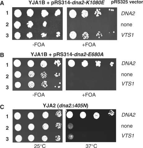
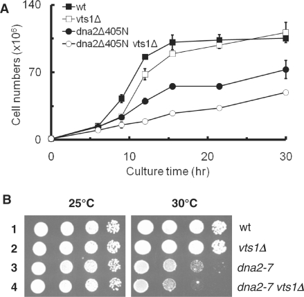
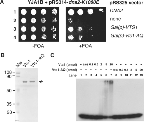
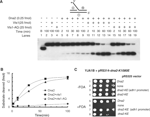
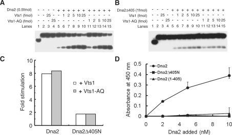
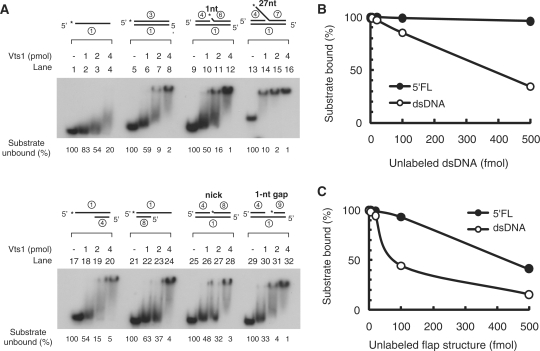
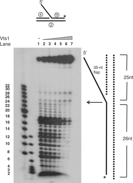
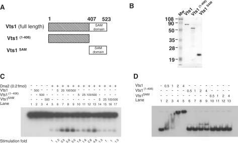
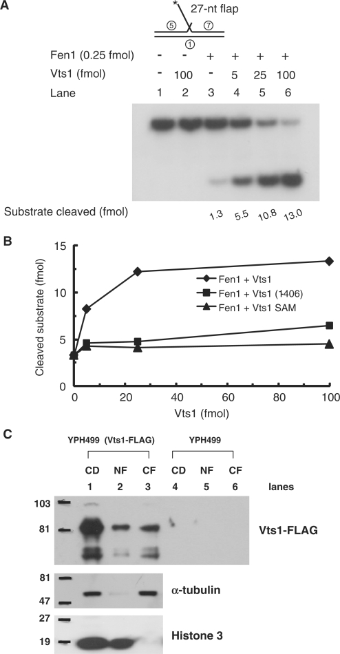
The non-essential VTS1 gene of Saccharomyces cerevisiae is highly conserved in eukaryotes and encodes a sequence- and structure-specific RNA-binding protein. The Vts1 protein has been implicated in post-transcriptional regulation of a specific set of mRNAs that contains its-binding site at their 3'-untranslated region. In this study, we identified VTS1 as a multi-copy suppressor of dna2-K1080E, a lethal mutant allele of DNA2 that lacks DNA helicase activity. The suppression was allele-specific, since overexpression of Vts1 did not suppress the temperature-dependent growth defects of dna2Delta405N devoid of the N-terminal 405-amino-acid residues. Purified recombinant Vts1 stimulated the endonuclease activity of wild-type Dna2, but not the endonuclease activity of Dna2Delta405N, indicating that the activation requires the N-terminal domain of Dna2. Stimulation of Dna2 endonuclease activity by Vts1 appeared to be the direct cause of suppression, since the multi-copy expression of Dna2-K1080E suppressed the lethality observed with its single-copy expression. We found that vts1Delta dna2Delta405N and vts1Deltadna2-7 double mutant cells displayed synergistic growth defects, in support of a functional interaction between two genes. Our results provide both in vivo and in vitro evidence that Vts1 is involved in lagging strand synthesis by modulating the Dna2 endonuclease activity that plays an essential role in Okazaki fragment processing.


Figures

Figure 1.
Figure 1.
VTS1 is a multi-copy suppressor specific for Dna2-K1080E, a Dna2 helicase-negative mutant. (A) Overexpression of VTS1 suppresses the dna2 helicase-negative mutant. The dna2Δ strain YJA1B harboring both pRS314-dna2-K1080E and pRS316-DNA2 plasmids was transformed with each of the three plasmids indicated at the right of the figure. Transformants were grown in liquid media and cells were spotted in 10-fold serial dilutions (105, 104, 103 and 102 cells) onto plates without (−FOA) or with (+FOA) 5-FOA, followed by incubation for 3 days at 30°C, as shown. (B) Overexpression of Vts1 does not suppress the dna2 endonuclease-deficient mutant. The same plasmids used in panel A were transformed into dna2Δ strain YJA1B harboring pRS314-dna2-E680A and pRS316-DNA2 plasmids. Cells were grown as shown in panel A. (C) Overexpression of Vts1 does not suppress dna2Δ405N that lacks the N-terminal 405-amino-acid domain. The same plasmids used in panel A were transformed into YJA2 (dna2Δ405N). Transformants were grown in liquid media and the cells were spotted in 10-fold serial dilutions (105, 104, 103 and 102 cells) onto SD-Leu plates. The plates were incubated for 3 days at 25°C or 37°C as indicated.
Figure 2.
Figure 2.
Synergistic growth defects associated with dna2 mutations and vts1Δ. (A) Cells were incubated in 32°C and the cell numbers were measured at the time point indicated using a hematocytometer. (B) Cells were spotted in serial 10-fold dilutions on YPD media and plates were incubated for 3 days at the temperatures indicated.
Figure 3.
Figure 3.
Purification of Vts1 and Vts1-A498Q. (A) The structure-specific RNA-binding activity of Vts1 is dispensable for suppression of the dna2-K1080E mutant. Plasmids containing genes indicated at the right of figure were introduced into dna2Δ strain YJA1B harboring pRS314-dna2-K1080E and pRS316-DNA2 plasmids. Cells were grown in SG-His-Trp-Leu (2% galactose without glucose) and 10-fold serial dilutions (105, 104, 103 and 102 cells) were spotted onto SG-His-Trp-Leu plates without (−FOA) or with (+FOA) 5-FOA. The plates were incubated for 5 days at 30°C as shown. (B) An SDS–PAGE analysis of wild-type Vts1 and mutant Vts1-A498Q (Vts1-AQ) proteins. Both proteins were purified as described in ‘Material and Methods’ section. Of each protein, 1 µg was subjected to electrophoresis in a 10% SDS–PAGE gel, which was then stained with Coomassie Blue G-250. (C) Vts1 binds to the SRE-RNA substrate, while Vts1-A498Q does not. Electrophoretic mobility shift assays were performed as described in ‘Materials and Methods’ section. The SRE-RNA substrate (5 fmol) was incubated in the presence of increasing amounts (0.5–20 pmol) of Vts1 or Vts1-A498Q at 4°C for 10 min, followed by additional 5-min incubation at 37°C. After incubation, reaction mixtures were subjected to electrophoresis in a 6% native polyacrylamide gel.
Figure 4.
Figure 4.
Vts1 stimulates the endonuclease activity of Dna2 in vitro. (A) Both Vts1 and Vts1-A498Q stimulate Dna2 endonuclease activity in vitro. Three sets of reaction mixtures were assembled on ice; one with Dna2 (0.25 fmol) only (lanes 5–9), the second with Dna2 (0.25 fmol) and Vts1 (25 fmol) and the third with Dna2 (0.25 fmol) and Vts1-A498Q (25 fmol). The reaction mixtures were then incubated at 37°C for varying periods of time (0, 2, 10, 30, 60 and 100 min). Cleavage products were analyzed on a 10% denaturing polyacrylamide gel as described in ‘Materials and Methods’ section. ‘B’ in lane 1 represents boiled. (B) The amount of product formed from reactions in panel A was plotted against the time of incubation. (C) Overexpression of Dna2-K1080E (Dna2-KE) suppresses its own growth defects. The experimental procedures are as described in Fig. 1A. Note that the expression of Dna2-K1080E was driven either by the ADH1 promoter or by its own promoter.
Figure 5.
Figure 5.
Vts1 stimulates Dna2 endonuclease via interaction with the DNA-binding domain of Dna2. (A) Vts1 stimulates Dna2 endonuclease in vitro. Reaction mixtures containing indicated amounts of Dna2 and either Vts1 wild-type or Vts1-A498Q (Vts1-AQ) were incubated for 15 min in 37°C. Products were analyzed on 10% denaturing polyacrylamide gel. (B) Vts1 does not stimulate Dna2Δ405N endonuclease in vitro. Experiments with Dna2Δ405N were done same as described in panel A. (C) The stimulation folds of Dna2 endonuclease activity by Vts1 (or Vts1-AQ) in reactions described in A and B were plotted. (D) Vts1-coated wells were incubated with increasing concentrations (2, 5 and 10 nM) of Dna2 derivatives indicated at the top of the figure. Wells were aspirated, washed three times and bound Dna2, Dna2Δ405N and Dna2(1–405) were detected by ELISA using rabbit polyclonal antibodies against Dna2. Absorbance readings at each point were corrected for background absorbance generated with BSA-coated wells.
Figure 6.
Figure 6.
Vts1 is a flap DNA-specific-binding protein. (A) Electrophoretic mobility shift assays were performed as described in ‘Materials and Methods’ section. Each substrate (15 fmol), shown at the top of the gel, was preincubated with increasing amounts (1, 2 and 4 pmol) of Vts1 at 4°C for 10 min, followed by an additional 5 min of incubation at 37°C. After incubation, samples were loaded on a 6% native polyacrylamide gel and the Vts1–DNA complexes formed were analyzed by autoradiography as described in ‘Materials and Methods’ section. The amount of substrate left was measured and the values are presented at the bottom of the gel. (B and C) Substrate challenge assays. The Vts1–DNA complex was first formed by incubating 5 fmol of labeled substrate (indicated at the top of the gel) with excess Vts1 (4 pmol) as described in panel A. Increasing amounts (5, 20, 100 and 500 fmol) of unlabeled competitor DNA were added to reactions containing the Vts1/labeled substrate complex. The amount of labeled substrate left was plotted against the amount of unlableled competitor DNA used.
Figure 7.
Figure 7.
DNase I protection analysis of Vts1 bound to 5′-flap DNA. DNase I protection assays were carried out with the DNA substrate indicated above the gel. The 3′-end of the strand indicated above the gel was 32P-labeled. Increasing amounts of Vts1 (0.5, 1, 2, 4, 8 and 16 pmol) were preincubated at 4°C for 10 min, followed by additional 5-min incubation at 37°C. DNase I (0.1U/reaction) was then added to the reaction mixtures, followed by 10-min incubation at 4°C. The reaction products were analyzed in a 15% high-resolution sequencing gel containing 7 M urea.
Figure 8.
Figure 8.
DNA-binding activity of Vts1 is required for the stimulation of Dna2 endonuclease activity. (A) A schematic diagram of Vts1 and its truncated fragments used in this experiment. Note that the C-terminal 117-amino-acid fragment contains the intact SAM domain. (B) Purified Vts1 (full-length), Vts1(1–406) and Vts1SAM proteins (0.5 µg each) were subjected to electrophoresis on a 12% SDS–PAGE. The gel was then Coomassie-stained. (C) The N-terminal fragment of Vts1 is able to stimulate Dna2 endonuclease activity. The reaction mixtures containing indicated amounts of Dna2 and Vts1 (or fragmented derivatives) were incubated for 15 min at 37°C. Cleavage products were analyzed in a 10% denaturing polyacrylamide gel. (D) The N-terminal fragment of Vts1 and Vts1 are able to bind 5′-flap DNA. Electrophoretic mobility shift assays were carried out as described in ‘Materials and Methods’ section. The 27-nt 5′-flap substrate (5 fmol) was incubated at 4°C for 10 min in the presence of increasing amounts (0.5, 1, 2 and 4 fmol) of Vts1, followed by additional 5 min of incubation at 37°C.
Figure 9.
Figure 9.
Vts1 stimulates yeast Fen1 endonuclease activity. (A) The reaction mixtures containing indicated amounts of Fen1 and wild-type Vts1 were incubated with 5′-flap substrate (15 fmol) for 15 min in 37°C. Cleavage products were analyzed in a 10% denaturing polyacrylamide gel. (B) The same reactions as panel A were repeated with Vts1 and its derivatives as indicated. The amount of product formed was plotted against the amount of Vts1 or its derivates added. (C) Cellular localization of Vts1 in S. cerevisiae. As described in ‘Materials and Methods’ section, nuclear and cytoplasmic fractions were prepared from spheroplasts of two strains, YPH499 (negative control) and YPH499 (Vts1-FLAG), that expresses endogenous FLAG-tagged Vts1. CD, cell debris fraction; NF, nuclear fraction; CF, cytoplasmic fraction. The fractions were subjected to western-blot analyses in a 10% SDS–PAGE with specific monoclonal antibodies against α-tubulin, histone 3 and the FLAG epitope. Proteins detected were as indicated at the right of each blot.

Similar articles

Cited by

References

    1. MacNeill SA. DNA replication: partners in the Okazaki two-step. Curr. Biol. 2001;11:R842–R844. - PubMed
    1. Hübscher U, Seo YS. Replication of the lagging strand: a concert of at least 23 polypeptides. Mol. Cells. 2001;12:149–157. - PubMed
    1. Liu Y, Kao HI, Bambara RA. Flap endonuclease 1: a central component of DNA metabolism. Annu. Rev. Biochem. 2004;73:589–615. - PubMed
    1. Garg P, Burgers PM. DNA polymerases that propagate the eukaryotic DNA replication fork. Crit. Rev. Biochem. Mol. Biol. 2005;40:115–128. - PubMed
    1. Rossi ML, Purohit V, Brandt PD, Bambara RA. Lagging strand replication proteins in genome stability and DNA repair. Chem. Rev. 2006;106:453–473. - PubMed

Publication types

MeSH terms