. 2010 Mar;38(5):1478-88.
doi: 10.1093/nar/gkp1143. Epub 2009 Dec 10.

Membrane association of mitochondrial DNA facilitates base excision repair in mammalian mitochondria

Affiliations

Membrane association of mitochondrial DNA facilitates base excision repair in mammalian mitochondria

Pierre Boesch et al. Nucleic Acids Res. 2010 Mar.

Abstract

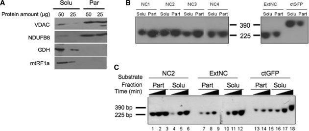
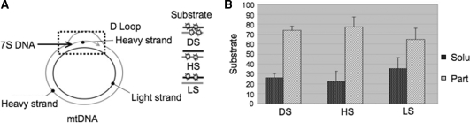
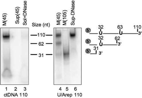
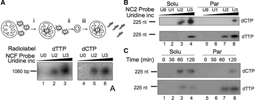
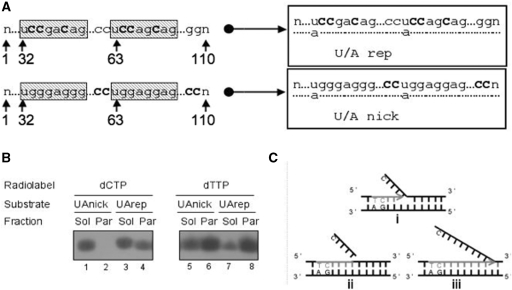
Mitochondrial DNA encodes a set of 13 polypeptides and is subjected to constant oxidative stress due to ROS production within the organelle. It has been shown that DNA repair in the mitochondrion proceeds through both short- and long-patch base excision repair (BER). In the present article, we have used the natural competence of mammalian mitochondria to import DNA and study the sub-mitochondrial localization of the repair system in organello. Results demonstrate that sequences corresponding to the mtDNA non-coding region interact with the inner membrane in a rapid and saturable fashion. We show that uracil containing import substrates are taken into the mitochondrion and are used as templates for damage driven DNA synthesis. After further sub-fractionation, we show that the length of the repair synthesis patch differs in the soluble and the particulate fraction. Bona fide long patch BER synthesis occurs on the DNA associated with the particulate fraction, whereas a nick driven DNA synthesis occurs when the uracil containing DNA accesses the soluble fraction. Our results suggest that coordinate interactions of the different partners needed for BER is only found at sites where the DNA is associated with the membrane.


Figures

Figure 1.
Figure 1.
Sequence-dependent association of imported DNA with the mitochondrial particulate fraction. (A) Mitochondrial sub-fractionation. Isolated rat liver mitochondria were disrupted by repeated freezing and thawing. Soluble (Solu) and particulate (Part) fractions were separated by centrifugation at 100 000g for 1 h and the indicated amount of protein from the different fractions was subjected to western blot analysis. Purity of fractions was assessed by blotting with antibodies against marker proteins. The VDAC and a component of respiratory chain complex I (NDUFB8) are outer and inner membrane markers, respectively; glutamate dehydrogenase (GDH) and the mitochondrial translation release factor (mtRF1a) are matrix soluble markers. (B) NCR corresponding sequences show a strong association with the particulate fraction after import into isolated rat mitochondria. Four probes ranging from 221 to 235 bp (NC1–NC4) spanning most of the NCR, an additional mtDNA probe from outside the NCR (ExtNC) and a 390 bp probe from the GFP coding sequence (ctGFP) were PCR amplified and internally labelled with [α32P]dCTP (primers and locations are given in Supplementary Table S1). Following a 45-min incubation with coupled rat mitochondria, non-imported DNA was DNase I digested, mitochondria sub-fractionated, protected DNA was extracted and subjected to non-denaturing gel electrophoresis prior to autoradiography and visualization as detailed in ‘Materials and Methods’ section. Typical results are shown from three repeats. Solu and Part refer to the soluble and particulate mitochondrial fractions, respectively. Markers refer to labelled probes (NC2 and ctGFP) of the indicated sizes. (C) Imported NCR DNA associates rapidly with the particulate fraction in a saturable process. A representative probe for the NCR, NC2 and the two additional probes ExtNC and ctGFP were internally labelled as in Figure 2B. Incubation of mitochondria with each probe was terminated after 15, 30 or 45 min (indicated as increasing time) before sub-fractionation, DNA extraction and visualization as for Figure 2B. Typical results are shown from three repeats.
Figure 2.
Figure 2.
Sequence selective association of imported DNA with the particulate fraction is not strand specific. (A) Schematic representation of the mitochondrial genome, with emphasis on the triplex displacement (D)-loop region. The short 7S species is indicated. (B) Association of imported NC DNA with the particulate fraction is not strand specific. Three different 225 bp NC2 probes were prepared, carrying radiolabel on both strands (DS), the heavy strand (HS) or the light strand (LS) as illustrated. Import experiments were performed with isolated rat mitochondria for 15 min before DNaseI treatment and sub-fractionation as described in the legend of Figure 2A. Radiolabelled probe from each different fraction was analysed after extraction and electrophoresis in non-denaturing conditions as in Figure 2B. The percentage of the DNA retrieved in each of the two fractions was calculated as described in ‘Materials and Methods’ section and are represented as a histogram. Bars show standard deviations from five independent repeats.
Figure 3.
Figure 3.
Imported uracil-containing DNA is cleaved at the uracils. Standard 110 nt oligodeoxynucleotide (ctDNA, lanes 1–3) or containing uracil residues at positions 32 and 63 (U/Arep 110; lanes 4–6) was γ32P-end-labelled, annealed to its complement and incubated with rat liver mitochondria in standard IM as described. Following import for 45 (lanes 1–4 and 6) or 105 min [M (105); lane 5], mitochondria were DNase treated (lanes 1, 2 and 4–6) or solubilized prior to DNase treatment (sol+DNase; lane 3). DNA was then extracted from the mitochondrial pellet (M; lanes 1, 4 and 5), solubilized mitochondria (lane 3) or supernatant (Sup; lanes 2 and 6), precipitated and separated by denaturing polyacrylamide gel electrophoresis before autoradiography. Markers were prepared by end-labelling oligomers of the indicated sizes and corresponded to the migration of the U/Arep DNA cleaved at the incorporated uracils as indicated.
Figure 4.
Figure 4.
DNA repair mechanisms differ dependent on their mitochondrial location. (A) Mitochondria repair uracil-containing DNA on import. In these import and repair experiments the isolated mitochondria were first (i) incubated with the free radionucleotide (dTTP lanes 1–3; dCTP lanes 4–6), (ii) harvested and (iii) washed twice to remove unincorporated radiolabel. Aliquots of 1060 bp probe were prepared with increasing levels of uracil incorporation (U0–U3) as detailed in ‘Materials and Methods’ section and incubated with the pre-loaded mitochondria in DNA repair buffer (RB) for 45 min before DNase I treatment, DNA extraction, separation through non-denaturing gels, transfer to nylon membranes and visualization. (B) Mitochondrial sub-fractions show differing DNA repair mechanisms. Aliquots of probe NC2 were prepared with increasing amounts of uridine prior to incubation with pre-loaded mitochondria in RB for 2 h. Following import, mitochondria were sub-fractionated and incorporation of radiolabel into the probe in each fraction was monitored after migration under denaturing conditions and autoradiography. Upper panel, mitochondria were pre-loaded with dCTP; lower panel, dTTP. (C) Repair driven DNA synthesis as a function of time. A similar experiment was performed with pre-loaded mitochondria incubated for the indicated times with probe NC2U3. Mitochondria were sub-fractionated, DNA isolated and analysed exactly as described in B.
Figure 5.
Figure 5.
DNA synthesis in the particulate fraction is limited to the long patch BER range. (A) Design of repair template. Two uracil-containing sequence-bias 110 nt oligonucleotides were designed to evaluate different DNA repair mechanisms in the mitochondrion, end-labelled and annealed to their complementary standard oligomer. Uracils were incorporated at positions 32 and 63 as indicated. To assess the mode of DNA repair, cytosines were added distal to the uracils and well beyond the 6–8 nt long patch region (U/A nick), or specifically within the long patch (U/A rep). (B) BER occurs at the mitochondrial membrane. Isolated mitochondria were preloaded with the indicated radionucleotide before 45 min incubation in RB with either U/Arep or U/Anick and sub-fractionation as described in ‘Materials and Methods’ section. DNA was extracted post-import and subjected to native gel electrophoresis prior to autoradiography. Images are accurate representations from one of three independent repeats. (C) Schematic representation to describe the modes of DNA repair observed. In (i) uracil is removed from the template. In (ii) enzymes coordinate uracil removal and nucleotides incorporation into a patch spanning six residues with the excision of the ‘flapping structure’, consistent with lpBER as seen only with U/Arep and only in the particulate fraction. In (iii) generic nick-directed DNA synthesis occurs on the oligomer, with cytosine replaced throughout the template, as seen with the U/Anick template.

Similar articles

Cited by

References

    1. Richter C, Park JW, Ames BN. Normal oxidative damage to mitochondrial and nuclear DNA is extensive. Proc. Natl Acad. Sci. USA. 1988;85:6465–6467. - PMC - PubMed
    1. Hudson EK, Hogue BA, Souza-Pinto NC, Croteau DL, Anson RM, Bohr VA, Hansford RG. Age-associated change in mitochondrial DNA damage. Free Radic. Res. 1998;29:573–579. - PubMed
    1. Yang JL, Weissman L, Bohr VA, Mattson MP. Mitochondrial DNA damage and repair in neurodegenerative disorders. DNA Repair. 2008;7:1110–1120. - PMC - PubMed
    1. Trifunovic A, Wredenberg A, Falkenberg M, Spelbrink JN, Rovio AT, Bruder CE, Bohlooly YM, Gidlof S, Oldfors A, Wibom R, et al. Premature ageing in mice expressing defective mitochondrial DNA polymerase. Nature. 2004;429:417–423. - PubMed
    1. Druzhyna NM, Wilson GL, LeDoux SP. Mitochondrial DNA repair in aging and disease. Mech. Ageing Dev. 2008;129:383–390. - PMC - PubMed

Publication types