. 2010 Feb 12;285(7):4298-306.
doi: 10.1074/jbc.M109.074609. Epub 2009 Dec 10.

Cdc42p is activated during vacuole membrane fusion in a sterol-dependent subreaction of priming

Affiliations

Cdc42p is activated during vacuole membrane fusion in a sterol-dependent subreaction of priming

Lynden Jones et al. J Biol Chem. .

Abstract

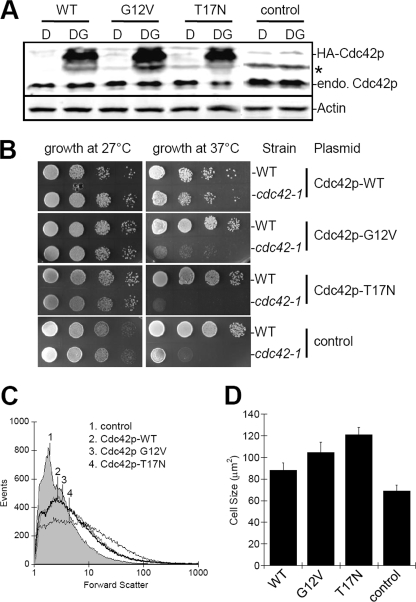
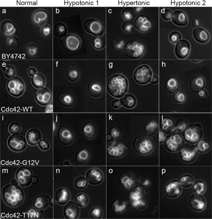
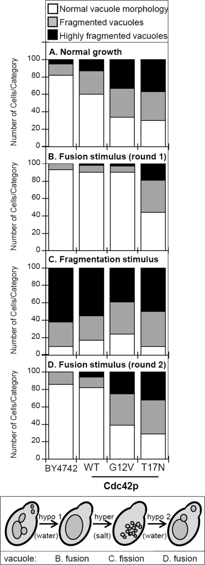
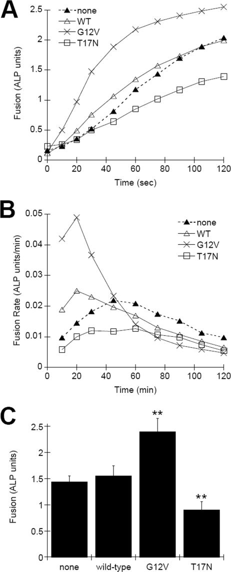
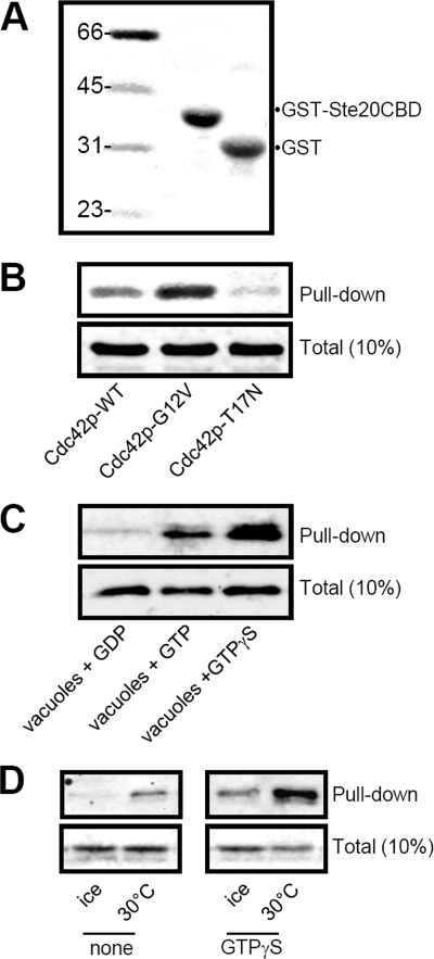
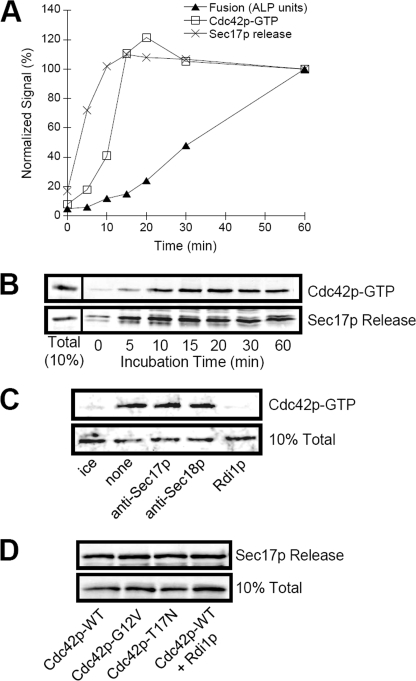
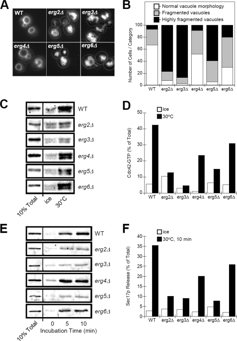
Cdc42p is a Rho GTPase that initiates signaling cascades at spatially defined intracellular sites for many cellular functions. We have previously shown that Cdc42p is localized to the yeast vacuole where it initiates actin polymerization during membrane fusion. Here we examine the activation cycle of Cdc42p during vacuole membrane fusion. Expression of either GTP- or GDP-locked Cdc42p mutants caused several morphological defects including enlarged cells and fragmented vacuoles. Stimulation of multiple rounds of fusion enhanced vacuole fragmentation, suggesting that cycles of Cdc42p activation, involving rounds of GTP binding and hydrolysis, are required to propagate Cdc42p signaling. We developed an assay to directly examine Cdc42p activation based on affinity to a probe derived from the p21-activated kinase effector, Ste20p. Cdc42p was rapidly activated during vacuole membrane fusion, which kinetically coincided with priming subreaction. During priming, Sec18p ATPase activity dissociates SNARE complexes and releases Sec17p, however, priming inhibitors such as Sec17p and Sec18p ligands did not block Cdc42p activation. Therefore, Cdc42p activation seems to be a parallel subreaction of priming, distinct from Sec18p activity. Specific mutants in the ergosterol synthesis pathway block both Sec17p release and Cdc42p activation. Taken together, our results define a novel sterol-dependent subreaction of vacuole priming that activates cycles of Cdc42p activity to facilitate membrane fusion.


Figures

FIGURE 1.
FIGURE 1.
Overexpression of HA-tagged Cdc42p. A, yeast strain BY4742 was transformed with plasmid pGREG-535 (PGAL1-7HA overexpression vector) containing WT, constitutively active (G12V), or dominant-negative (T17N) alleles of CDC42. Transformants were grown in YPD-kan (D) or YPDG-kan (DG), which contains 1% galactose to induce expression of cloned alleles. For control, BY4742 was transformed with empty vector and grown in YPD or YPDG. Equal amounts of lysate were analyzed. * indicates a background band. B, viability assay of strains overexpressing Cdc42p WT and mutant clones. Note that growth of the parental strain, TD4, is not affected whereas growth of the cdc42-1 temperature-sensitive strain, DJTD2-16A, is only rescued by expression of Cdc42p-WT. C, relative size of cells from strains in A, as determined by flow cytometry forward scatter measurements of 50,000 cells. Numbered lines indicate the mean for each sample. D, cell size quantitatively determined by cross-sectional area measurements of bright-field images imported into ImageJ (±S.E., n = 20 cells).
FIGURE 2.
FIGURE 2.
Characterization of vacuole morphology in CDC42 mutant strains. Strain BY4742 was transformed with the empty vector pGREG535 (BY4742), or with vectors that overexpress wild type (Cdc42p-WT), constitutively active (Cdc42p-G12V), or dominant-negative (Cdc42p-T17N) Cdc42p from the GAL1 galactose-inducible promoter. Cells were grown in YPDG-kan to ∼0.6 A600, vacuoles were stained with FM4-64, and cells were visualized by fluorescence microscopy (Normal). Vacuolar hypotonic and hypertonic stress response was examined by dilution in water (Hypotonic 1) or incubation in 0.4 m NaCl, (Hypertonic). Multiple rounds of vacuole fusion were examined after sequential hypotonic-hypertonic and hypotonic stresses (Hypotonic 2). Panels are 30 μm × 30 μm.
FIGURE 3.
FIGURE 3.
Quantification of vacuole fragmentation levels in strains expressing CDC42 mutant alleles. 100 cells for each strain depicted in Fig. 2 were categorized as having normal vacuole morphology (<4 vacuole lobes), fragmented vacuoles (4–10 vacuole lobes) or highly fragmented vacuoles (>10 vacuole lobes). Vacuole morphology was quantified in strains grown in YPDG-kan (A), after hypotonic stress via dilution in water (B), after hypertonic stress via addition of NaCl to 0.4 m (C), or after a second hypotonic stress treatment (D). The treatment scheme is shown in the lower box with the intended result on vacuole morphology indicated.
FIGURE 4.
FIGURE 4.
Effect of Cdc42p mutant protein expression on in vitro vacuole fusion. A, fusion of vacuoles isolated from BJ5459 and DKY6281 strains expressing wild-type Cdc42p-WT, constitutively active Cdc42p-G12V, or dominant-negative Cdc42p-T17N. At the indicated times, reactions were stopped by placing on ice and assayed for fusion signal via ALP enzyme assay. Shown are the average signals from two independent experiments. B, rate of fusion determined by integration of fusion curves in A. C, statistical analysis of fusion signals obtained after 1 h of incubation (± S.E., n = 4, **, p < 0.001).
FIGURE 5.
FIGURE 5.
Assay of Cdc42p activation in yeast. The CBD of yeast Ste20p was cloned as GST fusion proteins and expressed in E. coli. A, 30 μg of GST-CBD probe or GST was immobilized on glutathione beads and 10% was analyzed by immunoblot. B, incubation of GST-CBD probe with 200 μg of yeast whole-cell lysate prepared from strain overexpressing Cdc42p-WT, Cdc42p-G12V (GTP-bound), or Cdc42p-T17N (GDP-bound). C, incubation of GST-CBD probe with 50 μg of purified vacuoles subjected to GDP, GTP, or GTPγS nucleotide exchange as described under “Experimental Procedures.” D, determination of Cdc42p activation during membrane fusion. 50 μg of purified vacuoles were incubated in 200 μl of F.R.B., ATPreg, and cytosol in the absence (none) or presence of 40 μm GTPγS. All samples were incubated for 30 min on ice or at 30 °C, as indicated, and then 30 μg of GST-CBD beads were added to affinity isolate-activated (GTP-bound) Cdc42p (Pull-down). 10% Total refers to Cdc42p load controls.
FIGURE 6.
FIGURE 6.
Analysis of vacuole-priming subreactions. Standard vacuole fusion reactions were performed to assay for levels of fusion and Cdc42p activation and Sec17p release as described under “Experimental Procedures.” At the indicated times, reactions were stopped by placing on ice and assayed for fusion signal via ALP enzyme assay. Sec17p release and Cdc42p activation via pull-down with the GST-CBD probe were assayed in parallel reactions. A, quantification of fusion, Cdc42p activation, and Sec17p release, normalized to 60-min reactions. Shown are the average signals from at least three independent experiments. B, typical immunoblots showing the time course for Cdc42p activation and Sec17p release that were quantified by band densitometry. C, effect of incubation with priming inhibitors on Cdc42p activation. Reactions were incubated for 30 min at 30 °C (or on ice) in the presence of no inhibitor (none and ice), 150 μg/ml anti-Sec17p, anti-Sec18p antibodies (priming inhibitors), or Rdi1p (Rho GDP dissociation inhibitor). The upper panel shows the levels of Cdc42p-GTP via pull-down by GST-CBD probe that was added directly to reactions (i.e. without membrane reisolation due to potential Rdi1p extraction of Cdc42p). The lower panel shows 10% of the total Cdc42p in each reaction. D, effect of expressing Cdc42p mutant proteins, or incubation with 150 μg/ml Rdi1p, on Sec17p release. 10% Total refers to load controls for Cdc42p (A, upper panel; C) and Sec17p (A, lower panel; D).
FIGURE 7.
FIGURE 7.
Effect of ergosterol synthesis mutants on vacuole morphology and Cdc42p activation. A, normal vacuole morphology in BY4742 (WT) is compared with aberrant vacuole morphology of ERG-gene deletion strains. Vacuoles were stained by incubation with 3 μm FM4-64 for 1 h followed by a 5 h chase in dye-free medium. Panels are 20 μm × 20 μm. B, quantification of vacuole fragmentation of ERG-gene deletion strains as described in the legend to Fig. 3. C, assay for Cdc42p activation on vacuoles isolated from ERG-gene deletion strains. Vacuoles, isolated from BY4742 (WT) or ERG-deletions strains as indicated, were incubated in F.R.B., ATPreg, cytosol, and 40 μm GTPγS for 30 min at 30 °C or on ice. Levels of activated Cdc42p were determined by association with immobilized GST-CBD probe. D, quantification of Cdc42p activation as shown in panel C. E, assay for Sec17p released from vacuoles isolated from ERG-gene deletion strains. F, quantification of Sec17p release as shown in panel E. 10% Total refers to load controls for Cdc42p (C) and Sec17p (E).

Similar articles

Cited by

References

    1. Johnson D. I. (1999) Microbiol. Mol. Biol. Rev. 63, 54–105 - PMC - PubMed
    1. Hall A. (2005) Biochem. Soc. Trans. 33, 891–895 - PubMed
    1. Ridley A. J. (2006) Trends Cell Biol. 16, 522–529 - PubMed
    1. Eitzen G., Wang L., Thorngren N., Wickner W. (2002) J. Cell Biol. 158, 669–679 - PMC - PubMed
    1. Isgandarova S., Jones L., Forsberg D., Loncar A., Dawson J., Tedrick K., Eitzen G. (2007) J. Biol. Chem. 282, 30466–30475 - PubMed

Publication types

MeSH terms

Substances

LinkOut - more resources