

CT features of lobular capillary hemangioma of the nasal cavity
- PMID: 20007721
- PMCID: PMC7964208
- DOI: 10.3174/ajnr.A1908
CT features of lobular capillary hemangioma of the nasal cavity
Abstract
Background and purpose: Lobular capillary hemangioma is a benign capillary proliferation of unknown etiology. To our knowledge, no comprehensive review of imaging findings of LCHNC has been presented. Thus, we investigated characteristic CT features of LCHNC.
Materials and methods: This retrospective study included 6 patients (2 men and 4 women; age range, 30-65 years; mean age, 49.2 years) with histologically proved LCHNC. We evaluated the size, site of origin, attenuation on NECT, degree and pattern of enhancement, and bony changes.
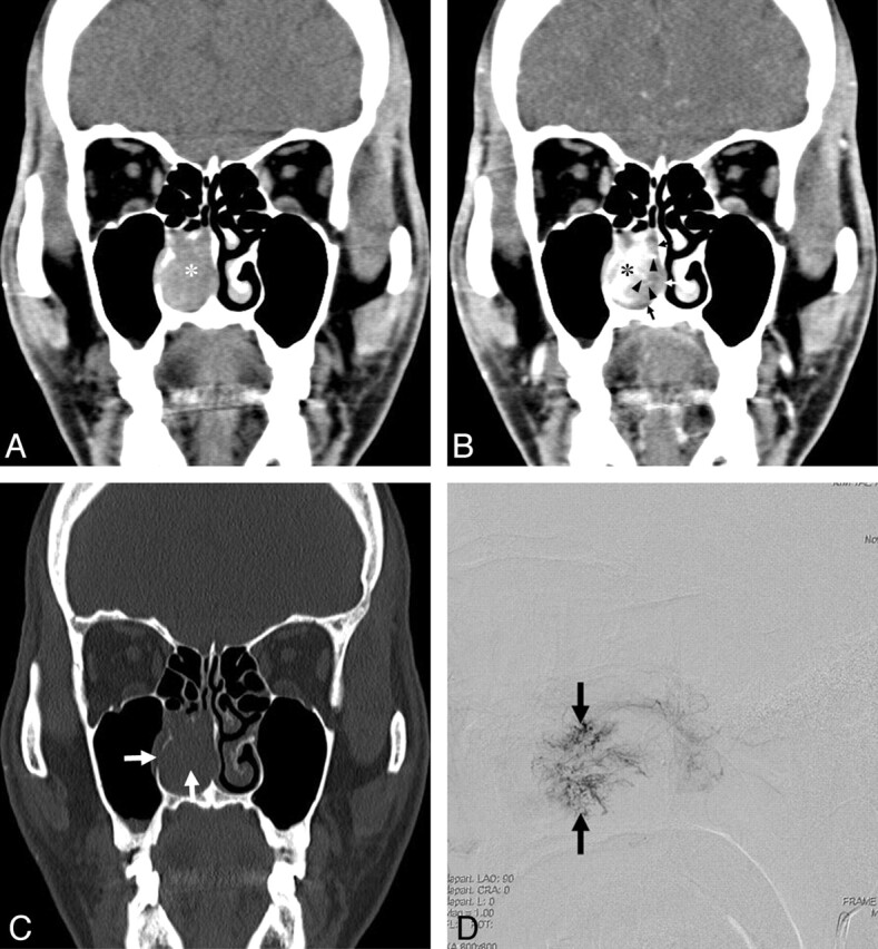
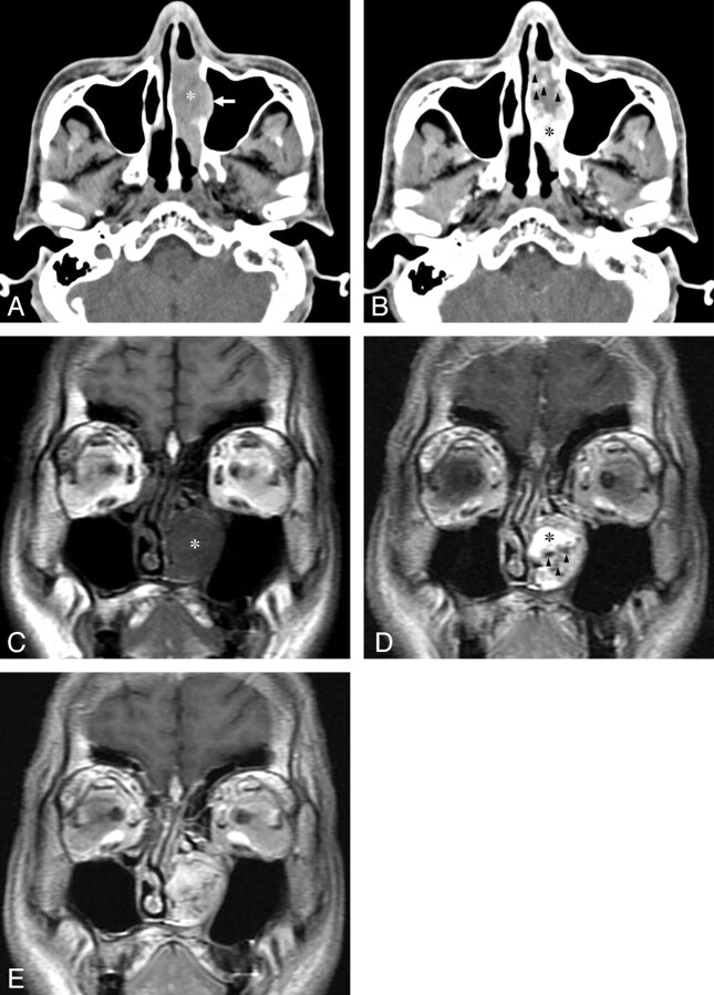
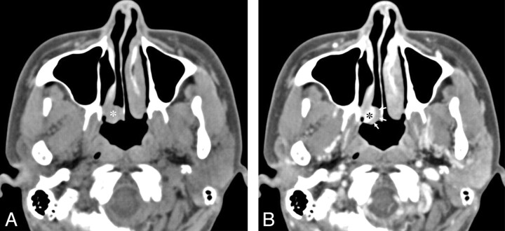
Results: The LCHNC lesion was 13.0-45.0 mm (average, 25.0 mm) in diameter. These lesions arose from the inferior turbinate in 5 (83.3%) patients and the anterior nasal septum in 1 (16.7%). Compared with the masticator muscles, the LCHNC lesion was hypoattenuating in 2 (33.3%) and isoattenuating on NECT in 4 (66.7%) patients. In 5 (83.3%) patients, the LCHNC lesion consisted of 2 distinct areas on CECT: a lobular intensely enhancing mass and an iso- or hypoattenuating cap of variable thickness around the intensely enhancing mass. Bony changes included erosion in 3 (50.0%) and displacement in 2 (33.3%) patients.
Conclusions: CT features of LCHNC consist of an intensely enhancing mass and an iso- or hypoattenuating cap on CECT. The inferior turbinate seems to be a common site of origin, and bony changes are not uncommon features of LCHNC. CT is useful not only in identifying the site of origin and assessing the extent but also in suggesting the nature of LCHNC.
Figures

Similar articles
-
Intraosseous cavernous hemangioma of the middle turbinate.Auris Nasus Larynx. 2011 Aug;38(4):516-8. doi: 10.1016/j.anl.2010.10.010. Epub 2011 Jan 21. Auris Nasus Larynx. 2011. PMID: 21256686
-
[Pre-operative effects of the administration of systemic corticosteroids combined with antibiotics on a lobular capillary hemangioma in the nasal cavity].Nihon Jibiinkoka Gakkai Kaiho. 2017 Jan;120(1):67. Nihon Jibiinkoka Gakkai Kaiho. 2017. PMID: 30035468 Japanese. No abstract available.
-
CT findings in two cases of lobular capillary haemangioma of the nasal cavity: focusing on the enhancement pattern.Dentomaxillofac Radiol. 2012 Feb;41(2):165-8. doi: 10.1259/dmfr/85015314. Epub 2011 Nov 10. Dentomaxillofac Radiol. 2012. PMID: 22074874 Free PMC article.
-
Bifid inferior turbinate: a case report.J Laryngol Otol. 2008 Jun;122(6):647-9. doi: 10.1017/S0022215107009188. Epub 2007 Jun 25. J Laryngol Otol. 2008. PMID: 17592660 Review.
-
Schwannoma of the nasal septum: a case report with review of the literature.Eur Arch Otorhinolaryngol. 2000;257(7):402-5. doi: 10.1007/s004050000242. Eur Arch Otorhinolaryngol. 2000. PMID: 11052256 Review.
Cited by
-
Computed tomography and magnetic resonance imaging findings of nasal cavity hemangiomas according to histological type.Korean J Radiol. 2015 May-Jun;16(3):566-74. doi: 10.3348/kjr.2015.16.3.566. Epub 2015 May 13. Korean J Radiol. 2015. PMID: 25995686 Free PMC article.
-
Middle turbinate angiofibroma in an adolescent boy.BMJ Case Rep. 2022 Feb 7;15(2):e246490. doi: 10.1136/bcr-2021-246490. BMJ Case Rep. 2022. PMID: 35131785 Free PMC article.
-
Endonasal Endoscopic Complete Excision of Unilateral Nasal Hemangioma in a Child: Case Report and Technical Note.Oman Med J. 2019 Mar;34(2):156-159. doi: 10.5001/omj.2019.28. Oman Med J. 2019. PMID: 30918610 Free PMC article.
-
Imaging features of benign mass lesions in the nasal cavity and paranasal sinuses according to the 2017 WHO classification.Jpn J Radiol. 2018 Jun;36(6):361-381. doi: 10.1007/s11604-018-0739-y. Epub 2018 Apr 25. Jpn J Radiol. 2018. PMID: 29696477 Review.
-
Recurrent Epistaxis and Unilateral Intranasal Mass in A Teenager.Am J Case Rep. 2021 Sep 13;22:e933075. doi: 10.12659/AJCR.933075. Am J Case Rep. 2021. PMID: 34511595 Free PMC article.
References
-
- Miller FR, D'Agostino MA, Schlack K. Lobular capillary hemangioma of the nasal cavity. Otolaryngol Head Neck Surg 1999; 120: 783–84 - PubMed
-
- Jones JE, Nguyen A, Tabaee A. Pyogenic granuloma (pregnancy tumor) of the nasal cavity: a case report. J Reprod Med 2000; 45: 749–53 - PubMed
-
- Ozcan C, Apa DD, Görür K. Pediatric lobular capillary hemangioma of the nasal cavity. Eur Arch Otorhinolaryngol 2004; 261: 449–51 - PubMed
-
- Puxeddu R, Berlucchi M, Ledda GP, et al. . Lobular capillary hemangioma of the nasal cavity: a retrospective study on 40 patients. Am J Rhinol 2006; 20: 480–84 - PubMed
-
- el-Sayed Y, al-Serhani A. Lobular capillary haemangioma (pyogenic granuloma) of the nose. J Laryngol Otol 1997; 111: 941–45 - PubMed
MeSH terms
LinkOut - more resources
Full Text Sources
Medical
Miscellaneous