. 2009 Dec 22;106(51):21500-4.
doi: 10.1073/pnas.0906636106. Epub 2009 Dec 10.

The role of seawater endocytosis in the biomineralization process in calcareous foraminifera

Affiliations

The role of seawater endocytosis in the biomineralization process in calcareous foraminifera

Shmuel Bentov et al. Proc Natl Acad Sci U S A. .

Abstract

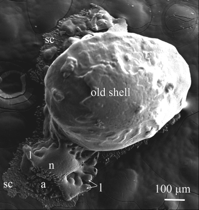
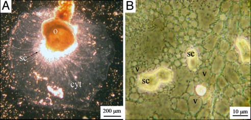
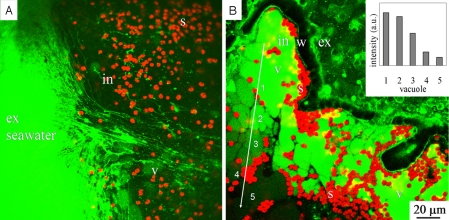
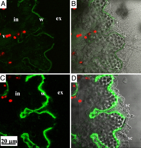
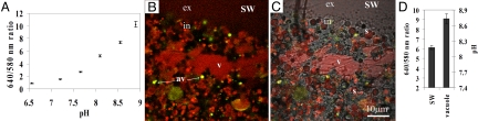
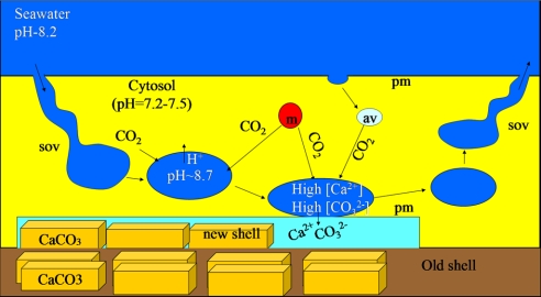
Foraminifera are unicellular organisms that inhabit the oceans in various ecosystems. The majority of the foraminifera precipitate calcitic shells and are among the major CaCO(3) producers in the oceans. They comprise an important component of the global carbon cycle and also provide valuable paleoceanographic information based on the relative abundance of stable isotopes and trace elements (proxies) in their shells. Understanding the biomineralization processes in foraminifera is important for predicting their calcification response to ocean acidification and for reliable interpretation of the paleoceanographic proxies. Most models of biomineralization invoke the involvement of membrane ion transporters (channels and pumps) in the delivery of Ca(2+) and other ions to the calcification site. Here we show, in contrast, that in the benthic foraminiferan Amphistegina lobifera, (a shallow water species), transport of seawater via fluid phase endocytosis may account for most of the ions supplied to the calcification site. During their intracellular passage the seawater vacuoles undergo alkalization that elevates the CO(3)(2-) concentration and further enhances their calcifying potential. This mechanism of biomineralization may explain why many calcareous foraminifera can be good recorders of paleoceanographic conditions. It may also explain the sensitivity to ocean acidification that was observed in several planktonic and benthic species.


Conflict of interest statement

The authors declare no conflict of interest.

Figures

Fig. 1.
Fig. 1.
SEM image of recalcified specimen of A. lobifera. The new chamber was built on the glass substrate instead of on the existing shell. Nevertheless, it displays all normal features of A. lobifera chamber. n = new chamber, a = aperture, l = lobes, sc = secondary crystals (consisting a layer of calcite, which is a part of the lamination process of these foraminifera).
Fig. 2.
Fig. 2.
The cytoplasm of a recalcifying A. lobifera. (A) The cytoplasmic layer (cyt) that delineates the calcification site. (B) Precipitation of secondary calcite crystals (sc) on the glass underneath the highly vacuolated cytoplasmic layer. Some of the vacuoles (v) are attached to the newly precipitated calcite aggregates.
Fig. 3.
Fig. 3.
FITC-dextran labeled seawater demonstrating endocytosis. (A) Labeled threads (green) associated with the inward cytoplasmic stream and the formation of new vacuoles (v). The symbiotic algae (s) with their red autofluorescence are intracellular (in). (B) Seawater vacuoles in the last peripheral chamber after 25 min of incubation. The yellow color represents combined signal of red (algae) and green (FITC in SWV) fluorescence. (Inset): The average fluorescence intensity of five marked vacuoles along the arrow, note the gradual inward decrease of the fluorescence. v = vacuoles, in = intracellular, ex = extracellular, w = chamber wall, s = algae symbionts.
Fig. 4.
Fig. 4.
Calcification from seawater vacuoles labeled with FITC-dextran: (A) At the end of a 2-h pulse, the labeled calcite represents calcification during the incubation period. (B) The same images as A, merged with transmitted light channel. (C) After 24 h of chase (in dye-free seawater) the fluorescence signal of the FITC-dextran has increased, while the only source for the dye is the intracellular seawater vacuoles. (D) The same images as C, merged with transmitted light channel. v = vacuole, in = intracellular, ex = extracellular, w = chamber wall, sc = secondary crystals.
Fig. 5.
Fig. 5.
Calcification from seawater vacuoles labeled with calcein: (A) At the end of a 1-h incubation with the dye (pulse) followed by washing with normal seawater (chase), the labeled outline of the chamber represents the calcification during the pulse incubation. (B) After 24 h of chase, the chamber wall is thicker, and the labeled calcite strip has expanded when the only source for the calcein is the labeled seawater vacuoles. v = vacuoles, in = intracellular, ex = extracellular, w = chamber wall, s = algae symbionts, sc = secondary crystals.
Fig. 6.
Fig. 6.
pH imaging of seawater vacuoles with SNARF-1 dextran. (A) In vitro calibration of SNARF-1 fluorescence in seawater (error bars = STD). (B) Alkaline seawater vacuoles. The image composed of green and red representing the 580- and 640-nm fluorescence, respectively. (C) The same image merged with transmitted light channel. (D) The emission ratio 640/580 nm, and the corresponding pH, of the large seawater vacuole (v) and the surrounding seawater. The small vesicles with bright yellow color (av) are acidic (pH <6) as indicated by their low 640/580 fluorescence ratio. v = vacuoles, av = acidic vesicles, in = intracellular, ex = extracellular, s = algae symbionts.
Fig. 7.
Fig. 7.
Model for temporal fusion of the SWV with the plasma membrane (pm) near the active calcification site. Seawater enters the cell through deep invaginations or semi open vacuoles (sov). A vacuole is pinched off and undergoes alkalization by one of the cellular proton transport mechanisms. This SWV concentrates inorganic carbon by diffusion of CO2(aq) from the acidic cytosol into the alkaline SWV. This process is enhanced by adjacent mitochondria (m) and by the acidic vesicles (av) that release CO2. The [Ca2+] and [CO32−]-enriched vacuoles fuse with the cell membrane and supply the ions for calcification. The vacuoles are then resealed and release their content apically (away from the growing crystals).

Similar articles

Cited by

References

    1. Erez J. The source of ions for biomineralization in foraminifera and their implications for paleoceanographic proxies. Rev Mineral Geochem. 2003;54:115–149.
    1. Lowenstam HA, Weiner S. On Biomineralization. Oxford: Oxford Univ Press; 1989.
    1. Simkiss K, Wilbur K. Biomineralization. Cell Biology and Mineral Deposition. San Diego: Academic, Inc.; 1989.
    1. Marshall AT, Clode PL. Effect of increased calcium concentration in sea water on calcification and photosynthesis in the scleractinian coral Galaxea fascicularis. J Exp Biol. 2002;205:2107–2113. - PubMed
    1. Wheatly MG, Zanotto FP, Hubbard MG. Calcium homeostasis in crustaceans: Subcellular Ca dynamics. Comp Biochem Phys B. 2002;132:163–178. - PubMed

Publication types