. 2009 Dec 29;106(52):22211-6.
doi: 10.1073/pnas.0910123106. Epub 2009 Dec 10.

A designer ligand specific for Kv1.3 channels from a scorpion neurotoxin-based library

Affiliations

A designer ligand specific for Kv1.3 channels from a scorpion neurotoxin-based library

Zoltan Takacs et al. Proc Natl Acad Sci U S A. .

Abstract

Venomous animals immobilize prey using protein toxins that act on ion channels and other targets of biological importance. Broad use of toxins for biomedical research, diagnosis, and therapy has been limited by inadequate target discrimination, for example, among ion channel subtypes. Here, a synthetic toxin is produced by a new strategy to be specific for human Kv1.3 channels, critical regulators of immune T cells. A phage display library of 11,200 de novo proteins is designed using the alpha-KTx scaffold of 31 scorpion toxin sequences known or predicted to bind to potassium channels. Mokatoxin-1 (moka1) is isolated by affinity selection on purified target. Moka1 blocks Kv1.3 at nanomolar levels that do not inhibit Kv1.1, Kv1.2, or KCa1.1. As a result, moka1 suppresses CD3/28-induced cytokine secretion by T cells without cross-reactive gastrointestinal hyperactivity. The 3D structure of moka1 rationalizes its specificity and validates the engineering approach, revealing a unique interaction surface supported on an alpha-KTx scaffold. This scaffold-based/target-biased strategy overcomes many obstacles to production of selective toxins.


Conflict of interest statement

Conflict of interest statement: Based in part on this work, a patent application (“Identification of Toxin Ligands,” PCT/US2008/013385) has been filed.

Figures

Fig. 1.
Fig. 1.
KcsA-1.3 channels bind KTX phage and isolate moka1 phage from an α-KTx scaffold library. Phage preparation, library construction, sorting and ELISA protocols, and KcsA, KcsA-1.3, and toxin synthesis and purification are described in SI Materials and Methods. Single-letter codes for amino acids are standard with Z for pyroglutamate. (A) ELISA shows KTX phage bind to KcsA-1.3 but not wild-type KcsA channels (Left) and phage expressing KTX bind to KcsA-1.3 channels, whereas those expressing DDD-KTX do not (Right). Ninety-six well plates coated with KcsA-1.3 or KcsA were incubated with phage (108–1010/well). Data are mean ± SE. for three wells. (B) Library construction and sorting. Thirty-one scorpion toxins that share the α-KTx scaffold were aligned to define three domains (A, B, and C). In KTX, domains were from residues Gly1-Pro12 (A), Leu15-Gly26 (B), and Asn30-Lys38 (C). Domains were linked by sharing of nucleotide codes for amino acids QC (A and B) or KCM (B and C), thereby conserving the important KTX residue Lys27. This yields 30, 22, and 17 unique A, B, and C domains, respectively, and a calculated library diversity of 11,220. Moka1 (GQ153941), a unique toxin isolated from the library, is composed of residues present in the natural toxins Ce3 (red), AgTx2 (yellow), and CTX (blue). Móka is a Hungarian word that translates into English as fun. Isolation of two or more identical clones in 20 by sorting was the basis for further study because the probability of this in the absence of enrichment (e.g., randomly) is smaller than 10−8.
Fig. 2.
Fig. 2.
Structure of moka1. Production of [U-13C, 15N]-moka1 and NMR spectroscopy are described in SI Materials and Methods. Statistics of the final structures are in Table S7 (A) Moka1 retains the α-KTx fold. NMR-derived solution structure of moka1 (PDB ID code 2kir). The Cα traces (gray) of 20 structures of moka1 are superimposed. The Cys residue side chains and the disulfide bonds are shown in yellow. (B Left) Superposition of the structure of moka1 (gray) with CTX (blue; PDB ID code 2crd) and AgTx2 (yellow, PDB ID code 1agt). (Right) Superposition of the structure of KTX (purple; PDB ID code 1ktx) and moka1 (gray). (C) The moka1 surface that is predicted to interact with the channel is shown en face in stick (Left) and surface representations (Right). Portions of moka1 originating from parental toxins are marked with different colors: Ce3 (red), AgTx2 (yellow), and CTX (blue). Residues common to all (QC and KCM) are shown in gray. Moka1 Lys24 equivalent to KTX Lys27 that is inserted in the channel pore is marked by an asterisk (*).
Fig. 3.
Fig. 3.
Moka1, a high-affinity and specific Kv1.3 channel blocker. Ion channel isoforms expressed in Xenopus laevis oocytes and studied by two electrode voltage clamp included rat Kv1.1, rat Kv1.2, human Kv1.3, and mouse KCa1.1. Bath solution was (in mM): 2 KCl, 96 NaCl, 1 MgCl2, 1.8 CaCl2, 5 Hepes (pH 7.5), and 0.1% bovine serum albumin (BSA). Equilibrium inhibition was determined by fitting dose-response curves for moka1 and calculated for other toxins from percent block. kon and koff were calculated. Methods are described in depth in SI Materials and Methods. (A) Representative current traces for human Kv1.3 channels before, in the presence of, and after washout of 3 nM moka1 at steady state. (Scale bars: 2 μA and 200 ms.) Voltage protocol: holding voltage −100 mV, 500 ms steps of 15 mV from −60 mV to 30 mV followed by a 200 ms step to −135 mV with a 30-s interpulse interval. (B) The time course for block and unblock of human Kv1.3 on acute application (bar) and washout of 3 nM moka1 during 100 ms steps to 0 mV from −100 mV followed by a 200-ms step to −135 mV every 2 s. (Inset) Voltage protocol. (C) Dose-response relationships for moka1 inhibition of human Kv1.3 (●), rat Kv1.1 (▴), rat Kv1.2 (◆), and mouse KCa1.1 (■), n = 3–11 cells. Kv1.1, Kv1.2, or Kv1.3 peak currents were recorded during a 500-ms step every 30 s to 0 mV from a holding voltage of −100 mV, followed by a 200-ms step to −135 mV. KCa1.1 currents were recorded with 50-ms steps every 3 s to 60 mV from −80 mV, followed by a 40-ms step to −100 mV. (D) Measured equilibrium inhibition (nM) for moka1, KTX, AgTx2, CTX on Kv1.3, Kv1.1, Kv1.2, and KCa1.1. Ce3 is reported to block human Kv1.3 with Ki of 366 nM (5). AgTx2 is reported to block KCa1.1 with Ki >1,000 nM (Table S8). Block of human Kv1.3 in human embryonic kidney (HEK293) cells showed inhibition with an affinity at equilibrium (Ki) of 4.4 ± 0.5 nM (Table S9).
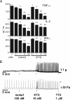
Fig. 4.
Fig. 4.
Moka1 inhibits secretion of effector cytokines by T cells and does not alter ileal motility and peristaltism. (A) Treatment with moka1 inhibits the secretion of effector cytokines by T cells. Human CD3+ T cells were purified from peripheral blood of healthy donors and 105 were treated with various doses of moka1 or KTX starting 1 h before stimulation for 16 h with anti-CD3/CD28 beads at a 1:1 ratio in triplicate. Supernatants were assessed for TNF-α, IL-2, and IFN-γ by ELISA. Data represent two independent experiments. Statistically significant differences between control (no toxin) and test conditions are indicated (*, P < 0.05; **,P < 0.01). Methods are described in-depth in SI Materials and Methods. (B) Effect of moka1 and KTX on isometric tension (Upper) and intraluminal pressure (Lower) of guinea pig ileum. Preparations were exposed to 100 nM moka1 which induced no discernable effect over 15–20 min. Subsequent addition of 10 nM KTX induced twitching and increased peristaltic activity—effects abolished by 1 μM tetrodotoxin (TTX). Ileal segments were from adult guinea pigs and studied in modified Krebs-Henseleit solution at 37 °C. For isometric tension assays, segments were mounted under tension and responses recorded using a force-displacement transducer coupled to a polygraph. Toxins were added after 60 min of equilibration (34). For assays of peristalsis, intraluminal perfusion from the oral end was continuous with pressure recording at the aboral end and the threshold for contraction used to quantify effects (12). Toxins were added after peristaltic activity was stable for >15 min. Methods are described further in SI Materials and Methods.

Similar articles

Cited by

References

    1. Price M, Lee SC, Deutsch C. Charybdotoxin inhibits proliferation and interleukin 2 production in human peripheral blood lymphocytes. Proc Natl Acad Sci. 1989;86:10171–10175. - PMC - PubMed
    1. Beeton C, et al. Kv1.3 channels are a therapeutic target for T cell-mediated autoimmune diseases. Proc Natl Acad Sci. 2006;103:17414–17419. - PMC - PubMed
    1. Leonard RJ, Garcia ML, Slaughter RS, Reuben JP. Selective blockers of voltage-gated K+ channels depolarize human T lymphocytes: Mechanism of the antiproliferative effect of charybdotoxin. Proc Natl Acad Sci USA. 1992;89:10094–10098. - PMC - PubMed
    1. Legros C, et al. Engineering-specific pharmacological binding sites for peptidyl inhibitors of potassium channels into KcsA. Biochemistry. 2002;41:15369–15375. - PubMed
    1. Olamendi-Portugal T, et al. Novel α-KTx peptides from the venom of the scorpion Centruroides elegans selectively blockade Kv1.3 over IKCa1 K+ channels of T cells. Toxicon. 2005;46:418–429. - PubMed

Publication types

MeSH terms