. 2009 Dec 22;106(51):21836-41.
doi: 10.1073/pnas.0909507106. Epub 2009 Dec 10.

Error-prone translesion replication of damaged DNA suppresses skin carcinogenesis by controlling inflammatory hyperplasia

Affiliations

Error-prone translesion replication of damaged DNA suppresses skin carcinogenesis by controlling inflammatory hyperplasia

Anastasia Tsaalbi-Shtylik et al. Proc Natl Acad Sci U S A. .

Abstract

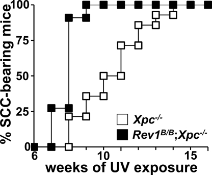
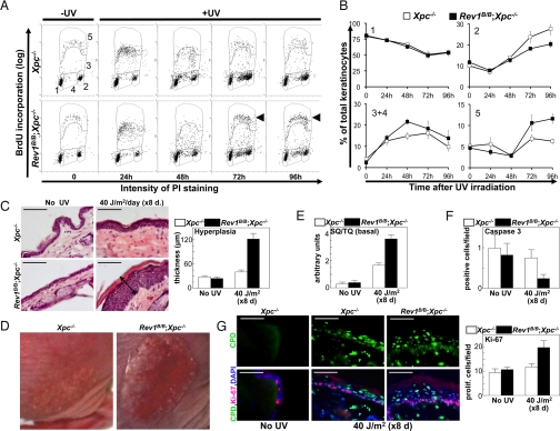
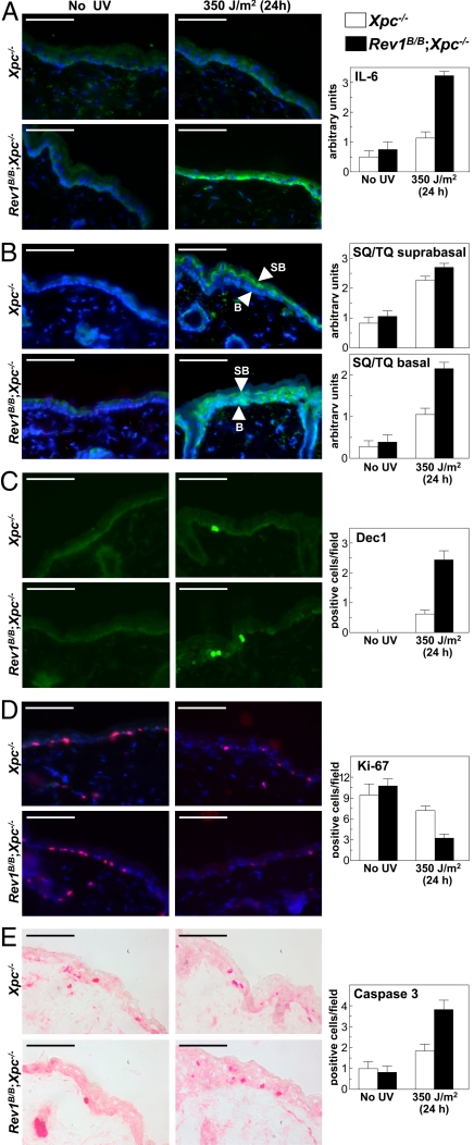
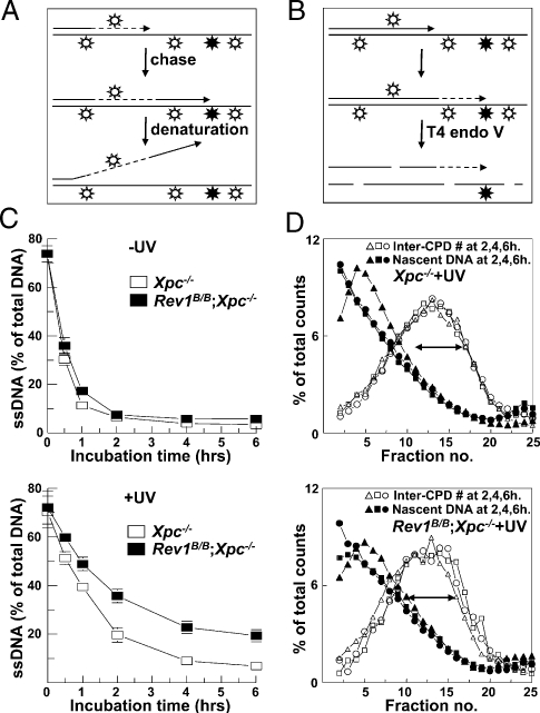
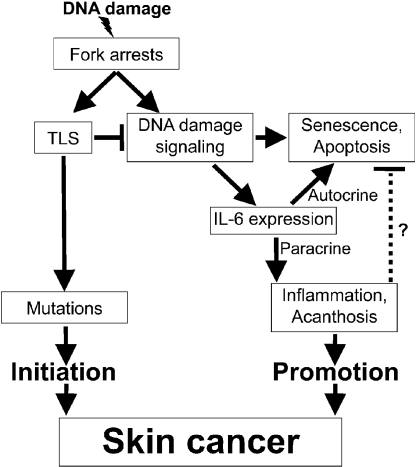
The induction of skin cancer involves both mutagenic and proliferative responses of the epidermis to ultraviolet (UV) light. It is believed that tumor initiation requires the mutagenic replication of damaged DNA by translesion synthesis (TLS) pathways. The mechanistic basis for the induction of proliferation, providing tumor promotion, is poorly understood. Here, we have investigated the role of TLS in the initiation and promotion of skin carcinogenesis, using a sensitive nucleotide excision repair-deficient mouse model that carries a hypomorphic allele of the error-prone TLS gene Rev1. Despite a defect in UV-induced mutagenesis, skin carcinogenesis was accelerated in these mice. This paradoxical phenotype was caused by the induction of inflammatory hyperplasia of the mutant skin that provides strong tumor promotion. The induction of hyperplasia was associated with mild and transient replicational stress of the UV-damaged genome, triggering DNA damage signaling and senescence. The concomitant expression of Interleukin-6 (IL-6) is in agreement with an executive role for IL-6 and possibly other cytokines in the autocrine induction of senescence and the paracrine induction of inflammatory hyperplasia. In conclusion, error-prone TLS suppresses tumor-promoting activities of UV light, thereby controlling skin carcinogenesis.


Conflict of interest statement

The authors declare no conflict of interest.

Figures

Fig. 1.
Fig. 1.
Latencies of UV-induced skin carcinogenesis. UV-induced skin carcinogenesis is significantly accelerated by the Rev1B/B mutation (χ2, P = 0.007).
Fig. 2.
Fig. 2.
Induction of inflammatory acanthosis in the Rev1B/B;Xpc−/− epidermis. (A) BrdU incorporation and cell cycle progression of keratinocytes after UV-irradiation in vivo (350 J/m2). The histogram was gated as indicated: 1: G0/G1 phase cells; 2: G2 phase cells; 3 and 4: poorly and nonreplicating (arrested) S phase cells, respectively; 5: actively replicating cells. Arrowheads indicate excessive mitogenic activity. PI, Propidium iodide. (B) Quantification of BrdU incorporation. Error bars, SEM. Significant differences between the genotypes: population 3 + 4 at 48 h: t test, P = 0.026; population 5 at 96 h: t test, P = 0.005. (C) HE-stained sections of the epidermis. The double arrow denotes the acanthotic epidermis of UV-treated Rev1B/B;Xpc−/− mice. Right: quantification of epidermal thickness. Error bars: SEM. Xpc−/−: pUV <0.05. Rev1B/B;Xpc−/−: pUV <0.001, pXpc <0.01. (Scale bars: 100 μm.) (D) Psoriasis-like plaques in Rev1B/B;Xpc−/− mice, 3 weeks after exposure to equitoxic high UV doses (1250 and 875 J/m2, for Xpc−/− and Rev1B/B;Xpc−/− mice, respectively). (E) DNA damage signaling in the basal layer of the epidermis after chronic low-dose UV treatment. Error bars: SEM. Xpc−/−:pUV <0.001. Rev1B/B;Xpc−/−:pUV <0.001, pXpc <0.001. (F) Apoptosis in the basal layer of the epidermis after chronic low-dose UV treatment. Error bars: SEM. Rev1B/B;Xpc−/−: pXpc <0.05. (G) Proliferation (red) and CPDs (green) in nuclei (blue) in the basal layer of the epidermis after chronic low-dose UV treatment. Right: quantification of the Ki-67 signal. Error bars: SEM. Rev1B/B;Xpc−/−: pUV <0.01, pXpc <0.01. (Scale bar, 100 μm.)
Fig. 3.
Fig. 3.
DNA damage responses of the epidermis, 24 h after acute subtoxic UV exposure. (Scale bar, 100 μm.) Error bars: SEM. (A) IL-6 in the epidermis and its quantification. Cytoplasmic granular IL-6 expression throughout the basal layer. Nuclei are stained blue (DAPI). Rev1B/B;Xpc−/−: pUV <0.001, pXpc <0.001. (B) Activation of DNA damage signaling and its quantification. SB: suprabasal layer; Xpc−/−: pUV <0.001. Rev1B/B;Xpc−/−: pUV <0.001, pXpc = 0.03. B: basal layer; Xpc−/−: pUV <0.001. Rev1B/B;Xpc−/−: pUV <0.001, pXpc <0.001. (C) Senescence and its quantification. Rev1B/B;Xpc−/−: pXpc <0.001. (D) Proliferation and its quantification. Rev1B/B;Xpc−/−: pUV <0.001, pXpc = 0.002. (E) Apoptosis and its quantification. Rev1B/B;Xpc−/−: pXpc = 0.012.
Fig. 4.
Fig. 4.
Perturbation of replication in Rev1B/B;Xpc−/− MEFs. (A) Measurement of replication fork progression by alkaline denaturation. Top: following UV treatment, nascent forks are pulse labeled with [3H]thymidine (interrupted lines). The persistence of label at DNA ends indicates fork stalling. (B) Measurement of global replication. Top: the template is uniformly labeled with [14C]thymidine (uninterrupted lines), followed by UV treatment. Middle: nascent DNA is labeled with [3H]thymidine (dashed line). Bottom: DNA is cleaved at CPDs and [14C]-labeled fragments serve as internal standards. Fragments are fractionated on alkaline sucrose gradients and a size increase of [3H]-labeled fragments indicates progression of replication at damaged templates. (C, Top) Fork progression at undamaged templates. Bottom: delayed TLS in Rev1B/B;Xpc−/− MEFs. (D) Transient perturbation of replication in Rev1B/B;Xpc−/− MEFs is illustrated by the double arrows that have the same length in both Top and Bottom. Open symbols: 14C-labeled internal standard.
Fig. 5.
Fig. 5.
Model for the role of error-prone TLS in regulating skin tumor initiation and promotion. See the Discussion for details.

Similar articles

Cited by

References

    1. Kundu JK, Surh YJ. Inflammation: Gearing the journey to cancer. Mutat Res. 2008;659:15–30. - PubMed
    1. Mueller MM. Inflammation in epithelial skin tumours: Old stories and new ideas. Eur J Cancer. 2006;42:735–744. - PubMed
    1. Melnikova VO, Ananthaswamy HN. Cellular and molecular events leading to the development of skin cancer. Mutat Res. 2005;571:91–106. - PubMed
    1. Lehmann AR, et al. Translesion synthesis: Y-family polymerases and the polymerase switch. DNA Repair. 2007;6:891–899. - PubMed
    1. Gan GN, Wittschieben JP, Wittschieben BØ, Wood RD. DNA polymerase zeta (pol zeta) in higher eukaryotes. Cell Res. 2008;18:174–183. - PubMed

Publication types