

Magnesium-aspartate-based crystallization switch inspired from shell molt of crustacean
- PMID: 20007788
- PMCID: PMC2799767
- DOI: 10.1073/pnas.0909040106
Magnesium-aspartate-based crystallization switch inspired from shell molt of crustacean
Abstract
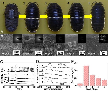
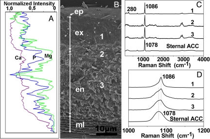
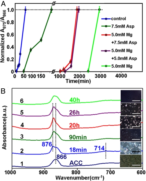
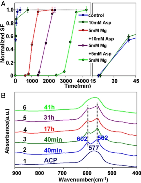
Many animals such as crustacean periodically undergo cyclic molt of the exoskeleton. During this process, amorphous calcium mineral phases are biologically stabilized by magnesium and are reserved for the subsequent rapid formation of new shell tissue. However, it is a mystery how living organisms can regulate the transition of the precursor phases precisely. We reveal that the shell mineralization from the magnesium stabilized precursors is associated with the presence of Asp-rich proteins. It is suggested that a cooperative effect of magnesium and Asp-rich compound can result into a crystallization switch in biomineralization. Our in vitro experiments confirm that magnesium increases the lifetime of amorphous calcium carbonate and calcium phosphate in solution so that the crystallization can be temporarily switched off. Although Asp monomer alone inhibits the crystallization of pure amorphous calcium minerals, it actually reduces the stability of the magnesium-stabilized precursors to switch on the transformation from the amorphous to crystallized phases. These modification effects on crystallization kinetics can be understood by an Asp-enhanced magnesium desolvation model. The interesting magnesium-Asp-based switch is a biologically inspired lesson from nature, which can be developed into an advanced strategy to control material fabrications.
Conflict of interest statement
The authors declare no conflict of interest.
Figures
Similar articles
-
Amorphous and crystalline calcium carbonate distribution in the tergite cuticle of moulting Porcellio scaber (Isopoda, Crustacea).J Struct Biol. 2011 Jul;175(1):10-20. doi: 10.1016/j.jsb.2011.03.019. Epub 2011 Mar 30. J Struct Biol. 2011. PMID: 21458575
-
Mineral deposition in bacteria-filled and bacteria-free calcium bodies in the crustacean Hyloniscus riparius (Isopoda: Oniscidea).PLoS One. 2013;8(3):e58968. doi: 10.1371/journal.pone.0058968. Epub 2013 Mar 12. PLoS One. 2013. PMID: 23554963 Free PMC article.
-
Biologically formed amorphous calcium carbonate.Connect Tissue Res. 2003;44 Suppl 1:214-8. Connect Tissue Res. 2003. PMID: 12952200 Review.
-
Testing the cation-hydration effect on the crystallization of Ca-Mg-CO3 systems.Proc Natl Acad Sci U S A. 2013 Oct 29;110(44):17750-5. doi: 10.1073/pnas.1307612110. Epub 2013 Oct 14. Proc Natl Acad Sci U S A. 2013. PMID: 24127571 Free PMC article.
-
Applications of Cryogenic Electron Microscopy in Biomineralization Research.J Dent Res. 2022 May;101(5):505-514. doi: 10.1177/00220345211053814. Epub 2021 Dec 17. J Dent Res. 2022. PMID: 34918556 Review.
Cited by
-
Convergent Evolution of Armor: Thermal Resistance in Deep-Sea Hydrothermal Vent Crustaceans.Biology (Basel). 2024 Nov 21;13(12):956. doi: 10.3390/biology13120956. Biology (Basel). 2024. PMID: 39765623 Free PMC article.
-
Biomineralisation by earthworms - an investigation into the stability and distribution of amorphous calcium carbonate.Geochem Trans. 2015 Apr 28;16:4. doi: 10.1186/s12932-015-0019-z. eCollection 2015. Geochem Trans. 2015. PMID: 26028991 Free PMC article.
-
Changes in the proximate and elemental composition of Alitropus typus (Crustacea: Flabellifera: Aegidae) exposed to lethal dose of bacterial consortium.J Parasit Dis. 2021 Sep;45(3):859-868. doi: 10.1007/s12639-021-01374-1. Epub 2021 Mar 17. J Parasit Dis. 2021. PMID: 34475669 Free PMC article.
-
Growth of second stage mineral in Lytechinus variegatus.Connect Tissue Res. 2018 Jul;59(4):345-355. doi: 10.1080/03008207.2017.1391233. Epub 2017 Oct 30. Connect Tissue Res. 2018. PMID: 29083939 Free PMC article.
-
Solubility investigations in the amorphous calcium magnesium carbonate system.CrystEngComm. 2019 Jan 7;21(1):155-164. doi: 10.1039/c8ce01596a. Epub 2018 Nov 20. CrystEngComm. 2019. PMID: 30760969 Free PMC article.
References
-
- Mann S. Oxford, UK: Oxford Univ Press; 2001. Biomineralization-principles and concepts in bioinorganic chemistry.
-
- Sanchez C, Arribart H, Guille MMG. Biomimetism and bioinspiration as tools for the design of innovative materials and systems. Nature Mat. 2005;4:277–288. - PubMed
-
- Orme CA, et al. Formation of chiral morphologies through selective binding of amino acids to calcite surface steps. Nature. 2001;411:775–779. - PubMed
-
- Addadi L, Raz S, Weiner S. Taking advantage of disorder: Amorphous calcium carbonate and its roles in biomineralization. Adv Mat. 2003;15:959–970.
-
- Politi Y, et al. Sea urchin spine calcite forms via a transient amorphous calcium carbonate phase. Science. 2004;306:1161–1164. - PubMed
Publication types
MeSH terms
Substances
LinkOut - more resources
Full Text Sources
