

Post-illumination pupil response in subjects without ocular disease
- PMID: 20007832
- PMCID: PMC2868495
- DOI: 10.1167/iovs.09-4717
Post-illumination pupil response in subjects without ocular disease
Abstract
Purpose: A sustained pupilloconstriction is often observed after the cessation of a bright visual stimulus. This post-illumination pupil response (PIPR) is produced by the intrinsically photosensitive retinal ganglion cells (ipRGCs). The present study was designed to examine the characteristics of the PIPR in a normal population without ocular disease.
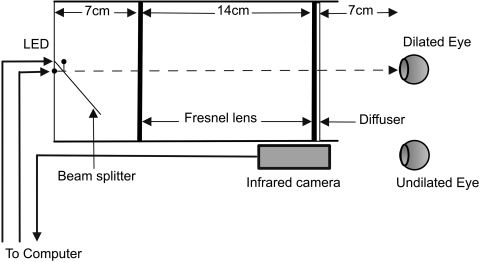
Methods: Thirty-seven subjects (mean age, 48.6 years) were tested by presenting a 60 degrees, 10-second light stimulus (13 log quanta/cm(2)/s retinal irradiance) and recording pupillary responses for 50 seconds after light cessation. The light stimuli (470 [blue] and 623 [red] nm) were presented by an optical system to one eye after dilation, while the consensual pupil response of the fellow, undilated eye was recorded by infrared pupillometry.
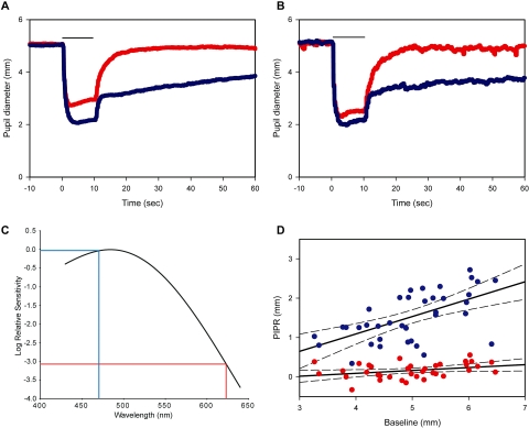
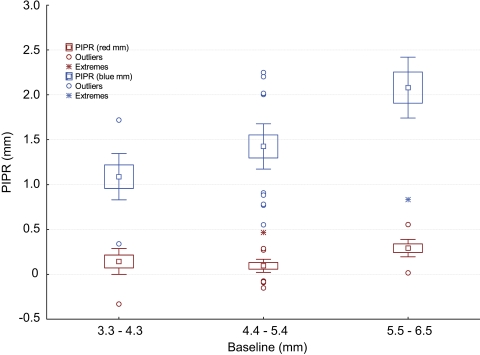
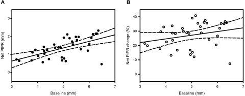
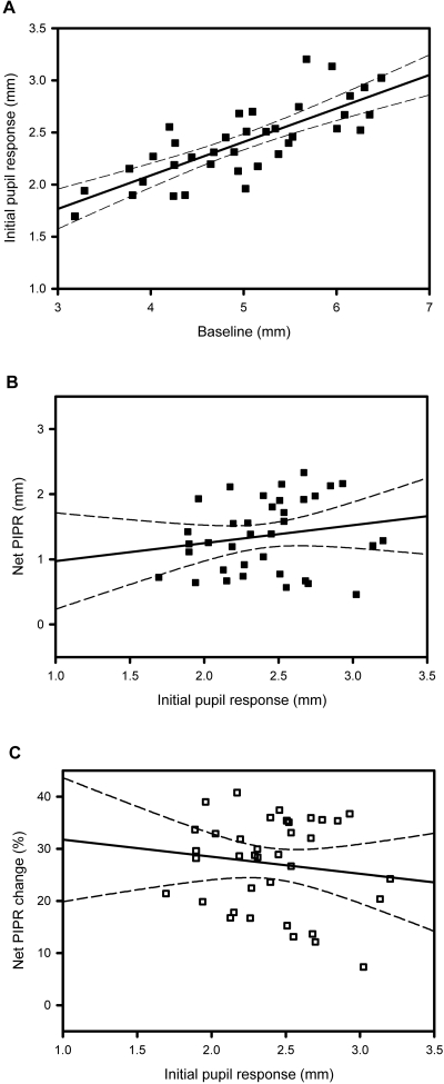
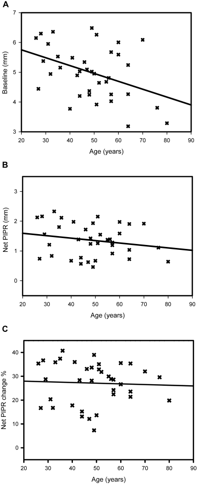
Results: A positive PIPR was seen in all subjects tested. The population average of the PIPR for 470-nm light was 1.5 mm (SEM 0.10, P < 0.05) and the net PIPR (blue PIPR minus red PIPR) was 1.4 mm (SEM 0.09, P < 0.0001). The net PIPR correlated positively with baseline pupil diameter (P < 0.05), but not significantly with age, race, or sex (P > 0.05) in the test population.
Conclusions: All normal subjects displayed a significant PIPR for a 10-second, 470-nm light stimulus, but not a 623-nm stimulus, which is consistent with the proposed melanopsin-mediated response. In most normal individuals, the amplitude of the PIPR was substantial. This test has the potential to be used as a tool in evaluating subjects with inner retinal dysfunction or melanopsin-related disorders.
Figures
Similar articles
-
The post-illumination pupil response is reduced in glaucoma patients.Invest Ophthalmol Vis Sci. 2011 Apr 8;52(5):2287-92. doi: 10.1167/iovs.10-6023. Print 2011 Apr. Invest Ophthalmol Vis Sci. 2011. PMID: 21212172 Free PMC article.
-
Rhodopsin and Melanopsin Contributions to the Early Redilation Phase of the Post-Illumination Pupil Response (PIPR).PLoS One. 2016 Aug 22;11(8):e0161175. doi: 10.1371/journal.pone.0161175. eCollection 2016. PLoS One. 2016. PMID: 27548480 Free PMC article.
-
Full-field chromatic pupillometry for the assessment of the postillumination pupil response driven by melanopsin-containing retinal ganglion cells.Invest Ophthalmol Vis Sci. 2014 Jun 12;55(7):4496-503. doi: 10.1167/iovs.14-14103. Invest Ophthalmol Vis Sci. 2014. PMID: 24925879
-
Intrinsically photosensitive retinal ganglion cells.J Neuroophthalmol. 2007 Sep;27(3):195-204. doi: 10.1097/WNO.0b013e31814b1df9. J Neuroophthalmol. 2007. PMID: 17895821 Review.
-
Intrinsically photosensitive melanopsin retinal ganglion cell contributions to the pupillary light reflex and circadian rhythm.Clin Exp Optom. 2010 May;93(3):137-49. doi: 10.1111/j.1444-0938.2010.00479.x. Clin Exp Optom. 2010. PMID: 20557555 Review.
Cited by
-
Selective stimulation of penumbral cones reveals perception in the shadow of retinal blood vessels.PLoS One. 2015 Apr 21;10(4):e0124328. doi: 10.1371/journal.pone.0124328. eCollection 2015. PLoS One. 2015. PMID: 25897842 Free PMC article.
-
Association Between Postillumination Pupil Response and Glaucoma Severity: A Cross-Sectional Analysis of the LIGHT Study.Invest Ophthalmol Vis Sci. 2022 Mar 2;63(3):24. doi: 10.1167/iovs.63.3.24. Invest Ophthalmol Vis Sci. 2022. PMID: 35333289 Free PMC article.
-
Light-Induced Pupillary Responses in Alzheimer's Disease.Front Neurol. 2019 Apr 12;10:360. doi: 10.3389/fneur.2019.00360. eCollection 2019. Front Neurol. 2019. PMID: 31031692 Free PMC article. Review.
-
The post-illumination pupil response is reduced in glaucoma patients.Invest Ophthalmol Vis Sci. 2011 Apr 8;52(5):2287-92. doi: 10.1167/iovs.10-6023. Print 2011 Apr. Invest Ophthalmol Vis Sci. 2011. PMID: 21212172 Free PMC article.
-
Pupillary Light Reflex Reveals Melanopsin System Alteration in the Background of Myopia-26, the Female Limited Form of Early-Onset High Myopia.Invest Ophthalmol Vis Sci. 2024 Jul 1;65(8):6. doi: 10.1167/iovs.65.8.6. Invest Ophthalmol Vis Sci. 2024. PMID: 38958970 Free PMC article.
References
-
- Loewenfeld IE, Lowenstein O. The Pupil Anatomy, Physiology, and Clinical Applications Vol. 1.Ames, IA: Iowa State University Press; 1993.
-
- Lucas RJ, Douglas RH, Foster RG. Characterization of an ocular photopigment capable of driving pupillary constriction in mice. Nat Neurosci 2001;4(6):621–626 - PubMed
Publication types
MeSH terms
Substances
Grants and funding
LinkOut - more resources
Full Text Sources
