Insulin-like growth factor-1 and cardiotrophin 1 increase strength and mass of extraocular muscle in juvenile chicken
- PMID: 20007833
- PMCID: PMC2868486
- DOI: 10.1167/iovs.09-4414
Insulin-like growth factor-1 and cardiotrophin 1 increase strength and mass of extraocular muscle in juvenile chicken
Abstract
Purpose: Insulin-like growth factor 1 (IGF1) and cardiotrophin 1 (CT1) are known to increase the strength of extraocular muscles in adult and embryonic animals, but no information is available for the early postnatal period, when strabismus treatment in humans is most urgent. Here the authors sought to determine whether these trophic factors strengthen juvenile maturing extraocular muscles and gain insight into mechanisms of force increase.
Methods: After two injections of IGF1, CT1, or both with different dosages in posthatch chickens, the authors quantified five parameters of the superior oblique extraocular muscle at 2 weeks of age: contractile force, muscle mass, total myofiber area, myofiber diameter, and number of proliferating satellite cells labeled by bromodeoxyuridine.
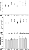
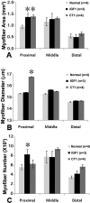
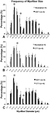
Results: Treatment with IGF1, CT1, and combination of IGF1 and CT1 significantly increased contractile force by 14% to 22%. CT1 and combination treatment significantly increased muscle mass by 10% to 24%. IGF1/CT1 combination treatment did not have additive effects on strengthening muscles, compared with single-drug treatments. Myofiber area increased significantly with IGF1 and CT1 treatment in proximal, but not distal, parts of the muscle and this was due to increased fiber numbers or length (IGF1) or increased diameters of global layer myofibers (CT1). Trophic factors increased the number of proliferating (bromodeoxyuridine-labeled) satellite cells in proximal and middle segments of muscles.
Conclusions: Exogenous IGF1 and CT1 strengthen extraocular muscles during maturation. They predominantly remodel the proximal segment of juvenile extraocular muscles. This information about muscle plasticity may aid the design of pharmacologic treatment of strabismus in children during the "critical period" of oculomotor maturation.
Figures


Similar articles
-
Role of exogenous and endogenous trophic factors in the regulation of extraocular muscle strength during development.Invest Ophthalmol Vis Sci. 2004 Oct;45(10):3538-45. doi: 10.1167/iovs.04-0393. Invest Ophthalmol Vis Sci. 2004. PMID: 15452060
-
How to make rapid eye movements "rapid": the role of growth factors for muscle contractile properties.Pflugers Arch. 2011 Mar;461(3):373-86. doi: 10.1007/s00424-011-0925-6. Epub 2011 Jan 29. Pflugers Arch. 2011. PMID: 21279379 Free PMC article.
-
Effects of sequential injections of hepatocyte growth factor and insulin-like growth factor-I on adult rabbit extraocular muscle.J AAPOS. 2012 Aug;16(4):354-60. doi: 10.1016/j.jaapos.2012.05.002. J AAPOS. 2012. PMID: 22929450 Free PMC article.
-
Classification and development of myofiber types in the superior oblique extraocular muscle of chicken.Anat Rec (Hoboken). 2007 Dec;290(12):1526-41. doi: 10.1002/ar.20614. Anat Rec (Hoboken). 2007. PMID: 17972279
-
Extraocular muscle: cellular adaptations for a diverse functional repertoire.Ann N Y Acad Sci. 2002 Apr;956:7-16. doi: 10.1111/j.1749-6632.2002.tb02804.x. Ann N Y Acad Sci. 2002. PMID: 11960789 Review.
Cited by
-
Effects of the sustained release of IGF-1 on extraocular muscle of the infant non-human primate: adaptations at the effector organ level.Invest Ophthalmol Vis Sci. 2012 Jan 5;53(1):68-75. doi: 10.1167/iovs.11-8356. Invest Ophthalmol Vis Sci. 2012. PMID: 22125277 Free PMC article.
-
Changing muscle function with sustained glial derived neurotrophic factor treatment of rabbit extraocular muscle.PLoS One. 2018 Aug 24;13(8):e0202861. doi: 10.1371/journal.pone.0202861. eCollection 2018. PLoS One. 2018. PMID: 30142211 Free PMC article.
-
Extraocular muscle regeneration in zebrafish requires late signals from Insulin-like growth factors.PLoS One. 2018 Feb 7;13(2):e0192214. doi: 10.1371/journal.pone.0192214. eCollection 2018. PLoS One. 2018. PMID: 29415074 Free PMC article.
-
Eye alignment changes caused by sustained GDNF treatment of an extraocular muscle in infant non-human primates.Sci Rep. 2020 Jul 17;10(1):11927. doi: 10.1038/s41598-020-68743-3. Sci Rep. 2020. PMID: 32681083 Free PMC article.
-
The effects of bupivacaine injection and oral nitric oxide on extraocular muscle in the rabbit.Graefes Arch Clin Exp Ophthalmol. 2013 Sep;251(9):2227-33. doi: 10.1007/s00417-013-2390-8. Epub 2013 Jun 4. Graefes Arch Clin Exp Ophthalmol. 2013. PMID: 23733036
References
-
- West CE, Asbury T. Strabismus. In: Riordan-Eva P, Whitcher JP. eds. Vaughan & Asbury's General Ophthalmology New York: McGraw-Hill Medical; 2007:229–248
-
- McNeer KW, Magoon EH, Scott AB. Chemodenervation therapy: technique and indications. In: Rosenbaum AL, Santiago PS. eds. Clinical Strabismus Management Philadelphia: WB Saunders; 1999:423–432
-
- McLoon LK, Christiansen SP. Pharmacological approaches of the treatment of strabismus. Drugs Future 2005;30:319–327
-
- Scott AB, Miller JM, Shieh KR. Bupivacaine injection of the lateral rectus muscle to treat esotropia. J AAPOS 2009;13:119–122 - PubMed
-
- von Bartheld CS, Croes SA, Johnson LA. Strabismus. In: Levin LA, Albert DM. eds. Ocular Disease: Mechanisms and Management San Diego: Elsevier; 2009:454–460
Publication types
MeSH terms
Substances
Grants and funding
LinkOut - more resources
Full Text Sources
Miscellaneous

