. 2010 Mar;298(3):G375-83.
doi: 10.1152/ajpgi.00343.2009. Epub 2009 Dec 10.

BMP2 promotes differentiation of nitrergic and catecholaminergic enteric neurons through a Smad1-dependent pathway

Affiliations

BMP2 promotes differentiation of nitrergic and catecholaminergic enteric neurons through a Smad1-dependent pathway

Mallappa Anitha et al. Am J Physiol Gastrointest Liver Physiol. 2010 Mar.

Abstract

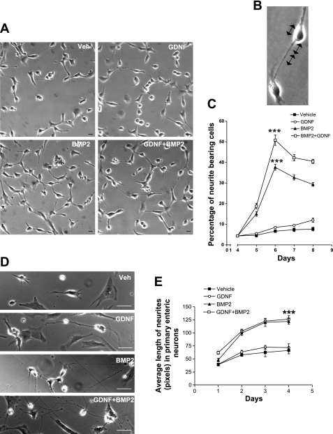
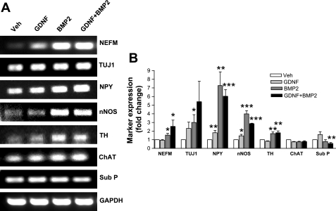
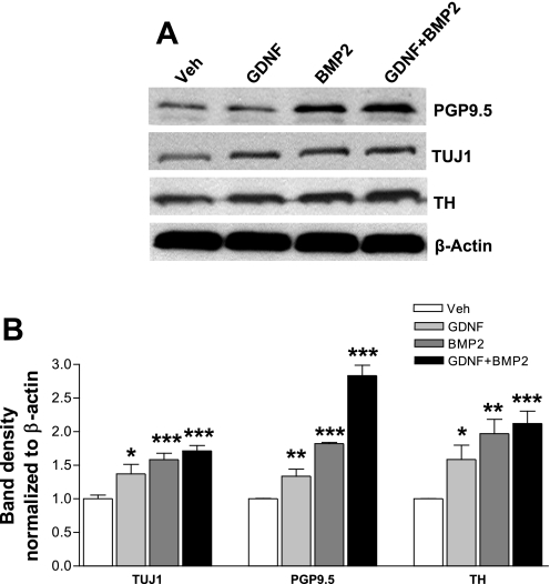
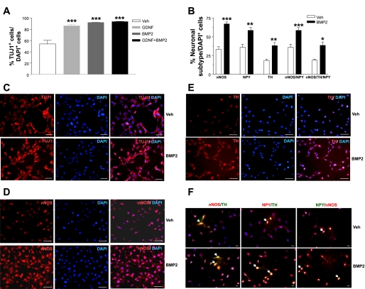
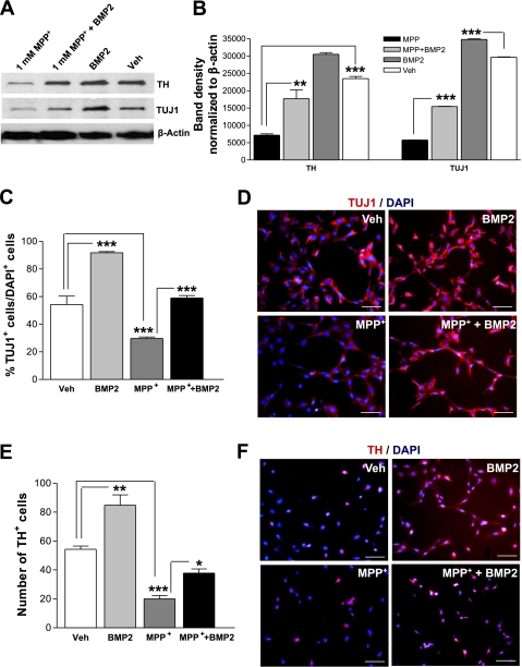
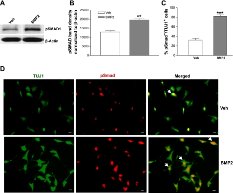
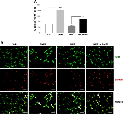
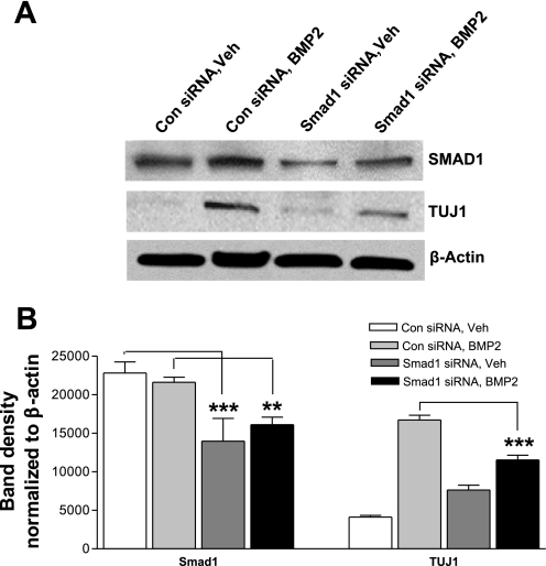
The bone morphogenetic protein (BMP) family is a class of transforming growth factor (TGF-beta) superfamily molecules that have been implicated in neuronal differentiation. We studied the effects of BMP2 and glial cell line-derived neurotrophic factor (GDNF) on inducing differentiation of enteric neurons and the signal transduction pathways involved. Studies were performed using a novel murine fetal enteric neuronal cell line (IM-FEN) and primary enteric neurons. Enteric neurons were cultured in the presence of vehicle, GDNF (100 ng/ml), BMP2 (10 ng/ml), or both (GDNF + BMP2), and differentiation was assessed by neurite length, markers of neuronal differentiation (neurofilament medium polypeptide and beta-III-tubulin), and neurotransmitter expression [neuropeptide Y (NPY), neuronal nitric oxide synthase (nNOS), tyrosine hydroxylase (TH), choline acetyltransferase (ChAT) and Substance P]. BMP2 increased the differentiation of enteric neurons compared with vehicle and GDNF-treated neurons (P < 0.001). BMP2 increased the expression of the mature neuronal markers (P < 0.05). BMP2 promoted differentiation of NPY-, nNOS-, and TH-expressing neurons (P < 0.001) but had no effect on the expression of cholinergic neurons (ChAT, Substance P). Neurons cultured in the presence of BMP2 have higher numbers of TH-expressing neurons after exposure to 1-methyl 4-phenylpyridinium (MPP(+)) compared with those cultured with MPP(+) alone (P < 0.01). The Smad signal transduction pathway has been implicated in TGF-beta signaling. BMP2 induced phosphorylation of Smad1, and the effects of BMP2 on differentiation of enteric neurons were significantly reduced in the presence of Smad1 siRNA, implicating the role of Smad1 in BMP2-induced differentiation. The effects of BMP2 on catecholaminergic neurons may have therapeutic implications in gastrointestinal motility disturbances.


Figures

Fig. 1.
Fig. 1.
Bone morphogenic protein 2 (BMP2) induces differentiation of a novel murine fetal enteric neuronal cell line (IM-FEN) and primary enteric neurons. A: phase-contrast photomicrographs of IM-FEN cells cultured in the presence of vehicle (Veh), glial cell line-derived neurotrophic factor (GDNF), BMP2, or both (GDNF + BMP2). B: cells bearing double the size of cell body length were considered as neurite-bearing cells. C: graph showing the percentage of neurite-bearing cells under different treatments. D: phase-contrast photomicrographs of primary enteric neurons cultured in the presence of vehicle, GDNF, BMP2, or both (GDNF + BMP2). E: graph showing the average length of neurite (pixels) in primary enteric neurons under different treatments. Results are means ± SE, n = 3, minimum of 40 neurons scored in each group, ***P < 0.001. Scale bar = 20 μm (A) and 100 μm (D).
Fig. 2.
Fig. 2.
BMP2 promotes differentiation of nitrergic and catecholaminergic neurons. A: RT-PCR was done using RNA from IM-FEN cells treated with vehicle, GDNF, BMP2, or both GDNF and BMP2 for 48 h and probed for different markers, n = 13. B: real-time PCR shows the fold increase of different marker expression in treated cells, n = 3. Results are means ± SE, *P < 0.05, **P < 0.01, ***P < 0.001. NEFM, neurofilament medium peptide; TUJ1, neuronal class III β-tubulin; NPY, neuropeptide Y; nNOS, neuronal nitric oxide synthase; TH, tyrosine hydroxylase; ChAT, choline acetyltransferase; Sub P, Substance P.
Fig. 3.
Fig. 3.
BMP2 increases neuronal differentiation and catecholaminergic marker expression in IM-FEN cells. Western blot analysis of cells cultured in the presence of vehicle, GDNF, BMP2, or both GDNF and BMP2 for 48 h and assessed for neuronal markers [protein gene product 9.5 (PGP9.5) and TUJ1], dopaminergic marker (TH), and β-actin as a loading control, n = 6. Results are means ± SE, *P < 0.05, **P < 0.01, ***P < 0.001.
Fig. 4.
Fig. 4.
BMP2 increases neuronal differentiation and nitrergic and catecholaminergic marker expression in IM-FEN cells. Immunostaining was done on the cells treated with vehicle, GDNF, BMP2, or both GDNF and BMP2 for 24 h. A and B: histogram shows the relative percentages of different marker-positive cells and percentage colocalization of nNOS/NPY/TH-expressing neurons with respect to total number of cells, n = 3. Results are means ± SE, *P < 0.05, **P < 0.01, ***P < 0.001. C, D, and E: representative images of IM-FEN cells treated with vehicle or BMP2, cultured for 24 h, and stained with different markers like TUJ1, TH, and nNOS. Nuclear stain is shown with 4,6-diamidino-2-phenylindole (DAPI) staining. Scale bar = 100 μm. F: merged images showing colocalization of nNOS-, NPY-, and TH-expressing neurons. Scale bar = 20 μm.
Fig. 5.
Fig. 5.
1-Methyl 4-phenylpyridinium (MPP+)-induced loss of dopaminergic neuronal is less in the presence of BMP2. A and B: Western blot analysis of cells treated with vehicle, BMP2, MPP+, or both BMP2 and MPP+ for neuronal marker (TUJ1) and dopaminergic marker (TH). β-actin was used as a loading control, n = 3. Results are means ± SE, **P < 0.01, ***P < 0.001. C and E: histogram shows the relative percentages of TUJ1 with respect to total number of cells and absolute number of TH-positive cells, n = 3. Results are means ± SE, *P < 0.05, **P < 0.01, ***P < 0.001. D and F: representative images of IM-FEN cells treated with vehicle, BMP2, MPP+, or both BMP2 and MPP+, cultured for 24 h, and stained for TUJ1 or TH. Nuclear stain is shown with DAPI staining. Scale bar = 100 μm.
Fig. 6.
Fig. 6.
BMP2 increases phosphorylation of Smad1 and pSmad nuclear translocation. A and B: Western blot analysis of IM-FEN cells treated with vehicle or BMP2 for 48 h and probed for pSmad, n = 3, **P < 0.01. C: histogram shows the percentage of pSmad-positive cells with respect to TUJ1-positive cells. Results are means ± SE, n = 3, ***P < 0.001. D: representative images of pSmad translocated in IM-FEN cells cultured for 24 h with or without BMP2. Scale bar = 20 μm.
Fig. 7.
Fig. 7.
BMP2 reduces loss of pSmad nuclear translocation in cells treated with MPP+. A: histogram shows the percentage of pSmad-positive cells with respect to TUJ1-positive cells. Results are means ± SE, n = 3, ***P < 0.001. B: representative images of pSmad translocated in IM-FEN cells cultured for 24 h with vehicle, BMP2, MPP+, or both BMP2 and MPP+, n = 2. Scale bar = 100 μm.
Fig. 8.
Fig. 8.
Smad1 knockdown prevents BMP2-mediated enteric neuronal differentiation. Western blot analysis (A) of IM-FEN cells transfected with Scrambled/Smad1 siRNA (30 nM) using lipofectamine RNAiMAX reagent for 24 h and treated with vehicle or BMP2 for another 24 h at 39°C was probed for Smad1 and TUJ1 (B). β-Actin was used as a loading control, n = 4. Results are means ± SE, **P < 0.01, ***P < 0.001.

Similar articles

Cited by

References

    1. Anderson G, Noorian AR, Taylor G, Anitha M, Bernhard D, Srinivasan S, Greene JG. Loss of enteric dopaminergic neurons and associated changes in colon motility in an MPTP mouse model of Parkinson's disease. Exp Neurol 207: 4–12, 2007 - PMC - PubMed
    1. Anderson RB, Newgreen DF, Young HM. Neural crest and the development of the enteric nervous system. Adv Exp Med Biol 589: 181–196, 2006 - PubMed
    1. Anitha M, Chandrasekharan B, Salgado JR, Grouzmann E, Mwangi S, Sitaraman SV, Srinivasan S. Glial-derived neurotrophic factor modulates enteric neuronal survival and proliferation through neuropeptide Y. Gastroenterology 131: 1164–1178, 2006 - PMC - PubMed
    1. Anitha M, Joseph I, Ding X, Torre ER, Sawchuk MA, Mwangi S, Hochman S, Sitaraman SV, Anania F, Srinivasan S. Characterization of fetal and postnatal enteric neuronal cell lines with improvement in intestinal neural function. Gastroenterology 134: 1424–1435, 2008 - PMC - PubMed
    1. Benavides-Piccione R, DeFelipe J. Different populations of tyrosine-hydroxylase-immunoreactive neurons defined by differential expression of nitric oxide synthase in the human temporal cortex. Cereb Cortex 13: 297–307, 2003 - PubMed

Publication types

MeSH terms

LinkOut - more resources