. 2010 Jan 22;327(5964):466-9.
doi: 10.1126/science.1179663. Epub 2009 Dec 10.

Tuberculous granuloma induction via interaction of a bacterial secreted protein with host epithelium

Affiliations

Tuberculous granuloma induction via interaction of a bacterial secreted protein with host epithelium

Hannah E Volkman et al. Science. .

Abstract

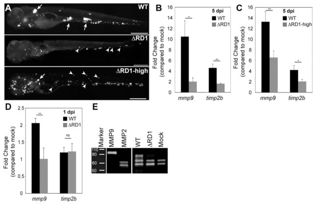
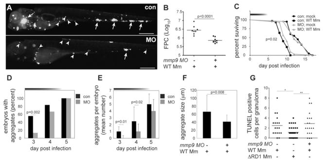
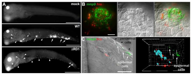
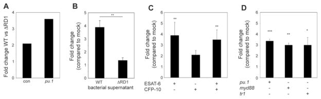
Granulomas, organized aggregates of immune cells, are a hallmark of tuberculosis and have traditionally been thought to restrict mycobacterial growth. However, analysis of Mycobacterium marinum in zebrafish has shown that the early granuloma facilitates mycobacterial growth; uninfected macrophages are recruited to the granuloma where they are productively infected by M. marinum. Here, we identified the molecular mechanism by which mycobacteria induce granulomas: The bacterial secreted protein 6-kD early secreted antigenic target (ESAT-6), which has long been implicated in virulence, induced matrix metalloproteinase-9 (MMP9) in epithelial cells neighboring infected macrophages. MMP9 enhanced recruitment of macrophages, which contributed to nascent granuloma maturation and bacterial growth. Disruption of MMP9 function attenuated granuloma formation and bacterial growth. Thus, interception of epithelial MMP9 production could hold promise as a host-targeting tuberculosis therapy.


Figures

Fig. 1
Fig. 1
RD1-dependent Mmp9 induction. (A) Representative fluorescence images of 5 dpi embryos used for gene expression studies in (B and C). Embryos in top and middle panels injected with similar doses of WT and ΔRD1, respectively (WT dose 193±36 and ΔRD1 dose 217±63), where ΔRD1 bacterial burdens are lower than WT at 5dpi. Embryo in bottom panel injected with ~ 5-fold more ΔRD1 (ΔRD1- high) to achieve similar bacterial burdens to WT at 5 dpi (5 dpi bacterial burdens were 1601±1071 for WT and 1531±1011 for ΔRD1-high, ns). Arrows, granulomas; arrowheads, single infected macrophages. Scale bars, 400 μm. (B-C) Relative gene expression levels (mean ± SEM of at least 3 biological replicates) of 5 dpi (B) WT- and ΔRD1-infected embryos and (C) WT and ΔRD1- high-infected embryos. Although there appears to be a dose-dependent induction of mmp9 by ΔRD1 (compare induction in panels B and C), the difference is not significant (p=0.2). (D) Relative gene expression levels (mean ± SEM of 3 biological replicates) 1 day after injection with 721±39 WT or 484±147 ΔRD1 (ns). *p < 0.05, **p < 0.01, ns, not significant (Student’s t test). (E) Gelatin zymography of embryos 5 dpi with 200 WT or 700 ΔRD1, or mock-infected. Controls are purified human MMP9 and MMP2.
Fig. 2
Fig. 2
Mmp9 promotes granuloma formation and virulence. (A) Fluorescence images of representative control (con), and mmp9 morphant (MO) embryos 4 dpi with 116 WT. Arrows, granulomas; arrowheads, single infected macrophages. Scale bars, 400 μm (B) Bacterial burdens of all 4dpi embryos determined by fluorescence pixel counts (FPC) (see supplementary methods). (C) Survival of con and MO embryos (n=30 each) infected with 150 CFU WT or mock-infected (n=20 each). Median survival 10 days for infected MO and 9 days for infected con (p=0.02; Log-rank test) and no different for uninfected MO and con. Top horizontal bar denotes duration of MO activity (see fig. S2). (D to F) Kinetics of granuloma formation in con and MO embryos infected with 101 WT. Data in (D) analyzed by Fisher’s exact test of a contingency table. Bars in (E) and (F) represent mean ± SEM (Student’s t test). (G) Median number of TUNEL-positive cells per con or MO granuloma 4dpi with 37 CFU WT and con granulomas 4 dpi with 585 ΔRD1. (ANOVA; One-Way Analysis of Variance p=0.003, with Dunnet’s Multiple Comparison Test).
Fig. 3
Fig. 3
mmp9 is selectively induced in epithelial cells neighboring infected macrophages. (A) mmp9 FISH images of embryos 5 days after mock infection or infection with 78 CFU WT or 130 CFU ΔRD1. Arrows, mmp9 expression corresponding to granulomas, arrowheads, single mmp9-expressing cells. Scale bars, 400μm. (B and C) Images of WT granulomas after dual mmp9 and fms FISH. (B) Fluorescence (left), DIC (middle), and overlay (right) images. e, epidermal cell; p, peridermal cell; MΦ, macrophage. Scale bar, 20μm. Also see movie S1. (C) Fluorescence and DIC overlay of nascent WT muscle granuloma (left panel). Dotted white circles outline bacterial clusters discerned by DIC microscopy. Fluorescence data has been deconvolved. Right panel represents 3D reconstruction from fluorescence image of same lesion with bacterial locations pseudocolored blue, showing complete absence of mmp9 expression in adjacent muscle, and strong mmp9 expression in nearest neighboring epidermal cells. Scale bar, 20μm. Also see movie S2.
Fig. 4
Fig. 4
Mycobacterial ESAT-6 is sufficient to induce mmp9 in epithelial cells independent of Myd88 and TNF signaling. (A-D) Relative mmp9 expression analyzed by qRT-PCR of (A) con or pu.1 morphant embryos 3 dpi with 84 WT or 126 ΔRD1 (represents one biological replicate), (B) 34 hpf embryos 4 hours post-injection with WT or ΔRD1 bacterial supernatant. Bars represent means ± SEM of three biological replicates. (C) 34 hpf con embryos 4 hours post-injection with 4.8×10⁻¹⁷ moles of purified ESAT-6 or CFP-10, or 4.9×10⁻¹⁷ moles of ESAT-6 plus 5.0×10⁻¹⁷ moles of CFP-10. Bars represent means (± SEM) of five biological replicates. (D) 34 hpf con embryos, myd88 morphants, or tr1 morphants 4 hours post-injection with 5.7×10⁻¹⁷ moles of purified ESAT-6. Bars represent means ± SEM of four biological replicates (pu.1 morphant), or three biological replicates (myd88 and tr1 morphants).

Comment in

References

    1. Clay H, et al. Cell Host & Microbe. 2007;2:29. - PMC - PubMed
    1. Wolf AJ, et al. J Immunol. 2007;179:2509. - PubMed
    1. Adams DO. Am J Pathol. 1976;84:164. - PMC - PubMed
    1. Ulrichs T, Kaufmann SH. J Pathol. 2006;208:261. - PubMed
    1. Volkman HE, et al. PLoS Biol. 2004;2:e367. - PMC - PubMed

Publication types

MeSH terms