. 2010 Jan 15;327(5963):302-5.
doi: 10.1126/science.1182213. Epub 2009 Dec 10.

Adaptive evolution of pelvic reduction in sticklebacks by recurrent deletion of a Pitx1 enhancer

Affiliations

Adaptive evolution of pelvic reduction in sticklebacks by recurrent deletion of a Pitx1 enhancer

Yingguang Frank Chan et al. Science. .

Abstract

The molecular mechanisms underlying major phenotypic changes that have evolved repeatedly in nature are generally unknown. Pelvic loss in different natural populations of threespine stickleback fish has occurred through regulatory mutations deleting a tissue-specific enhancer of the Pituitary homeobox transcription factor 1 (Pitx1) gene. The high prevalence of deletion mutations at Pitx1 may be influenced by inherent structural features of the locus. Although Pitx1 null mutations are lethal in laboratory animals, Pitx1 regulatory mutations show molecular signatures of positive selection in pelvic-reduced populations. These studies illustrate how major expression and morphological changes can arise from single mutational leaps in natural populations, producing new adaptive alleles via recurrent regulatory alterations in a key developmental control gene.


Figures

Figure 1
Figure 1
Alleles of Pitx1 from pelvic-complete (FRIL, LITC) and pelvic-reduced populations (PAXB) were combined in F1 hybrids, and brain and pelvic tissues were isolated to compare the expression of either the LITC or PAXB allele normalized to the level of expression of the FRIL allele in the same trans-acting environment. Expression of the PAXB Pitx1 allele is greatly reduced in the pelvis but not the head of F1 hybrids (two-tailed t-test, P < 0.0001), indicating a tissue-specific, cis-regulatory change in the Pitx1 locus.
Figure 2
Figure 2
(A) VISTA/mLAGAN alignment of Pitx1 candidate region from pelvic-complete stickleback (SALR), medaka and zebrafish. Red peaks, >40% sequence identity in 20bp sliding windows; grey bars at top, repetitive sequence; formula image 
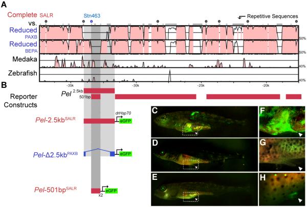
symbols, microsatellite markers used in association mapping in Fig. S1). (B) Reporter gene expression in transgenic animals. (C) Pel-2.5kbSALR from a marine population drives tissue-specific EGFP (green) expression in the developing pelvic bud of Swarup stage 32 larvae (36); (F) detail. (D and G) Altered Pel-Δ2.5kbPAXB sequence from pelvic-reduced PAXB stickleback fails to drive pelvic EGFP expression. (E and H) A smaller fragment from marine fish, Pel-501bpSALR, also drives EGFP expression in the developing pelvic bud of multiple St. 30 larvae. This region is completely missing in PAXB.
Fig. 3
Fig. 3
(A) Juvenile pelvic-reduced BEPA stickleback expressing a Pitx1 transgene driven by the Pel-2.5kbSALR enhancer, compared to (B) uninjected sibling. External spines form only in transgenic fish (black arrowhead) (C and D) Alizarin red stained pelvic structures of adult transgenic fish compared to BEPA parental phenotype. BEPA fish normally develop only a small ovoid vestige (OV) of the anterior pelvic process (AP). Transgenic fish show clear development of the anterior process (AP), ascending branch (AB), and posterior process (PP) of the pelvis, and a prominent serrated pelvic spine. Pectoral fin (PF) rays develop in both fish.
Fig. 4
Fig. 4
(A) SNP genotyping in additional pelvic-reduced populations identifies nine different deletions that overlap in a 488bp region. Triangles, SNP markers; grey bars, putative deleted regions flanked by two failed SNP genotypes; dark blue bars, markers flanked by two successful SNP genotypes; light blue bars, markers with successful genotypes only on one side; red bars, positions of Pel-2.5kb and Pel-501bp enhancers. Apparent deletions were confirmed by sequencing in Populations 4, 6 and 7, with the size of deletions indicated on the right, and micro-homologies of two to three base pairs at deletion junctions shown in red. (B) Location of populations surveyed. (C) TwistFlex prediction of highly flexible DNA regions (red circles) in Pitx1 locus (Pel region score: 3263) compared to frequency distribution of flexibility scores in rest of stickleback genome (median score: 265). Area of red circles is proportional to flexibility score.
Fig. 5
Fig. 5
(A and B) Fay and Wu’s H and relative heterozygosity (θπ) statistics across the Pitx1 region. Blue (freshwater pelvic-reduced) and green (freshwater pelvic-complete) data points and LOESS smoothed (α=0.2) line indicates the behavior in each group. The Pel-containing regulatory region of Pitx1 (grey candidate region from Fig. S1B) shows both negative H values, indicating an excess of derived alleles; and reduced heterozygosity in pelvic-reduced fish, consistent with positive selection (see text). θπ values are plotted relative to the grouped marine mean (per SNP) to control for variation in ascertainment between SNPs. (C) Heterozygosity (θπ) from different genomic regions, grouped by population type. Freshwater fish show a general decrease in heterozygosity across both Pitx1 and control loci compared to marine fish (red bars), as expected from founding of new freshwater populations from marine ancestors. In the Pel enhancer region, but not in Pitx1-flanking regions, or in control loci, pelvic-reduced freshwater populations (blue bars) show even lower heterozygosity than pelvic-complete freshwater populations (green bars) (**, P < 0.01).

Comment in

Similar articles

Cited by

References

    1. Orr HA. Nat. Rev. Genet. 2005;6:119. - PubMed
    1. Hoekstra HE, Coyne JA. Evolution. 2007;61:995. - PubMed
    1. Carroll SB. Cell. 2008;134:25. - PubMed
    1. Stern DL, Orgogozo V. Evolution. 2008;62:2155. - PMC - PubMed
    1. Hinchliffe JR, Johnson DR. The development of the vertebrate limb. Clarendon Press; Oxford: 1980.

Publication types