. 2010 Feb 12;285(7):4629-36.
doi: 10.1074/jbc.M109.063073. Epub 2009 Dec 11.

Pattern recognition receptors require N-glycosylation to mediate plant immunity

Affiliations

Pattern recognition receptors require N-glycosylation to mediate plant immunity

Heidrun Häweker et al. J Biol Chem. .

Abstract

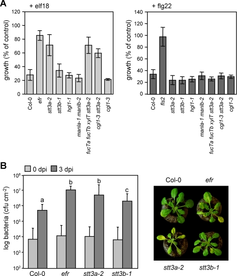
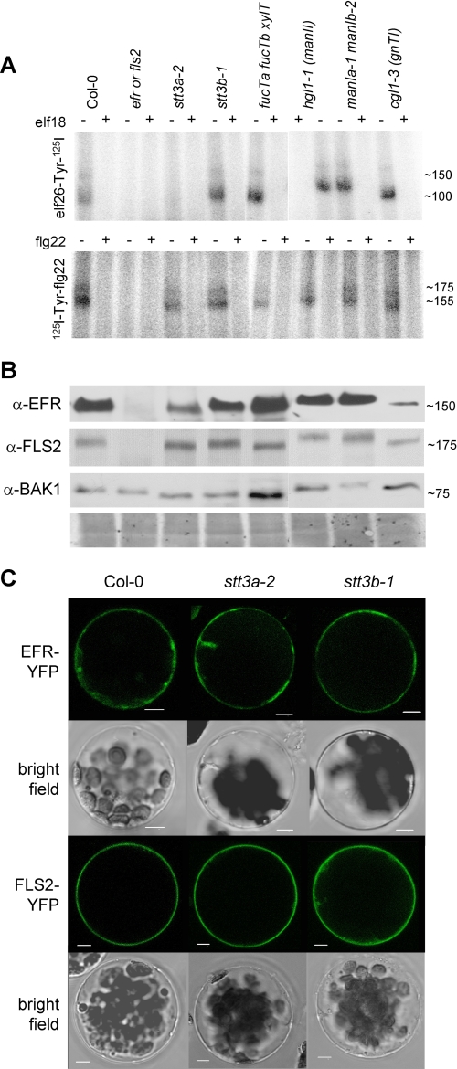
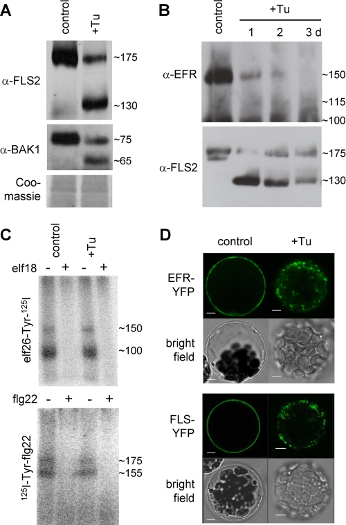
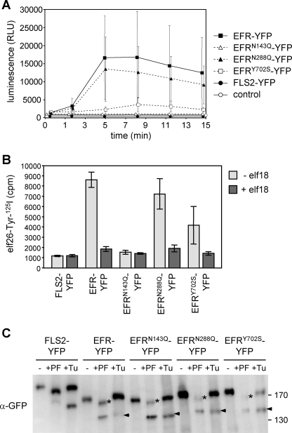
N-Glycans attached to the ectodomains of plasma membrane pattern recognition receptors constitute likely initial contact sites between plant cells and invading pathogens. To assess the role of N-glycans in receptor-mediated immune responses, we investigated the functionality of Arabidopsis receptor kinases EFR and FLS2, sensing bacterial translation elongation factor Tu (elf18) and flagellin (flg22), respectively, in N-glycosylation mutants. As revealed by binding and responses to elf18 or flg22, both receptors tolerated immature N-glycans induced by mutations in various Golgi modification steps. EFR was specifically impaired by loss-of-function mutations in STT3A, a subunit of the endoplasmic reticulum resident oligosaccharyltransferase complex. FLS2 tolerated mild underglycosylation occurring in stt3a but was sensitive to severe underglycosylation induced by tunicamycin treatment. EFR accumulation was significantly reduced when synthesized without N-glycans but to lesser extent when underglycosylated in stt3a or mutated in single amino acid positions. Interestingly, EFR(N143Q) lacking a single conserved N-glycosylation site from the EFR ectodomain accumulated to reduced levels and lost the ability to bind its ligand and to mediate elf18-elicited oxidative burst. However, EFR-YFP protein localization and peptide:N-glycosidase F digestion assays support that both EFR produced in stt3a and EFR(N143Q) in wild type cells correctly targeted to the plasma membrane via the Golgi apparatus. These results indicate that a single N-glycan plays a critical role for receptor abundance and ligand recognition during plant-pathogen interactions at the cell surface.


Figures

FIGURE 1.
FIGURE 1.
ArabidopsisN-glycosylation mutants display different immune reactions. A, seedling growth arrest in response to MAMP treatment. Growth reduction upon 100 nm elf18 or flg22 treatment is shown as percentage of control growth (without the peptides). Similar results were obtained in three independent experiments. Bars represent means ± S.D. (n = 6). B, pathogen infection. Plants were spray-inoculated with PtoDC3000. Bacterial inoculation and growth were determined at 0 and 3 days post infection, respectively, and photographs were taken at 4 dpi. Bars represent means ± S.D. of two biological independent experiments each of six replicates (p < 0.0001 indicated by letters).
FIGURE 2.
FIGURE 2.
Ligand binding, protein accumulation, and localization of PRRs in selected N-glycosylation mutants. A, equal amounts of ground seedling material were incubated with elf26-Tyr-125I or 125I-Tyr-flg22 in the absence (−) or presence (+) of 10 μm elf18 or flg22 peptide, respectively. Similar results were obtained in at least three independent experiments. B, accumulation of EFR, FLS2, and BAK1 proteins. Equal amounts of the lines indicated in A were loaded for immunoblotting and revealed with specific antibodies (α-FLS2, α-EFR, or α-BAK1). As controls, efr mutants were included for elf26 binding and EFR and BAK1 accumulation, and fls2 mutants were included for flg22 binding and FLS2 accumulation. Representative Coomassie staining is shown as loading reference below. C, EFR-YFP and FLS2-YFP were expressed in protoplasts of Col-0 wild type and indicated glycosylation mutants. Confocal and bright field images of representative transfected protoplasts are shown. Scale bars, 5 μm.
FIGURE 3.
FIGURE 3.
Effect of interference with receptor N-glycosylation on PRR ligand binding. A, untreated (control) and tunicamycin-treated (+Tu) Col-0 wild type plants were analyzed by immunoblotting with specific antibodies (α-FLS2, α-EFR, or α-BAK1). Coomassie staining is shown for equal loading. B, time course of tunicamycin treatment (10 μg/ml for 3 days). C, cross-linking with elf18 and flg22 peptides was performed with untreated (control) or tunicamycin-treated (Tu) Col-0 seedlings. D, effect of 8–10-h tunicamycin incubation on localization of EFR- and FLS2-YFP in Col-0 wild type protoplasts. Scale bars, 5 μm.
FIGURE 4.
FIGURE 4.
Analysis of EFR mutant variants. EFR-YFP and derived mutant variations were expressed in N. benthamiana by transient transformation. A, EFR function was tested by elf18-triggered oxidative burst (control = not transformed). Data points are means ± S.D. (n = 15). Similar results were obtained in two independent experiments. RLU, relative light units. B, quantitative elf18-ligand binding. Bars represent means ± S.D. (n = 3). C, samples for PNGase F incubation (−/+PF) were taken at day 3 after transformation. Tunicamycin (+Tu, 10 μm) was infiltrated and harvested 5 h later. Note that PNGase F-resistant bands (+PF, asterisks) are indicative of post-Golgi transport of the chimeric glycoprotein-YFP fusions. Also, compared with wild type EFR-YFP, all mutant EFR variants are apparently more stable in the ER (+Tu, arrows). The immunoblot was revealed with green fluorescent protein antibodies (α-green fluorescent protein (α-GFP)).

Similar articles

Cited by

References

    1. Elbein A. D. (1987) Annu. Rev. Biochem. 56, 497–534 - PubMed
    1. Martínez I. M., Chrispeels M. J. (2003) Plant Cell 15, 561–576 - PMC - PubMed
    1. Iwata Y., Koizumi N. (2005) Proc. Natl. Acad. Sci. U.S.A. 102, 5280–5285 - PMC - PubMed
    1. Koiwa H., Li F., McCully M. G., Mendoza I., Koizumi N., Manabe Y., Nakagawa Y., Zhu J., Rus A., Pardo J. M., Bressan R. A., Hasegawa P. M. (2003) Plant Cell 15, 2273–2284 - PMC - PubMed
    1. Kang J. S., Frank J., Kang C. H., Kajiura H., Vikram M., Ueda A., Kim S., Bahk J. D., Triplett B., Fujiyama K., Lee S. Y., von Schaewen A., Koiwa H. (2008) Proc. Natl. Acad. Sci. U.S.A. 105, 5933–5938 - PMC - PubMed

Publication types

MeSH terms