. 2010 Mar 1;19(5):774-89.
doi: 10.1093/hmg/ddp543. Epub 2009 Dec 11.

Prdm16 is required for normal palatogenesis in mice

Affiliations

Prdm16 is required for normal palatogenesis in mice

Bryan C Bjork et al. Hum Mol Genet. .

Abstract

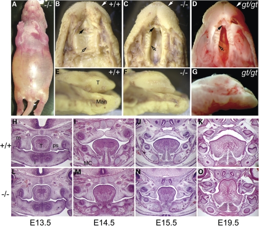
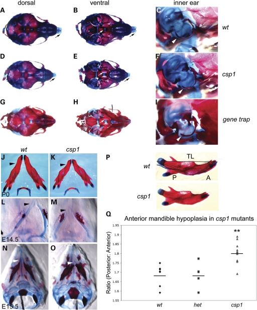
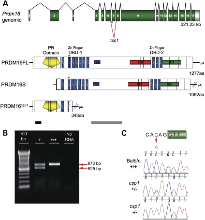
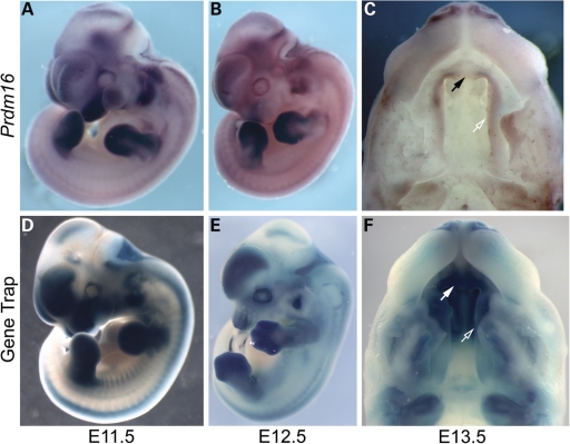
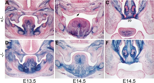
Transcriptional cofactors are essential to the regulation of transforming growth factor beta (TGFbeta) superfamily signaling and play critical and widespread roles during embryonic development, including craniofacial development. We describe the cleft secondary palate 1 (csp1) N-ethyl-N-nitrosourea-induced mouse model of non-syndromic cleft palate (NSCP) that is caused by an intronic Prdm16 splicing mutation. Prdm16 encodes a transcriptional cofactor that regulates TGFbeta signaling, and its expression pattern is consistent with a role in palate and craniofacial development. The cleft palate (CP) appears to be the result of micrognathia and failed palate shelf elevation due to physical obstruction by the tongue, resembling human Pierre Robin sequence (PRS)-like cleft secondary palate. PRDM16 should be considered a candidate for mutation in human clefting disorders, especially NSCP and PRS-like CP.


Figures

Figure 1.
Figure 1.
The csp1 and Prdm16Gt683Lex mutants exhibit CP. (AG) Newborn csp1 and Prdm16Gt683Lex mutant pups die shortly after birth with a distended abdomen (A). Gross examination of mutant embryos identified the presence of a wide CP (open arrows) and pointed snout (white arrows) (B–D) accompanied by abnormal tongue position and morphology (E–G). Black arrows show the unaffected primary palate. (HO) Histological analysis shows normal palate shelves at E13.5 that fail to elevate, remaining at the sides of the tongue at late E14.5 (I and M). Palate shelves remain in this position after normal palate fusion occurs by E15.5 (J and N) and do not undergo delayed elevation as evidenced by the failure of elevation at E19.5 (K and O) and postnatally (data not shown). T, tongue; Man, mandible; m, molar; i, incisor; MC, Meckel's cartilage.
Figure 2.
Figure 2.
csp1 and Prdm16Gt683Lex mutants exhibit craniofacial skeleton defects with anterior-specific mandibular hypoplasia. (AI) Abnormalities in the craniofacial skeleton in newborn csp1 mutants are evident in dorsal (A, D and G) and ventral (B, E and H) views of Alcian blue/Alazarin-stained heads. In addition, hypoplasia of the mutant tympanic rings is evident (C, F and I). Newborn mutants show failure of palatine bone fusion (white arrows in B, E and H), variably shortened frontonasal region (D and G) and abnormal nasal cartilage formation (black arrows in A, B, D and E) and shortening and abnormal curvature of the anterior mandible (black arrowheads in J and K). Anterior shortening of Meckel's cartilage is evident early during craniofacial bone formation at E14.5 (L and M) and E15.5 (N and O). In addition, csp1 mutant mandibles appear smaller than wild-type counterparts, and ossification appears to be more robust. Morphometric analysis using measurements of the posterior (P in P) and anterior (A in P) aspects of newborn csp1 (n = 14), heterozygous (n = 5) and wild-type (n = 7) mandible bones, followed by calculation of the posterior:anterior (P/A) ratios, detects an anterior-specific mandibular hypoplasia (Q). P/A ratios were significantly greater in csp1 mutants (mean: 1.802, standard deviation: 0.050) compared with wild-type (mean: 1.687, standard deviation: 0.056) and heterozygous pups (mean: 1.688, standard deviation: 0.066) using ANOVA (P < 0.0001). In the chart (Q), the double asterisk designates statistical significance, and horizontal lines denote the means for each genotype class.
Figure 3.
Figure 3.
A splicing mutation in Prdm16 causes the csp1 mutant phenotype. (A) Prdm16 genomic structure and the domain structures of wild-type PR-containing (FL), PR-minus (S) and csp1 mutant PRDM16 protein structure. Green numbered boxes depict exons, and black splicing lines show the intervening introns roughly to scale. Positive regulatory (PR), zinc finger DNA-binding domains 1 and 2 (DBD-1 and DBD-2) and repressor (RD), acidic (AD) and proline-rich (PRR) domains of PRDM16 with homology to EVI1 are shown. The intronic exon 7 splice acceptor mutation (red asterisk and splicing lines), identified in csp1 mutants, causes variable skipping of exon 7. The putative truncated PRDM16csp1 mutant protein is depicted. Exons (verticle black lines) are superimposed upon the PRDM16 protein. The PRDM16 peptides used to generate N-terminal and middle anti-PRDM16 polyclonal antibodies are depicted by black and gray bars, respectively. (B) RT–PCR amplification of Prdm16 from mutant and wild-type postnatal day 0 (P0) heads. An aberrant short splice product (red arrow) produced by exon 7 skipping is observed in csp1 mutants, but not in wild-type pups, in addition to the wild-type splice product (red arrow). (C) A recessive C-to-A mutation in the base pair preceding the AG-dinucleotide of the exon 7 SA is permissive for normal splicing with variably reduced efficiency in csp1 mutants.
Figure 4.
Figure 4.
Comparison of whole-mount in situ hybridization analysis of endogenous Prdm16 expression with reporter expression visualized by whole-mount X-gal staining in Prdm16Gt683Lex heterozygous gene trap embryos. (A, B, D and E) Overlapping expression of endogenous Prdm16 and reporter expression in E11.5 (A and D) and E12.5 (B and E) is observed in developing orofacial structures including the nasal, maxillary and mandibular prominences, forebrain, choroid plexi, cranial nerves, dorsal root ganglia, dermomyotome, forelimb and hindlimb mesenchymes, otic vesicle, the eye and ventricles of the heart. Prdm16 expression in Prdm16Gt683Lex embryos appears more intense in all structures. Craniofacial expression becomes more restricted to frontonasal region by E12.5. (C and F) Palate shelf expression in whole-mount E13.5 embryonic heads via in situ hybridization in csp1-FVB/NJ strain background (C) and X-gal staining of Prdm16Gt683Lex-mixed 129/SvEvBrd × C57BL6/J strain background (F) Prdm16 expression is visible in the secondary palate (open arrows), primary palate (solid arrows), incisor teeth mesenchyme and nasal cartilage.
Figure 5.
Figure 5.
The Prdm16Gt683Lex mutation causes recessive CP, and reporter expression in craniofacial structures is consistent with this phenotype. (AF) Histological analysis of coronal sections through the medial aspect of the secondary palate and the primary palate in heterozygous control and homozygous Prdm16Gt683Lex E13.5 and E14.5 embryos. Reporter expression is dose-sensitive. (A and D) At E13.5, before palate shelf elevation and fusion, Prdm16 reporter expression is strongest in the mesenchyme at the oral side of the palate shelves (ps), although it is visible throughout the palate shelf and hinge region. Expression within the tongue (T) musculature, molar tooth mesenchyme (m) and within Meckel's cartilage (MC) and surrounding perichondrium is also detected. Expression in palate epithelia is difficult to detect. (B and E) This expression pattern is maintained at E14.5, after palate shelf elevation and fusion, although it appears weaker. Black arrow, shown in B, depicts epithelial seam at the point of fusion between the palate shelves. Prdm16 expression levels are highest in the primary palate (C and F).
Figure 6.
Figure 6.
The csp1 and Prdm16Gt683Lex mutations are loss-of-function alleles that affect normal PRDM16 function in the negative regulation of TGFβ signaling in a TGFβ-responsive luciferase assay. (A) Western blot analysis using a novel polyclonal anti-PRDM16 antibody (Mid) (Fig. 3A, gray). Comparison of protein products detected in nuclear lysates isolated from HepG2 cells transfected with N-terminal V5-tagged PRDM16 FL and S isoform- and LacZ control-expressing plasmids using an anti-V5 antibody (black arrows on left) with the protein products detected from the same lysates using the anti-PRDM16 Mid antibody (red arrows, right) shows that the anti-PRDM16 antibody binds PRDM16 specifically. Western blot analysis of PRDM16 expression in Prdm16 loss-of-function mutants shows loss of endogenous PRDM16 in Prdm16GT683Lex and csp1 homozygous mutants in nuclear lysates isolated from wild-type and mutant embryonic head tissue. The doublet (black arrows on right) present in wild-type samples that disappears in the mutant samples is most consistent with the PRDM16 FL isoform and may represent post-translational modification of PRDM16 in vivo. Strong bands in the middle of this blot are cross-reacting background bands consistently observed when using this antibody. (B) V5-tagged LacZ, Prdm16FL, Prdm16S and Prdm16csp1 expression plasmids transfected into Mv1Lu cells show localization of PRDM16 isoforms to the nucleus, but this subcellular localization is abolished in the truncated CSP1 mutant protein. HepG2 cells (C) and Mv1 Lu cells (D) were co-transfected with pRL-TK, p3TP-Lux and plasmids expressing LacZ (control), Prdm16FL, Prdm16S or the Prdm16csp1 mouse mutant. Relative luciferase activity (RLA) was measured (Firefly luciferase: Renilla luciferase ratio) in the presence (black) or absence (gray) of TGFβ1. In both cell lines, both wild-type PRDM16 isoforms strongly inhibited TGFβ signaling, whereas CSP1 fails to abrogate TGFβ signaling. RLA values and error bars represent the means and standard deviations, respectively, for three separate experiments.
Figure 7.
Figure 7.
Loss of Prdm16 expression perturbs in vivo TGFβ signaling in the mandible. Immunofluorescent detection of TGFβ2, Phospho-SMAD2 and SMAD7 protein expression in wild-type (wt) (AF) versus Prdm16Gt683Lex gene trap null mutant (gt) (GI) E13.5 embryos. Expression of each protein in wild-type embryos is evident in secondary palate shelves (ps), tongue (T), Meckel's cartilage (white ovals in A–I), molar teeth (white arrows) and the undifferentiated mesenchyme between the oral sulci (white asterisks) at the sides of the tongue and the perichodrium surrounding Meckel's cartilage which contains the salivary ducts (sd) (A–F). A dramatic reduction in protein expression levels for each protein is observed in Prdm16Gt683Lex mutants in and around Meckel's cartilage and the region near the base of the tongue, consistent with the mandibular and salivary gland hypoplasia and gross tongue abnormalities observed in these mutants.
Figure 8.
Figure 8.
Additional phenotypic abnormalities in csp1 and Prdm16Gt683Lex mutants correlate with Prdm16 expression. Prdm16 is expressed in choroid plexi epithelium (A), and csp1 mutants exhibit dramatic choroid plexi hypoplasia (B and C). Prdm16 is expressed in the developing salivary glands (black arrowheads in D), and csp1 mutants have severe salivary gland hypoplasia (E and F). csp1 mutants exhibit abnormal retinal folds of variable severity (HJ). Although in situ hybridization shows that endogenous Prdm16 is expressed in the retina in increasingly high levels as embryos progress to birth (data not shown), the Prdm16 gene trap reporter expression is only very weak in the retina, and strong in the pigmented layer of the retina (rpe) (G). We cannot explain this inconsistency. We observe histologic abnormalities in lungs of late embryonic (E19.5) mutants embryos (L and M) and general lung hypoplasia (O and P) when compared with wild-type embryos accompanied by Prdm16 expression in the lung and airway epithelium (K and N). Prdm16 expression in the heart is primarily restricted to the left ventricle as early as E10.5 (N, E13.5 ventral view) and gradually becomes expressed throughout the ventricles, evident postnatally (Q, P3 shown). This expression pattern correlates with gross cardiac ventricular hypoplasia (S), which is more severe in Prdm16Gt683Lex mutants that can exhibit a severe cleft between the ventricles as shown (white arrowhead in T). Lu (lung), LV (left ventricle), RV (right ventricle).

References

    1. Mossey P.A., Little J. Epidemiology of oral clefts: an international perspective. In: Wyszynski D.F., editor. Cleft Lip and Palate. From Origin to Treatment. New York: Oxford University Press; 2002. pp. 127–158.
    1. Wyszynski D.F. Cleft Lip and Palate: From Origin to Treatment. 1st edn. Oxford: Oxford University Press; 2002.
    1. Shiang R., Lidral A.C., Ardinger H.H., Buetow K.H., Romitti P.A., Munger R.G., Murray J.C. Association of transforming growth-factor alpha gene polymorphisms with nonsyndromic cleft palate only (CPO) Am. J. Hum. Genet. 1993;53:836–843. - PMC - PubMed
    1. FitzPatrick D.R., Carr I.M., McLaren L., Leek J.P., Wightman P., Williamson K., Gautier P., McGill N., Hayward C., Firth H., et al. Identification of SATB2 as the cleft palate gene on 2q32–q33. Hum. Mol. Genet. 2003;12:2491–2501. - PubMed
    1. Marcano A.C., Doudney K., Braybrook C., Squires R., Patton M.A., Lees M.M., Richieri-Costa A., Lidral A.C., Murray J.C., Moore G.E., et al. TBX22 mutations are a frequent cause of cleft palate. J. Med. Genet. 2004;41:68–74. - PMC - PubMed

Publication types

MeSH terms