

Myricetin inhibits UVB-induced angiogenesis by regulating PI-3 kinase in vivo
- PMID: 20008033
- PMCID: PMC2864405
- DOI: 10.1093/carcin/bgp221
Myricetin inhibits UVB-induced angiogenesis by regulating PI-3 kinase in vivo
Abstract
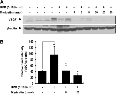
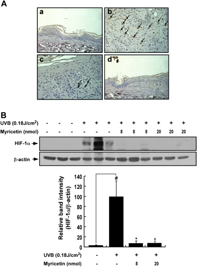
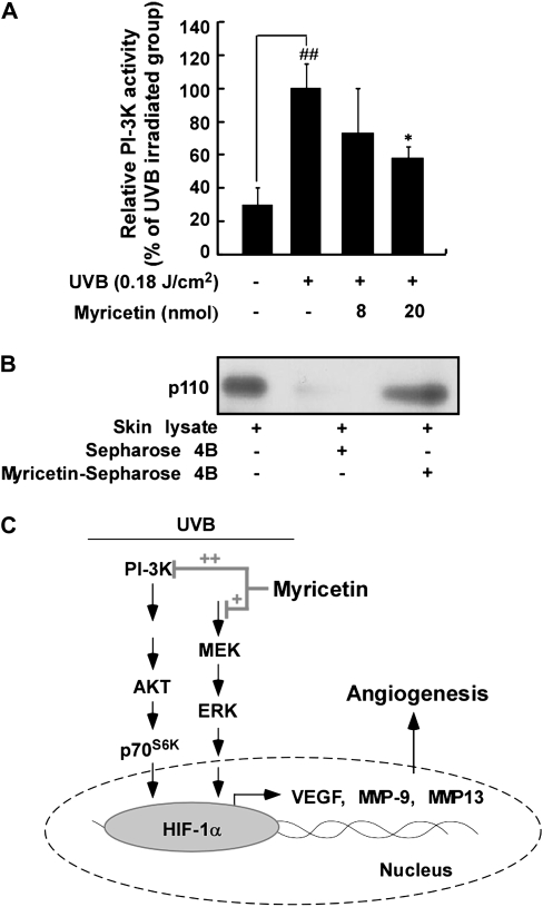
Myricetin is one of the principal phytochemicals in onions, berries and red wine. Previous studies showed that myricetin exhibits potent anticancer and chemopreventive effects. The present study examined the effect of myricetin on ultraviolet (UV) B-induced angiogenesis in an SKH-1 hairless mouse skin tumorigenesis model. Topical treatment with myricetin inhibited repetitive UVB-induced neovascularization in SKH-1 hairless mouse skin. The induction of vascular endothelial growth factor, matrix metalloproteinase (MMP)-9 and MMP-13 expression by chronic UVB irradiation was significantly suppressed by myricetin treatment. Immunohistochemical and western blot analyses revealed that myricetin inhibited UVB-induced hypoxia inducible factor-1alpha expression in mouse skin. Western blot analysis and kinase assay data revealed that myricetin suppressed UVB-induced phosphatidylinositol-3 (PI-3) kinase activity and subsequently attenuated the UVB-induced phosphorylation of Akt/p70(S6K) in mouse skin lysates. A pull-down assay revealed the direct binding of PI-3 kinase and myricetin in mouse skin lysates. Our results indicate that myricetin suppresses UVB-induced angiogenesis by regulating PI-3 kinase activity in vivo in mouse skin.
Figures


Similar articles
-
Hesperidin Inhibits UVB-Induced VEGF Production and Angiogenesis via the Inhibition of PI3K/Akt Pathway in HR-1 Hairless Mice.Biol Pharm Bull. 2021;44(10):1492-1498. doi: 10.1248/bpb.b21-00367. Biol Pharm Bull. 2021. PMID: 34602557
-
Myricetin suppresses UVB-induced wrinkle formation and MMP-9 expression by inhibiting Raf.Biochem Pharmacol. 2010 May 15;79(10):1455-61. doi: 10.1016/j.bcp.2010.01.004. Epub 2010 Jan 20. Biochem Pharmacol. 2010. PMID: 20093107 Free PMC article.
-
Myricetin is a potent chemopreventive phytochemical in skin carcinogenesis.Ann N Y Acad Sci. 2011 Jul;1229:124-32. doi: 10.1111/j.1749-6632.2011.06122.x. Ann N Y Acad Sci. 2011. PMID: 21793847
-
Myricetin suppresses UVB-induced skin cancer by targeting Fyn.Cancer Res. 2008 Jul 15;68(14):6021-9. doi: 10.1158/0008-5472.CAN-08-0899. Cancer Res. 2008. PMID: 18632659
-
Luteolin suppresses UVB-induced photoageing by targeting JNK1 and p90 RSK2.J Cell Mol Med. 2013 May;17(5):672-80. doi: 10.1111/jcmm.12050. Epub 2013 Apr 4. J Cell Mol Med. 2013. PMID: 23551430 Free PMC article.
Cited by
-
The Promising Role of Polyphenols in Skin Disorders.Molecules. 2024 Feb 15;29(4):865. doi: 10.3390/molecules29040865. Molecules. 2024. PMID: 38398617 Free PMC article. Review.
-
Melanoma Cellular Signaling Transduction Pathways Targeted by Polyphenols Action Mechanisms.Antioxidants (Basel). 2023 Feb 7;12(2):407. doi: 10.3390/antiox12020407. Antioxidants (Basel). 2023. PMID: 36829966 Free PMC article. Review.
-
Molecular targets of phytochemicals for cancer prevention.Nat Rev Cancer. 2011 Mar;11(3):211-8. doi: 10.1038/nrc3017. Epub 2011 Feb 10. Nat Rev Cancer. 2011. PMID: 21326325 Review.
-
Quantum Biology and the Potential Role of Entanglement and Tunneling in Non-Targeted Effects of Ionizing Radiation: A Review and Proposed Model.Int J Mol Sci. 2023 Nov 17;24(22):16464. doi: 10.3390/ijms242216464. Int J Mol Sci. 2023. PMID: 38003655 Free PMC article. Review.
-
The P110 subunit of PI3-K is a therapeutic target of acacetin in skin cancer.Carcinogenesis. 2014 Jan;35(1):123-30. doi: 10.1093/carcin/bgt266. Epub 2013 Aug 2. Carcinogenesis. 2014. PMID: 23913940 Free PMC article.
References
-
- Folkman J. Seminars in Medicine of the Beth Israel Hospital. Boston. Clinical applications of research on angiogenesis. N. Engl. J. Med. 1995;333:1757–1763. - PubMed
-
- Bergers G, et al. Tumorigenesis and the angiogenic switch. Nat. Rev. Cancer. 2003;3:401–410. - PubMed
-
- Carmeliet P, et al. Angiogenesis in cancer and other diseases. Nature. 2000;407:249–257. - PubMed
-
- Liekens S, et al. Angiogenesis: regulators and clinical applications. Biochem. Pharmacol. 2001;61:253–270. - PubMed
-
- Detmar M, et al. Increased microvascular density and enhanced leukocyte rolling and adhesion in the skin of VEGF transgenic mice. J. Invest. Dermatol. 1998;111:1–6. - PubMed
Publication types
MeSH terms
Substances
Grants and funding
LinkOut - more resources
Full Text Sources
Other Literature Sources
Molecular Biology Databases
Research Materials
Miscellaneous
