. 2010 Mar;192(6):1479-86.
doi: 10.1128/JB.01457-09. Epub 2009 Dec 11.

ExsD inhibits expression of the Pseudomonas aeruginosa type III secretion system by disrupting ExsA self-association and DNA binding activity

Affiliations

ExsD inhibits expression of the Pseudomonas aeruginosa type III secretion system by disrupting ExsA self-association and DNA binding activity

Evan D Brutinel et al. J Bacteriol. 2010 Mar.

Abstract

Pseudomonas aeruginosa utilizes a type III secretion system (T3SS) to damage eukaryotic host cells and evade phagocytosis. Transcription of the T3SS regulon is controlled by ExsA, a member of the AraC/XylS family of transcriptional regulators. ExsA-dependent transcription is coupled to type III secretory activity through a cascade of three interacting proteins (ExsC, ExsD, and ExsE). Genetic data suggest that ExsD functions as an antiactivator by preventing ExsA-dependent transcription, ExsC functions as an anti-antiactivator by binding to and inhibiting ExsD, and ExsE binds to and inhibits ExsC. T3SS gene expression is activated in response to low-calcium growth conditions or contact with host cells, both of which trigger secretion of ExsE. In the present study we reconstitute the T3SS regulatory cascade in vitro using purified components and find that the ExsD.ExsA complex lacks DNA binding activity. As predicted by the genetic data, ExsC addition dissociates the ExsD.ExsA complex through formation of an ExsD.ExsC complex, thereby releasing ExsA to bind T3SS promoters and activate transcription. Addition of ExsE to the purified system results in formation of the ExsE.ExsC complex and prevents ExsC from dissociating the ExsD.ExsA complex. Although purified ExsA is monomeric in solution, bacterial two-hybrid analyses demonstrate that ExsA can self-associate and that ExsD inhibits self-association of ExsA. Based on these data we propose a model in which ExsD regulates ExsA-dependent transcription by inhibiting the DNA-binding and self-association properties of ExsA.


Figures
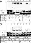
FIG. 1.
FIG. 1.
Reconstitution of the ExsADCE regulatory cascade in vitro. Electrophoretic mobility shift assays were performed by incubating ExsAHis (18 nM; lanes 3 to 5) or the ExsD·ExsAHis complex (18 nM; lanes 6 to 8) alone or with ExsCHis (180 nM; lanes 2, 4, 5, 7, and 8) and/or ExsEHis (720 nM; lanes 5 and 8) for 20 min at 4°C. DNA binding activity was then examined by adding radiolabeled nonspecific (NS) and specific probes derived from the PexsC (A) or PexoT (B) promoters for 15 min. Samples were subjected to electrophoresis and phosphorimaging. Shift products 1 and 2 for each of the promoter fragments are indicated.
FIG. 2.
FIG. 2.
Binding affinity of ExsAHis and the ExsD·ExsAHis complex to the PexoT promoter. (A) The PexoT promoter fragment and a nonspecific probe (0.25 nM each) were incubated with increasing concentrations of ExsAHis (2 to 70 nM) or ExsD·ExsAHis complex (25 to 400 nM) for 15 min followed by electrophoresis and phosphorimaging. (B) Binding curve for ExsAHis and ExsD·ExsAHis to the PexoT promoter. The percentage of shifted probe (y axis) was plotted as a function of the protein concentration (x axis). The reported values are the averages of three independent experiments.
FIG. 3.
FIG. 3.
ExsCHis dissociates ExsD·ExsAHis by forming a complex with ExsD. ExsCHis (lanes 1), the ExsD·ExsAHis complex (lanes 2), the ExsD·ExsCHis complex (lanes 3), or the ExsD·ExsAHis complex and ExsCHis (lanes 4) were incubated for 20 min at 4°C under the conditions used for the EMSAs presented in Fig. 1. Reaction mixtures were electrophoresed through a nondenaturing polyacrylamide gel and subjected to silver staining (A) or Western blotting with antibodies directed against ExsA (B), ExsD (C), or ExsC (D).
FIG. 4.
FIG. 4.
ExsD inhibits the DNA binding activity of ExsA in vivo. (A and B) An exsA mutant or an exsA exsD double mutant carrying the PexsD-lacZ reporter was transformed with a vector control (pJN105) or an expression plasmid (p2UY95, labeled pExsA in the figure) that constitutively expresses low levels of ExsA. The resulting strains were grown under noninducing conditions for T3SS gene expression and assayed for β-galactosidase activity (A) or protein expression levels (B) by performing immunoblotting of whole-cell lysates using the indicated antibodies. The reported values are the averages of three independent experiments, and error bars indicate the standard errors of the means. (C) ChIP assays performed in the presence or absence of ExsD. Cells were treated with formaldehyde to cross-link ExsA to the DNA and processed for ChIP assays using polyclonal anti-ExsA antibody. The immunoprecipitate was then used in a PCR with primers designed to amplify 200-bp regions of the PexsC, PexsD, and PfleQ promoters. The PCRs were programmed to run for 25 or 27 extension cycles, as indicated on the figure. The resulting PCR products were separated on an agarose gel and stained with ethidium bromide. P. aeruginosa chromosomal DNA was used as a positive control (lane 1) for the PCR, and reaction mixtures lacking antibody served as negative controls (lane 2) for chromosomal contamination.
FIG. 5.
FIG. 5.
ExsD dissociated from the ExsD·ExsCHis complex does not bind to ExsAHis. (A) ExsAHis (18 nM) was incubated with the ExsD·ExsCHis complex (180 nM) (lanes 4, 5, 9, and 10) and/or ExsEHis (720 nM) (lane 3, 5, 8, and 10) for 20 min at 4°C. The protein mixes were incubated for 15 min with radiolabeled nonspecific (NS) and specific probes derived from the PexsC (lanes 1 to 5) or PexoT (lanes 6 to 10) promoters. Samples were subjected to electrophoresis and phosphorimaging. Shift products 1 and 2 for each of the promoter fragments are indicated. (B) The DNA binding reaction mixtures from panel A were incubated with Ni-NTA agarose and washed, and the unbound (U) and bound (B) fractions were separated on an SDS-PAGE gel and stained with silver. A standard consisting of all four proteins is included in the left lane.
FIG. 6.
FIG. 6.
ExsD inhibits the self-association activity of ExsA. (A) Detection of ExsA self-association in the LexA monohybrid assay. E. coli SU101 (a reporter strain with a LexA-repressible lacZ reporter) was transformed with an IPTG-inducible plasmid expressing LexA1-87 lacking a dimerization domain (residues 1 to 87) or LexA1-87 fused to chloramphenicol acetyltransferase (LexA-CAT), ExsA (LexA-ExsA), or the amino-terminal domain of ExsA (LexA-NTD). The resulting strains were grown in the presence of 50 or 1,000 μM IPTG and assayed for β-galactosidase expression (reported in Miller units). (B) ExsD inhibits ExsA self-association. The strains from panel A were transformed with either a vector control (V) or an arabinose-inducible ExsD expression plasmid (pJNexsDΔα, labeled as pExsD in the figure), grown in the presence of 50 μM IPTG and 0.5% arabinose, and assayed for β-galactosidase activity. Whole-cell lysates from the same strains were analyzed by immunoblotting using anti-LexA or anti-ExsD antiserum. We presume that LexA1-87, which lacks a dimerization domain, is not stably expressed. The reported values for the data in both panels are the averages of three independent experiments, and error bars indicate the standard errors of the means.

Similar articles

Cited by

References

    1. Bernhards, R. C., X. Jing, N. J. Vogelaar, H. Robinson, and F. D. Schubot. 2009. Structural evidence suggests that antiactivator ExsD from Pseudomonas aeruginosa is a DNA binding protein. Protein Sci. 18:503-513. - PMC - PubMed
    1. Brutinel, E. D., C. A. Vakulskas, K. M. Brady, and T. L. Yahr. 2008. Characterization of ExsA and of ExsA-dependent promoters required for expression of the Pseudomonas aeruginosa type III secretion system. Mol. Microbiol. 68:657-671. - PubMed
    1. Brutinel, E. D., C. A. Vakulskas, and T. L. Yahr. 2009. Functional domains of ExsA, the transcriptional activator of the Pseudomonas aeruginosa type III secretion system. J. Bacteriol. 191:3811-3821. - PMC - PubMed
    1. Brutinel, E. D., and T. L. Yahr. 2008. Control of gene expression by type III secretory activity. Curr. Opin. Microbiol. 11:128-133. - PMC - PubMed
    1. Chai, Y., J. Zhu, and S. C. Winans. 2001. TrlR, a defective TraR-like protein of Agrobacterium tumefaciens, blocks TraR function in vitro by forming inactive TrlR:TraR dimers. Mol. Microbiol. 40:414-421. - PubMed

Publication types

MeSH terms

LinkOut - more resources