. 2010 Feb;192(4):994-1010.
doi: 10.1128/JB.01390-09. Epub 2009 Dec 11.

Genetic analysis of the regulation of type IV pilus function by the Chp chemosensory system of Pseudomonas aeruginosa

Affiliations

Genetic analysis of the regulation of type IV pilus function by the Chp chemosensory system of Pseudomonas aeruginosa

Jacob J Bertrand et al. J Bacteriol. 2010 Feb.

Abstract

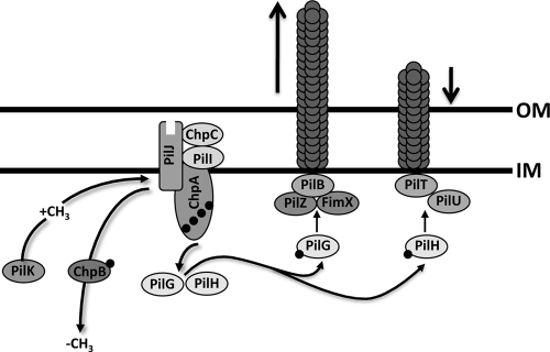
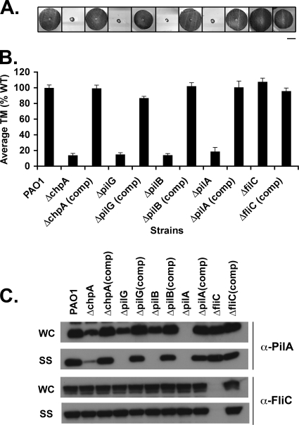
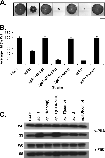
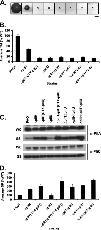
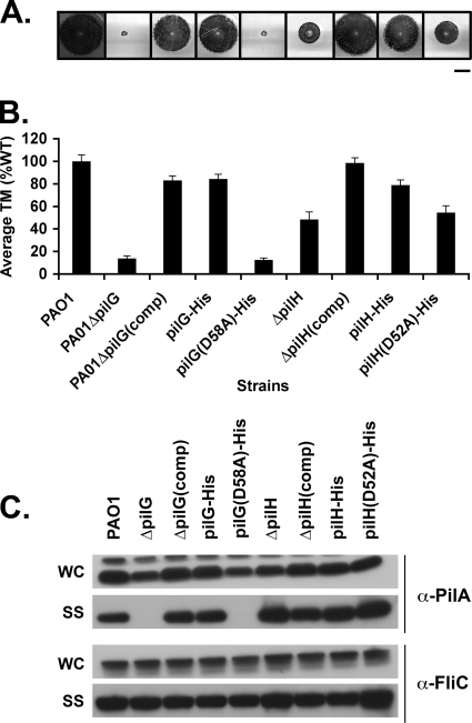
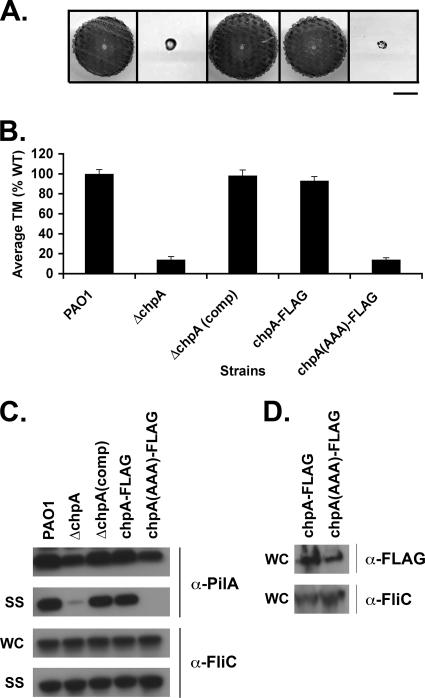
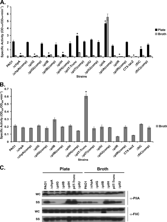
The virulence of the opportunistic pathogen Pseudomonas aeruginosa involves the coordinate expression of many virulence factors, including type IV pili, which are required for colonization of host tissues and for twitching motility. Type IV pilus function is controlled in part by the Chp chemosensory system, which includes a histidine kinase, ChpA, and two CheY-like response regulators, PilG and PilH. How the Chp components interface with the type IV pilus motor proteins PilB, PilT, and PilU is unknown. We present genetic evidence confirming the role of ChpA, PilG, and PilB in the regulation of pilus extension and the role of PilH and PilT in regulating pilus retraction. Using informative double and triple mutants, we show that (i) ChpA, PilG, and PilB function upstream of PilH, PilT, and PilU; (ii) that PilH enhances PilT function; and (iii) that PilT and PilB retain some activity in the absence of signaling input from components of the Chp system. By site-directed mutagenesis, we demonstrate that the histidine kinase domain of ChpA and the phosphoacceptor sites of both PilG and PilH are required for type IV pilus function, suggesting that they form a phosphorelay system important in the regulation of pilus extension and retraction. Finally, we present evidence suggesting that pilA transcription is regulated by intracellular PilA levels. We show that PilA is a negative regulator of pilA transcription in P. aeruginosa and that the Chp system functionally regulates pilA transcription by controlling PilA import and export.


Figures

FIG. 1.
FIG. 1.
Model for the regulation of type IV pilus function by the Chp chemosensory system. The hybrid histidine kinase ChpA, likely associated with the inner membrane, is coupled to a methyl-accepting chemotaxis protein receptor, PilJ, by one of two CheW adaptor protein homologues, PilI and ChpC. Upon receipt of a yet-to-be-elucidated signal, PilJ undergoes a conformational change causing ChpA to autophosphorylate. Phosphate groups are transferred from ChpA to two CheY-like response regulator proteins, PilG and PilH. PilG-P interacts with a motor complex including PilZ, the diguanylate cyclase FimX, and ATPase PilB to mediate pilus extension. PilH-P interacts with ATPases PilT and/or PilU to mediate pilus retraction. Adaptation to the chemical signal is mediated through methylation of PilJ by the competing activities of the methyltransferase PilK and the methylesterase PilB.
FIG. 2.
FIG. 2.
Assays of pilus function for mutants defective in pilus extension. (A) Subsurface TM assay of PAO1, the indicated in-frame deletion mutants, and complemented (comp) strains in which the wild-type gene was reintroduced at its endogenous locus. TM assays were performed by the subsurface stab method, followed by Coomassie blue staining, as described previously (1). Bar, 1 cm. (B) Graph depicting TM zone diameters for the indicated strains. The diameter is expressed as a percentage of PAO1 TM. Shown are the means ± the standard deviation (SD, n = 5). The residual zones observed in ΔpilA, ΔchpA, ΔpilG, and ΔpilB strains represent colony growth. (C) Intracellular and surface pilin and flagellin levels. For the indicated strains, surface structures (SS) including pili and flagella were sheared by vigorous vortexing of bacteria cultured on solid media and separated from cells (WC) by centrifugation. The sheared pili and flagella were precipitated. WC (15 μg of total protein) and SS (5% of the total resuspended volume of the precipitate) samples were separated by SDS-PAGE and immunoblotted with a polyclonal antibody to PilA (α-pilA) or to FliC (α-FliC).
FIG. 3.
FIG. 3.
Assays of pilus function for mutants defective in pilus retraction. (A) Subsurface TM assays were performed as described in Fig. 1. The bar represents 1 cm. (B) Graph depicting average twitching zone diameters for the indicated strains. Shown are the means ± the SD (n = 5) The residual zone observed in the ΔpilT and ΔpilB strains represents colony growth and is similar to that of the ΔpilA strain. (C) Surface structures (SS) and intracellular (WC) preparations of the indicated strains were immunoblotted with a polyclonal antibody to PilA (α-pilA) or to FliC (α-FliC), as described in Fig. 2.
FIG. 4.
FIG. 4.
chpA, pilG, and pilB function upstream of pilH, pilT, and pilU. (A) Subsurface TM assays were performed as described in Fig. 1. Bar, 1 cm. (B) Graph depicting average TM zone diameters for the indicated strains. Shown are the means ± the SD (n = 5). The residual zone of TM in all of the mutants represents colony growth and is similar to of the ΔpilA mutant. (C) Surface structures (SS) and intracellular (WC) preparations of the indicated strains were immunoblotted with a polyclonal antibody to PilA (α-pilA) or to FliC (α-FliC), as described in Fig. 2.
FIG. 5.
FIG. 5.
PilH enhances the function of PilT but not PilU. (A) Subsurface TM assays were performed as described in Fig. 1. Bar, 1 cm. (B) Graph depicting average TM zone diameters for the indicated strains. Shown are the means ± the SD (n = 5). The residual zone of TM in all of the mutants represents colony growth and is similar to that of the ΔpilA mutant. (C) A 1:5 dilution of surface structures (SS) and cell intracellular (WC) preparations of the indicated strains were immunoblotted with a polyclonal antibody to PilA (α-pilA) or to FliC (α-FliC), as described in Fig. 2. (D) Quantification of surface piliation levels relative to a FliC loading control. Surface piliation levels are expressed as a percentage of PAO1. Shown are the means ± the SD (n = 3).
FIG. 6.
FIG. 6.
The phosphoacceptor sites of PilG and PilH are required for function. (A) Subsurface TM assays of PAO1 (WT) and the ΔpilG, ΔpilG(comp), ΔpilH, ΔpilH(comp), His-tagged pilG (pilG-His), and pilH (pilH-His) mutants, as well as the His-tagged pilG and pilH point mutant strains PAO1ΔpilG(D58A)-His and PAO1ΔpilH(D52A)-His. Bar, 1 cm. (B) Graph depicting average TM zone diameters for the indicated strains. Shown are the means ± the SD (n = 5). The residual zone of TM in the pilG mutants represents colony growth and is similar to that of the ΔpilA mutant. (C) Surface structures (SS) and intracellular (WC) preparations of the indicated strains were immunoblotted with a polyclonal antibody to PilA (α-pilA) or to FliC (α-FliC), as described in Fig. 2.
FIG. 7.
FIG. 7.
The histidine kinase domain of ChpA is required for function. (A) Subsurface TM assay of PAO1 (WT) and the ΔchpA, ΔchpA(comp), and FLAG-tagged chpA (chpA-FLAG) mutants, as well as the FLAG-tagged chpA histidine kinase mutant [chpA(AAA)-FLAG]. Bar, 1 cm. (B) Graph depicting average TM zone diameters for the indicated strains. Shown are the means ± the SD (n = 5). The residual zone of TM in the ChpA mutants represents colony growth and is similar to that of the ΔpilA mutant. (C) Surface structures (SS) and intracellular (WC) preparations of the indicated strains were immunoblotted with a polyclonal antibody to PilA (α-pilA) or to FliC (α-FliC), as described in Fig. 2. (D) Levels of ChpA-FLAG and ChpA(AAA)-FLAG. To prepare protein samples, cells were harvested from 2 ml of culture grown shaking for 16 h at 37°C, resuspended in 400 μl of SDS-PAGE sample buffer, passaged through a 27.5 gauge needle three times, and boiled for 5 min. Then, 10% of the total sample volumes were separated by SDS-PAGE and immunoblotted with 2.5 μg of α-FLAG M2 monoclonal antibody/ml.
FIG. 8.
FIG. 8.
The Chp system regulates pilA transcription by mediating intracellular levels of PilA. (A) The indicated strains harboring the PpilA-lacZ fusion and PAO1 harboring a promoterless lacZ gene (CTX-lacZ) integrated into the chromosome at the attB site were cultured in liquid (A and B) or on solid (A) medium for 16 h. The β-galactosidase activity of samples was measured, and the specific activities relative to the OD600 were calculated. Shown are the means ± the SD (n = 3). *, P < 0.05. (C) Surface structures (SS) and intracellular (WC) preparations of the indicated strains cultured in liquid (broth) or on solid (plate) medium were immunoblotted with a polyclonal antibody to PilA (α-pilA) or to FliC (α-FliC), as described in Fig. 2. The higher-molecular-weight band above pilin in the whole-cell lysates represents a cross-reactive band present in all samples.

References

    1. Alm, R., and J. S. Mattick. 1995. Identification of a gene, pilV, required for type 4 fimbrial biogenesis in Pseudomonas aeruginosa, whose product possesses a pre-pilin-like leader sequence. Mol. Microbiol. 16:485-496. - PubMed
    1. Baker, M. D., P. M. Wolanin, and J. B. Stock. 2006. Signal transduction in bacterial chemotaxis. Bioessays 28:9-22. - PubMed
    1. Barken, K. B., S. J. Pamp, L. Yang, M. Gjermansen, J. J. Bertrand, M. Klausen, M. Givskov, C. B. Whitchurch, J. N. Engel, and T. Tolker-Nielsen. 2008. Roles of type IV pili, flagellum-mediated motility and extracellular DNA in the formation of mature multicellular structures in Pseudomonas aeruginosa biofilms. Environ. Microbiol. 10:2331-2343. - PubMed
    1. Becher, A., and H. P. Schweizer. 2000. Integration-proficient Pseudomonas aeruginosa vectors for isolation of single-copy chromosomal lacZ and lux gene fusions. Biotechniques 29:948-952. - PubMed
    1. Bhaya, D., N. R. Bianco, D. Bryant, and A. Grossman. 2000. Type IV pilus biogenesis and motility in the cyanobacterium Synechocystis sp. PCC6803. Mol. Microbiol. 37:941-951. - PubMed

Publication types

MeSH terms

LinkOut - more resources