. 2010 Mar;298(3):L315-23.
doi: 10.1152/ajplung.00089.2009. Epub 2009 Dec 11.

Bone marrow-derived angiogenic cells restore lung alveolar and vascular structure after neonatal hyperoxia in infant mice

Affiliations

Bone marrow-derived angiogenic cells restore lung alveolar and vascular structure after neonatal hyperoxia in infant mice

Vivek Balasubramaniam et al. Am J Physiol Lung Cell Mol Physiol. 2010 Mar.

Abstract

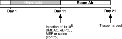
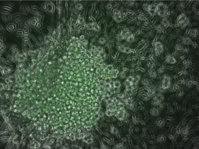
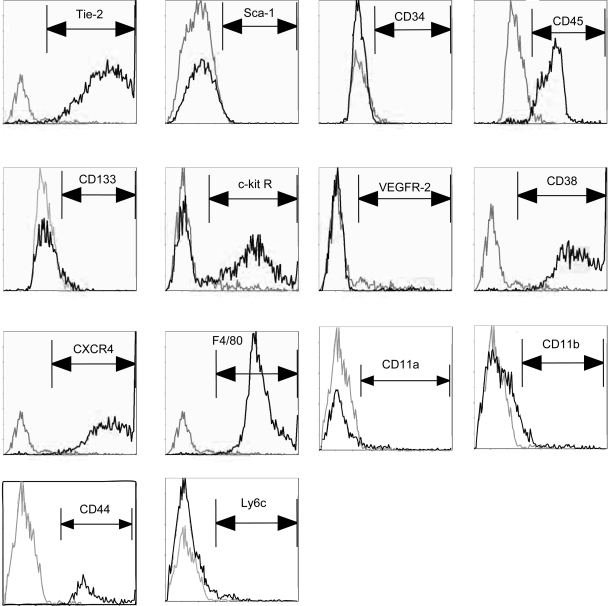
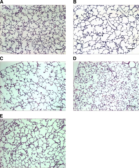
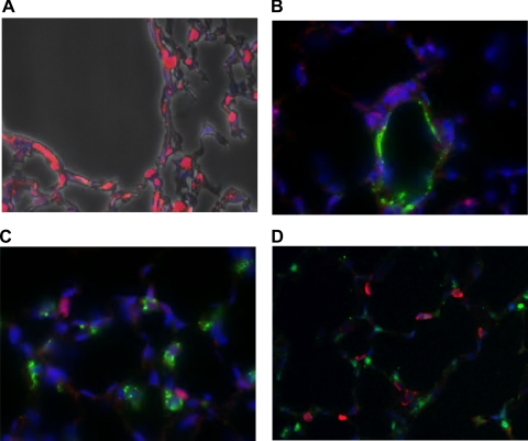
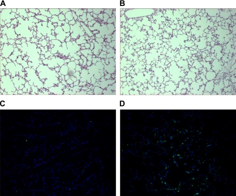
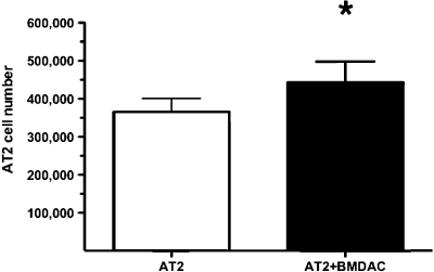
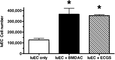
Neonatal hyperoxia impairs vascular and alveolar growth in mice and decreases endothelial progenitor cells. To determine the role of bone marrow-derived cells in restoration of neonatal lung structure after injury, we studied a novel bone marrow myeloid progenitor cell population from Tie2-green fluorescent protein (GFP) transgenic mice (bone marrow-derived angiogenic cells; BMDAC). We hypothesized that treatment with BMDAC would restore normal lung structure in infant mice during recovery from neonatal hyperoxia. Neonatal mice (1-day-old) were exposed to 80% oxygen for 10 days. BMDACs (1 x 10(5)), embryonic endothelial progenitor cells, mouse embryonic fibroblasts (control), or saline were then injected into the pulmonary circulation. At 21 days of age, saline-treated mice had enlarged alveoli, reduced septation, and a reduction in vascular density. In contrast, mice treated with BMDAC had complete restoration of lung structure that was indistinguishable from room air controls. BMDAC comprised 12% of distal lung cells localized to pulmonary vessels or alveolar type II (AT2) cells and persist (8.8%) for 8 wk postinjection. Coculture of AT2 cells or lung endothelial cells (luEC) with BMDAC augmented AT2 and luEC cell growth in vitro. We conclude that treatment with BMDAC after neonatal hyperoxia restores lung structure in this model of bronchopulmonary dysplasia.


Figures

Fig. 1.
Fig. 1.
Study design. Neonatal mice are exposed to hyperoxia for 10 days. At the end of hyperoxia exposure, 1 × 10⁵ bone marrow-derived angiogenic cells (BMDAC), embryonic endothelial progenitor cells (eEPC), mouse embryonic fibroblasts (MEF), or saline is injected into the pulmonary circulation via the right ventricle. Lung tissue was collected after 10 days of recovery in room air.
Fig. 2.
Fig. 2.
Colony of BMDACs from Tie2-green fluorescent protein (GFP) mouse on a feeder layer of MEF.
Fig. 3.
Fig. 3.
FACS characterization of BMDAC. After isolation and before injection, BMDACs were subjected to single-stain FACS. Gray line represents isotype antibody control, and black line represents stained cells. R, receptor.
Fig. 4.
Fig. 4.
BMDACs enhance alveolar growth during recovery in room air after exposure to neonatal hyperoxia. An example of lung histology of 11-day-old mouse exposed to room air (A) and hyperoxia (B). Improved alveolar structure is seen in 3-wk-old mice after injection of bone marrow-derived cells during recovery from neonatal hyperoxia (D) compared with saline-treated animals (C). Animals treated with eEPCs (E) have a persistence of enlarged air spaces compared with saline controls (C). n = 8 animals in each group. Scale bar = 100 μm.
Fig. 5.
Fig. 5.
Immunofluorescence staining of BMDACs-injected lungs at 3 wk of age. A: staining for GFP (red) reveals engraftment of injected cell in both perivascular regions and the alveolar interstitium. B: colocalization for GFP (red) and von Willebrand factor (green; endothelial cells) reveals that injected cells reside in a subendothelial location. C: colocalization for GFP (red) and prosurfactant protein B [pro-SP-B; green; alveolar type II (AT2) cells] shows that injected cells are in close association with type II cells in the alveolar interstitium. D: colocalization with the hematopoietic marker CD45 (red) and GFP (green) show that the injected cells do not express CD45. n = 8 animals in each group.
Fig. 6.
Fig. 6.
Long-term engraftment of BMDAC. Abnormal lung structure persists in 8-wk-old mice treated with saline after exposure to neonatal hyperoxia (A). Improved alveolar structure persists in 8-wk-old mice treated with BMDACs after neonatal hyperoxia (B). In the BMDAC-treated mice, the BMDAC remain detectable in the lung based on their expression of GFP (green; D) compared with saline-treated mice (C). n = 4 animals in each group.
Fig. 7.
Fig. 7.
BMDAC promotes AT2 cell growth in vitro. Isolated mouse lung AT2 cell growth increases after culture with BMDACs. n = 4 replicates in each group. *P < 0.01.
Fig. 8.
Fig. 8.
BMDAC promotes lung endothelial cell (luEC) growth in vitro. Mouse luEC growth increases after culture with BMDACs to levels equivalent to growth with endothelial cell growth supplement (ECGS). n = 4 replicates in each group. *P < 0.01 compared with luEC only.

Similar articles

Cited by

References

    1. Abe S, Boyer C, Liu X, Wen FQ, Kobayashi T, Fang Q, Wang X, Hashimoto M, Sharp JG, Rennard SI. Cells derived from the circulation contribute to the repair of lung injury. Am J Respir Crit Care Med 170: 1158–1163, 2004 - PubMed
    1. Abman S. Pulmonary hypertension in chronic lung disease of infancy: pathogenesis, pathophysiology, and treatment. In: Chronic Lung Disease in Early Infancy, edited by Bland RD, Coalson JJ. New York: Dekker, 1999, p. 619–668
    1. Anghelina M, Krishnan P, Moldovan L, Moldovan NI. Monocytes/macrophages cooperate with progenitor cells during neovascularization and tissue repair: conversion of cell columns into fibrovascular bundles. Am J Pathol 168: 529–541, 2006 - PMC - PubMed
    1. Asahara T, Masuda H, Takahashi T, Kalka C, Pastore C, Silver M, Kearne M, Magner M, Isner JM. Bone marrow origin of endothelial progenitor cells responsible for postnatal vasculogenesis in physiological and pathological neovascularization. Circ Res 85: 221–228, 1999 - PubMed
    1. Asahara T, Murohara T, Sullivan A, Silver M, van der Zee R, Li T, Witzenbichler B, Schatteman G, Isner JM. Isolation of putative progenitor endothelial cells for angiogenesis. Science 275: 964–967, 1997 - PubMed

Publication types

LinkOut - more resources