Review
. 2010 Jan;51(1):23-41.
doi: 10.1194/jlr.R001941-JLR200.

Bile acids: analysis in biological fluids and tissues

Affiliations
Review

Bile acids: analysis in biological fluids and tissues

William J Griffiths et al. J Lipid Res. 2010 Jan.

Abstract

The formation of bile acids/bile alcohols is of major importance for the maintenance of cholesterol homeostasis. Besides their functions in lipid absorption, bile acids/bile alcohols are regulatory molecules for a number of metabolic processes. Their effects are structure-dependent, and numerous metabolic conversions result in a complex mixture of biologically active and inactive forms. Advanced methods are required to characterize and quantify individual bile acids in these mixtures. A combination of such analyses with analyses of the proteome will be required for a better understanding of mechanisms of action and nature of endogenous ligands. Mass spectrometry is the basic detection technique for effluents from chromatographic columns. Capillary liquid chromatography-mass spectrometry with electrospray ionization provides the highest sensitivity in metabolome analysis. Classical gas chromatography-mass spectrometry is less sensitive but offers extensive structure-dependent fragmentation increasing the specificity in analyses of isobaric isomers of unconjugated bile acids. Depending on the nature of the bile acid/bile alcohol mixture and the range of concentration of individuals, different sample preparation sequences, from simple extractions to group separations and derivatizations, are applicable. We review the methods currently available for the analysis of bile acids in biological fluids and tissues, with emphasis on the combination of liquid and gas phase chromatography with mass spectrometry.


Figures

Scheme. 1.
Scheme. 1.
Structures of some human bile acids and precursors. (I) cholesterol, (II) R1=OH, R2=H in 7α-hydroxycholesterol; R1=H, R2=OH in 27-hydroxycholesterol; R1=OH, R2=OH in 7α,27-dihydroxycholesterol (some confusion may arise concerning the nomenclature of 27-hydroxycholesterol and related compounds. According to rules of priority of numbering, the correct description of 27-hydroxycholesterol is 25R,26-hydroxycholesterol. However, the common name is 27-hydroxycholesterol, which will be used here). (III) R1=H in 3-oxocholest-4-enoic acid; R1=OH in 7α-hydroxy-3-oxocholest-4-enoic acid. (IV) R1=H, R2=H, in lithocholic acid (LCA); R1=OH, R2=H in chenodeoxycholic acid (CDCA); R1=H, R2=OH in deoxycholic acid (DCA); R1=OH, R2=OH in cholic acid (CA). (V) glycochenodeoxycholic acid (GCDCA). (VI) taurochenodeoxycholic acid (TCDCA). (VII) taurochenodeoxycholic acid 3α-sulfate. (VIII) taurochenodeoxycholic acid 3α-glucuronide. (IX) tauroursodeoxycholic acid 7β-N-acetylglucosaminide. (X) deoxycholic acid 24-glucuronide.
Fig. 1.
Fig. 1.
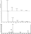
Negative-ion FAB-MS (upper panel) and ESI-MS (lower panel) spectra of a urine extract from an infant with cholestatic liver disease. Major peaks correspond to di-, tri-, and tetrahydroxycholanoylglycine (m/z 448, 464, and 480, respectively), di-, tri-, and tetrahydroxycholanoyltaurine (m/z 498, 514, and 530, respectively), sulfated dihydroxycholanoylglycine (m/z 528), and -taurine (m/z 578 in FAB spectrum and doubly charged at m/z 288.6 in ESI spectrum), glycine- and taurine-conjugated 7-hydroxy- (m/z 444 and 494, respectively), and 7,12-dihydroxy-3-oxochol-4-enoic acids (m/z 460 and 510, respectively). Glucuronides of tri- and tetrahydroxycholest-4-en-3-ones are seen at m/z 607 and 623, respectively, and m/z 657 corresponds to the 3-sulfate, 24-glucuronide of 24-hydroxycholesterol (see Tables 1 and 2 in ref. 38). Reproduced with permission from (38).
Fig. 2.
Fig. 2.
Theoretical mass and isotopic pattern for [M-H] ions of a disulfate of a trihydroxycholanoyltaurine ([M-H] m/z 674.1980) (upper panel), and (center panel) a glucuronide of a dihydroxycholanoyltaurine ([M-H] m/z 674.3216). Resolution is 30,000 FWHM. Negative-ion ESI-MS spectrum utilizing "in-source" CID of a mixture of a disulfate of a trihydroxycholanoyltaurine ([M-H] m/z 674, [M-H-80] m/z 594, [M-H-2 × 80] m/z 514) and a monoglucuronide of a dihydroxycholanoyltaurine ([M-H] m/z 674, [M-H-176] m/z 498] (lower panel). The compounds were isolated from urine by solid phase extraction from an infant with cholestatic liver disease (as in Fig. 1).
Fig. 3.
Fig. 3.
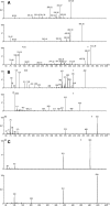
Low-energy MS/MS spectra of ESI-generated [M-H] ions of (a) cholic acid m/z 407, glycocholic acid m/z 464, taurocholic acid m/z 514: (b) taurolithocholic acid m/z 482, lithocholic acid 3-sulfate m/z 455, lithocholic acid 3-glucuronide m/z 551: (c) hyodeoxycholic acid 3-glucoside m/z 553, ursodeoxycholic acid 7β-N-acetylglucosaminide m/z 594. Spectra were recorded on beam instruments.
Fig. 4.
Fig. 4.
Total-ion current chromatograms obtained in the GLC-MS analyses of bile acid profiles of different (selected) groups of conjugates isolated by ion exchange chromatography of a solid phase extract of urine from a healthy pregnant woman in the 38th week of gestation. Samples equivalent to 60 μl, 100 μl, 120 μl, and 200 μl of urine were injected from fractions BA-3 (sulfates with or without aminoacyl amidation), BA-1 (aminoacyl amidated only), BA-2 (glucuronidated only), and BA-4 (doubly conjugated with sulfate and glucuronide) following removal of the conjugating groups and derivatisation [from Meng and Sjövall (77), with permission]. Further subfractionation of conjugate groups is possible if needed. However, the figure clearly illustrates the differences in bile acid profiles between the groups. The numbers above the peaks refer to identified or partially characterized bile acids listed in Table 5 of the cited paper. C36 is the peak of hexatriacontane added before injection. The figure (and Table 5 in the cited paper) also illustrates the complexity of bile acid profiles in urine which would be difficult to elucidate by direct LC-MS analysis of the intact conjugates.

Similar articles

Cited by

References

    1. Motola D. L., Cummins C. L., Rottiers V., Sharma K. K., Li T., Li Y., Suino-Powell K., Xu H. E., Auchus R. J., Antebi A., et al. 2006. Identification of ligands for DAF-12 that govern dauer formation and reproduction in C. elegans. Cell. 124: 1209–1223. - PubMed
    1. Maneerat S., Nitoda T., Kanzaki H., Kawai F. 2005. Bile acids are new products of a marine bacterium, Myroides sp. strain SM1. Appl. Microbiol. Biotechnol. 67: 679–683. - PubMed
    1. Sobotka H. (1937) Physiological chemistry of the bile. Ballière, Tyndall & Cox, London.
    1. Sobotka H. (1938) Chemistry of the sterids. Ballière, Tyndall & Cox, London.
    1. Bergström S., Danielsson H., Samuelsson B. (1960) Formation and metabolism of bile acids. Lipide Metabolism. Block K., editor John Wiley & Sons, New York: 291–366.

Publication types

LinkOut - more resources