

Fate tracing reveals the pericyte and not epithelial origin of myofibroblasts in kidney fibrosis
- PMID: 20008127
- PMCID: PMC2797872
- DOI: 10.2353/ajpath.2010.090517
Fate tracing reveals the pericyte and not epithelial origin of myofibroblasts in kidney fibrosis
Abstract
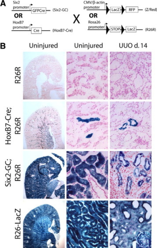
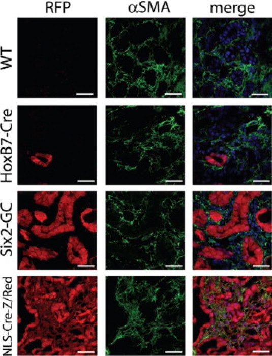
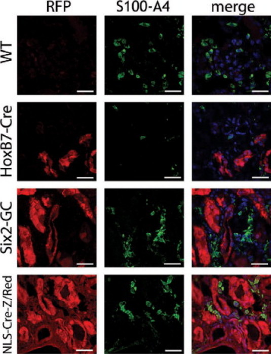
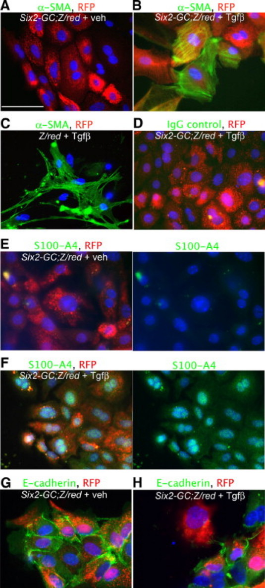
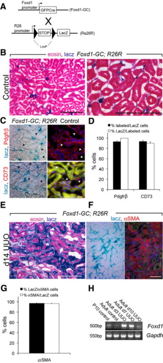
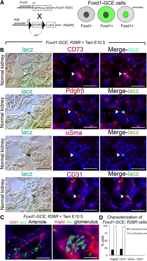
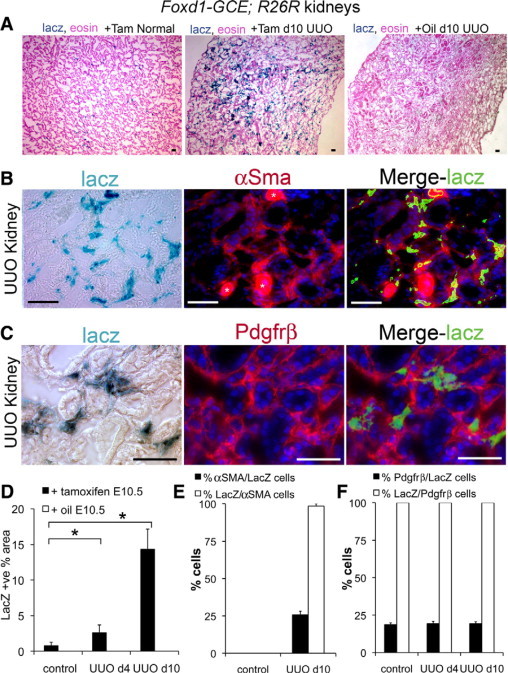
Understanding the origin of myofibroblasts in kidney is of great interest because these cells are responsible for scar formation in fibrotic kidney disease. Recent studies suggest epithelial cells are an important source of myofibroblasts through a process described as the epithelial-to-mesenchymal transition; however, confirmatory studies in vivo are lacking. To quantitatively assess the contribution of renal epithelial cells to myofibroblasts, we used Cre/Lox techniques to genetically label and fate map renal epithelia in models of kidney fibrosis. Genetically labeled primary proximal epithelial cells cultured in vitro from these mice readily induce markers of myofibroblasts after transforming growth factor beta(1) treatment. However, using either red fluorescent protein or beta-galactosidase as fate markers, we found no evidence that epithelial cells migrate outside of the tubular basement membrane and differentiate into interstitial myofibroblasts in vivo. Thus, although renal epithelial cells can acquire mesenchymal markers in vitro, they do not directly contribute to interstitial myofibroblast cells in vivo. Lineage analysis shows that during nephrogenesis, FoxD1-positive((+)) mesenchymal cells give rise to adult CD73(+), platelet derived growth factor receptor beta(+), smooth muscle actin-negative interstitial pericytes, and these FoxD1-derivative interstitial cells expand and differentiate into smooth muscle actin(+) myofibroblasts during fibrosis, accounting for a large majority of myofibroblasts. These data indicate that therapeutic strategies directly targeting pericyte differentiation in vivo may productively impact fibrotic kidney disease.
Figures
Comment in
-
The origin of renal fibroblasts and progression of kidney disease.Am J Pathol. 2010 Jan;176(1):22-4. doi: 10.2353/ajpath.2010.090898. Epub 2009 Dec 11. Am J Pathol. 2010. PMID: 20008128 Free PMC article.
References
-
- Friedman SL, Roll FJ, Boyles J, Arenson DM, Bissell DM. Maintenance of differentiated phenotype of cultured rat hepatic lipocytes by basement membrane matrix. J Biol Chem. 1989;264:10756–10762. - PubMed
-
- Ivarsson M, Sundberg C, Farrokhnia N, Pertoft H, Rubin K, Gerdin B. Recruitment of type I collagen producing cells from the microvasculature in vitro. Exp Cell Res. 1996;229:336–349. - PubMed
-
- Schlondorff D. The glomerular mesangial cell: an expanding role for a specialized pericyte. FASEB J. 1987;1:272–281. - PubMed
-
- Campagnoli C, Roberts IA, Kumar S, Bennett PR, Bellantuono I, Fisk NM. Identification of mesenchymal stem/progenitor cells in human first-trimester fetal blood, liver, and bone marrow. Blood. 2001;98:2396–2402. - PubMed
-
- Johnson RJ, Floege J, Yoshimura A, Iida H, Couser WG, Alpers CE. The activated mesangial cell: a glomerular “myofibroblast”? J Am Soc Nephrol. 1992;2:S190–S197. - PubMed
Publication types
MeSH terms
Substances
Grants and funding
- R01 DK088923/DK/NIDDK NIH HHS/United States
- R01 DK054364/DK/NIDDK NIH HHS/United States
- DK073628/DK/NIDDK NIH HHS/United States
- R01 DK072381/DK/NIDDK NIH HHS/United States
- R01 DK084077/DK/NIDDK NIH HHS/United States
- DK87389/DK/NIDDK NIH HHS/United States
- DK054364/DK/NIDDK NIH HHS/United States
- DK073299/DK/NIDDK NIH HHS/United States
- R37 DK054364/DK/NIDDK NIH HHS/United States
- DK084077/DK/NIDDK NIH HHS/United States
- K08 DK073299/DK/NIDDK NIH HHS/United States
- K08 DK073628/DK/NIDDK NIH HHS/United States
- RC1 DK087389/DK/NIDDK NIH HHS/United States
- R03 DK084316/DK/NIDDK NIH HHS/United States
LinkOut - more resources
Full Text Sources
Other Literature Sources
Molecular Biology Databases
Research Materials
