. 2010 Jan;176(1):246-58.
doi: 10.2353/ajpath.2010.090488. Epub 2009 Dec 11.

Antigen-specific induction of osteopontin contributes to the chronification of allergic contact dermatitis

Affiliations

Antigen-specific induction of osteopontin contributes to the chronification of allergic contact dermatitis

Anne M Seier et al. Am J Pathol. 2010 Jan.

Abstract

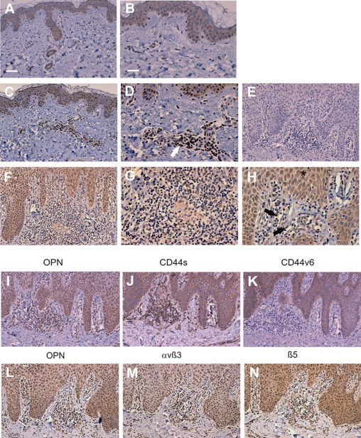
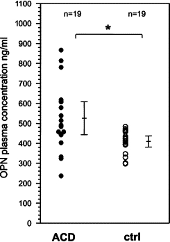
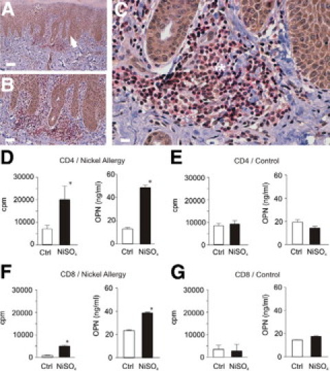
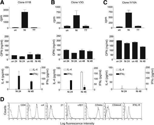
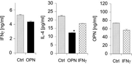
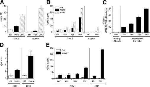
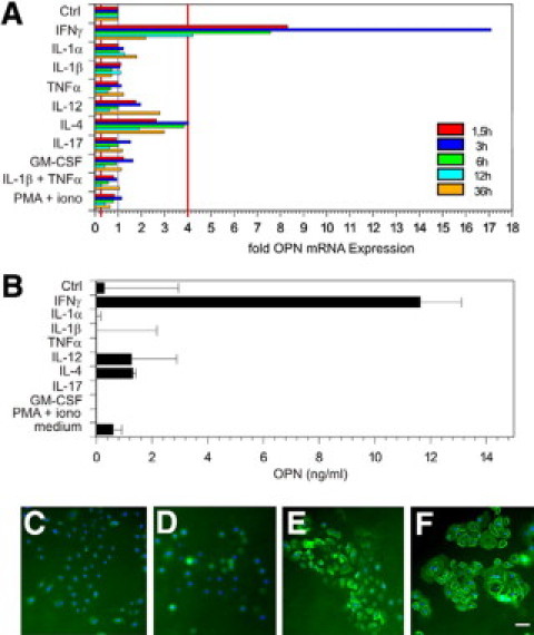
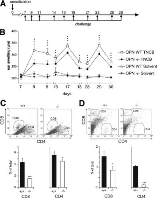
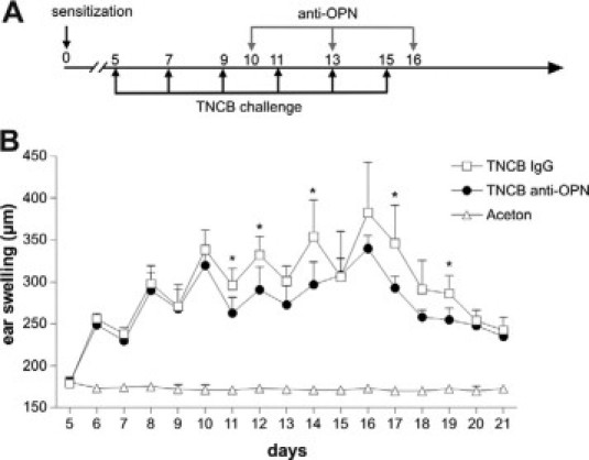
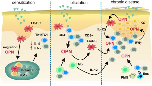
Allergic contact dermatitis is a T cell-mediated immune response, which in its relapsing chronic form is of high socioeconomic impact. The phosphoglycoprotein osteopontin (OPN) has chemotactic and Th1 cytokine functions and in various models is essential for robust T cell-mediated immunity. Here we demonstrate that OPN is abundantly expressed by both effector T cells and keratinocytes in allergic contact dermatitis lesions. T cells from nickel-allergic donors secrete high levels of OPN following antigen-specific stimulation. OPN may substitute for missing IFN-gamma secretion in T effector cells because low IFN-gamma-producing T cell clones secrete high levels of OPN, and OPN down-modulates their interleukin-4 expression. Furthermore, interferon-gamma from T effector cells augments OPN in allergic contact dermatitis by inducing OPN in keratinocytes, which in turn polarizes dendritic cells and attracts inflammatory cells. In the murine contact hypersensitivity (CHS) model for allergic contact dermatitis, OPN is strongly induced in antigen-specific proliferating T cells, and OPN null mice display a reduced chronic CHS inflammatory response due to a decreased influx of effector T cells. Importantly, because of its function for chronic allergic contact dermatitis, OPN may well be a therapeutic target, because anti-OPN antibody treatment in part suppresses established chronic CHS.


Figures

Figure 1
Figure 1
OPN and its receptors are abundantly expressed in chronic allergic contact dermatitis by infiltrating inflammatory cells and keratinocytes. Immunohistochemical staining of skin biopsies was performed with mAb MPIIIB10 against OPN (brown stain) in normal skin (A and B). A similar staining pattern was also observed for the OPN antibody 10A16 (IBL, data not shown). C and D: Acute allergic contact dermatitis (arrow). OPN-expressing infiltrating inflammatory cells. F–H: Chronic allergic contact dermatitis,(arrow) OPN-expressing endothelial cells, and (asterisk) OPN-expressing keratinocytes. E: Chronic allergic contact dermatitis stained with primary isotype control antibody. Serial sections of skin biopsies from donors with chronic allergic contact dermatitis were stained with anti OPN antibody (I and L), anti CD44 antibody (J), anti-CD44v6 antibody (K), anti-αvβ3 antibody (M), or anti-β5 integrin antibody (N). A, C, F, E, I, and J–N: Representative bar in A corresponds to 80 μm. B, D, E, G, and H: Representative bar in B corresponds to 40 μm. Representative data from 5 donors each with chronic and acute ACD and 10 healthy donors.
Figure 2
Figure 2
Patients with chronic allergic contact dermatitis have elevated OPN plasma concentrations. OPN concentration was measured in plasma from 19 healthy volunteers (10 male, 9 female, average age 45.8 years) and from 19 donors with allergic contact dermatitis (6 male, 13 female, average age 42.7 years) by OPN ELISA (IBL). Arrow bars indicate the SD The differences between both groups are statistically significant: *P = 0.007, Mann-Whitney rank-sum test.
Figure 3
Figure 3
Antigen-specific proliferating CD4+ and CD8+ memory T cells from nickel-allergic donors secrete OPN. Immunohistochemical double staining of paraffin sections from biopsies of chronic allergic contact dermatitis was performed with antibodies specific for OPN (MPIIB10, brown, Envision; Dako) and CD45RO (red, APAAP; Dako) for memory T cells. In chronic allergic contact dermatitis CD45RO+ memory T cells (asterisk) and keratinocytes (arrow) express OPN. Magnifications: ×10 (A), ×20 (B), and ×40 (C). Representative data from five donors. Scale bars correspond to 80 μm in A, 40 μm in B, and 25 μm in C. PBMCs from nickel-allergic donors (D and F) with positive patch test to NiSO4 and healthy donors (E and G) with negative patch test were generated by gradient centrifugation and CD4+ (D and E) or CD8+ T cells (F and G) were purified by MACS negative depletion (purity >95%). Antigen-specific proliferation was determined by [3H]thymidine incorporation (D–G). Forty-eight hours after stimulation with NiSO4 supernatants were obtained and subjected to OPN-specific ELISA. Representative results for eight donors.
Figure 4
Figure 4
OPN expression by memory T cell clones correlates with their cytokine expression pattern. CD4+CD45RO+ T cell clones were generated from peripheral blood of nickel-allergic donors, identified by NiSO4 patch testing, as described in Materials and Methods. NiSO4 specific proliferation was measured by [3H]thymidine incorporation. Following antigen-specific stimulation, the secretion of OPN, IFN-γ, and IL-4 was measured by ELISA. Abbreviations: un, unstimulated; Ni, stimulated with NiSO4; TT, stimulated with tetanus toxoid. Cytokine expression was analyzed 24 and 48 hours after NiSO4 stimulation. Data for three representative clones (A–C) of a total of six analyzed clones established from two donors are shown. D: FACS analysis of the indicated OPN receptors on a CD4+ T cell clone IV10A. Representative data for three clones.
Figure 5
Figure 5
OPN modulates the expression of IL-4 in nickel-specific T cell clones. CD4+CD45RO+ T cell clones were generated from peripheral blood of nickel-allergic donors, identified by NiSO4 patch-testing, as described in Materials and Methods. Cells were washed and then stimulated with OPN (2 μg/ml) or IFN-γ (200 U/ml) for 48 hours. Cytokine secretion was measured by IL-4, IFN-γ, or OPN-specific ELISA. A representative experiment from independent experiments with three clones is shown. (Statistically significant, paired t-test: *P < 0.05.)
Figure 6
Figure 6
Antigen-specific stimulation of CHS effector T cells from sensitized mice induces their OPN secretion. On day 0, C57BL/6 mice were sensitized on the abdomen with 7% TNCB and challenged with 1% TNCB on both ears on day 5. Twenty-four hours after challenge, lymph node single-cell suspensions were prepared (A–C). Lymph node cells from challenged mice were stimulated with untreated or TNBS-loaded splenic cells or concavalin A (ConA) was added to untreated cells. Proliferation was measured by [3H]thymidine uptake (A), secretion of OPN was detected by ELISA (B), and OPN mRNA expression was analyzed by real-time PCR. Data are given as ratio of OPN mRNA to glyceraldehyde-3-phosphate dehydrogenase housekeeping gene (GAPDH) (C). D and E: CD4+ and CD8+ T cells were isolated by MACS-negative depletion. Cells were cultured with 30-Gy irradiated, TNBS-loaded spleen cells or unloaded spleen cells as controls (Ctrl). Proliferation was measured by [3H]thymidine uptake (D), and secretion of OPN was detected by ELISA (E). The data represent the results of three independent experiments.
Figure 7
Figure 7
Among proinflammatory cytokines expressed in contact dermatitis, IFN-γ specifically induces OPN expression in keratinocytes. Human-cultured keratinocytes were stimulated with the indicated proinflammatory cytokines and OPN mRNA expression was measured by real-time PCR at time points 1.5, 3, 6, 12, and 36 hours after cytokine stimulation (A) or OPN secretion was measured 48 hours after cytokine stimulation (B). Immunohistochemistry was performed with layers of untreated keratinocytes (D) or with IFN-γ (48 hours) stimulated keratinocytes (C, E, and F). Cells were stained with primary isotype control antibody (C) or antibodies against OPN (D and E) or CD44s (F). Cy2 (green fluorescent) labeled secondary antibody was used to visualize staining (C–F). Cell nuclei are stained in blue (bisBenzidineH 33342 trihydrochloride; Sigma-Aldrich); bar = 15 μm. The data represent the results of three independent experiments with keratinocytes from three different donors.
Figure 8
Figure 8
OPN null mice have impaired chronic CHS response. A and B: OPN wild-type (WT) and OPN null (−/−) mice were sensitized on the abdominal skin with 100 μl of TNCB (7% in acetone). On day 7, the mice were challenged with 20 μl of TNCB on both sides of the ear (1% in acetone). To induce chronification of CHS, the challenge was repeated three times per week for 3 weeks. Control animals were treated by solvent alone. Ear swelling was quantified by dial thickness gauge (Mitutoyo) on days 7 (before first challenge), 8, 9, 16, 17, 18, 28, 29, and 30. OPN wild-type TNCB: n = 12, OPN−/− TNCB: n = 5, OPN wild-type solvent: n = 12, OPN−/− solvent: n = 8 mice. C: OPN wild-type (+/+) and OPN null (−/−) mice were sensitized on the abdominal skin with 100 μl of 7% TNCB. After 5 days, the mice were challenged with 20 μl of 1% TNCB on both sides of the ear, and single-cell suspension of ear skin was analyzed for infiltrating CD4+ and CD8+ cells by flow cytometry after 24 hours (n = 4 mice). D: Pan T cells were purified from lymph nodes and spleens of OPN wild-type (+/+) and OPN null (−/−) donor mice and adoptively transferred i.v. into syngeneic Rag2−/− recipient mice. Twelve hours after transfer, recipient mice were sensitized on the abdominal skin with 100 μl of 7% TNCB in acetone or acetone alone. After 5 days, the mice were challenged with 20 μl of TNCB (1% in acetone) on both sides of the ear. Twenty-four hours after challenge, single-cell suspension of ear skin was analyzed by flow cytometry (60,000 to 100,000 events) to assess infiltration of CD4+ and CD8+ (n = 3 recipient mice). (Statistically significant, unpaired t-test: P < 0.05, P < 0.01, P < 0.001). The data represent the results of two independent experiments.
Figure 9
Figure 9
Anti-OPN antibodies suppress established chronic CHS. Mice were sensitized on day 0 with 7% TNCB in acetone or acetone as control. CHS was elicited with 1% TNCB or acetone as indicated in A. TNCB sensitized animals were injected i.p. with 400 μg of anti-OPN monoclonal antibody or isotype matched control antibody on days 10, 13, and 16 (A and B). Ear swelling was measured daily beginning on day 5. TNCB IgG: n = 5, TNCB anti-OPN: n = 5, acetone: n = 8 mice. (Statistically significant, unpaired t-test: P < 0.05.)
Figure 10
Figure 10
A model for the role of OPN in the chronification of allergic contact dermatitis. During the sensitization-phase OPN interacts with CD44 and αv integrins on LC/DC to guide them from the skin into skin draining lymph nodes. Simultaneously OPN induces the polarization of LC/DC toward a Th1-modulating phenotype by inducing their IL-12 secretion. During the effector phase, OPN is secreted by activated memory T cells, and as a chemokine, it induces the influx of additional T effector cells, neutrophils, and mast cells. When chronification occurs after repeated application of the allergen, the strong expression of OPN can stabilize the inflammatory response by inducing IL-12 in macrophages and DCs to maintain IFN-γ-dominated Th1 immune response. Simultaneously, IFN-γ secreted by effector cells induces the production of OPN by keratinocytes. Keratinocyte OPN may directly affect the polarization and activation of LCs (epidermal DCs).

Similar articles

Cited by

References

    1. Diepgen TL, Coenraads PJ. The epidemiology of occupational contact dermatitis. Int Arch Occup Environ Health. 1999;72:496–506. - PubMed
    1. Kimber I, Dearman RJ. Allergic contact dermatitis: the cellular effectors. Contact Dermatitis. 2002;46:1–5. - PubMed
    1. Cavani A, Girolomoni G. Immune mechanisms in allergic contact dermatitis. Landes Bioscience; Georgetown, TX: 2005. p. 1.
    1. Hogan DJ, Dannaker CJ, Maibach HI. The prognosis of contact dermatitis. J Am Acad Dermatol. 1990;23:300–307. - PubMed
    1. Thyssen JP, Johansen JD, Menne T. Contact allergy epidemics and their controls. Contact Dermatitis. 2007;56:185–195. - PubMed

Publication types

MeSH terms