. 2010 Jan;176(1):259-69.
doi: 10.2353/ajpath.2010.081117. Epub 2009 Dec 11.

Platelets control leukocyte recruitment in a murine model of cutaneous arthus reaction

Affiliations

Platelets control leukocyte recruitment in a murine model of cutaneous arthus reaction

Toshihide Hara et al. Am J Pathol. 2010 Jan.

Abstract

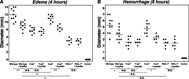
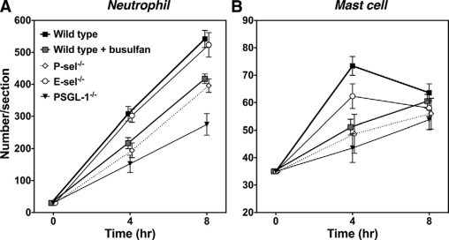
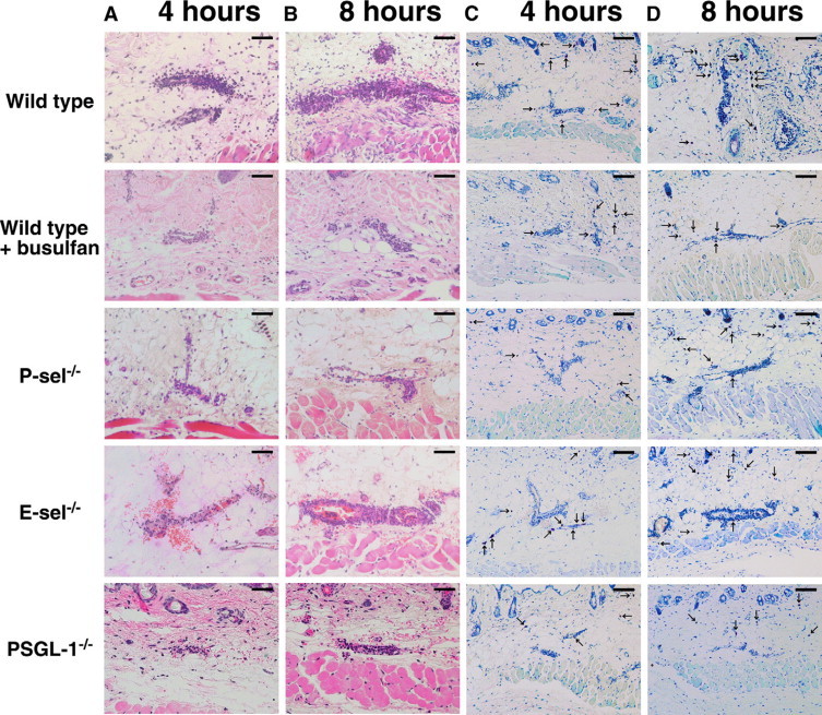
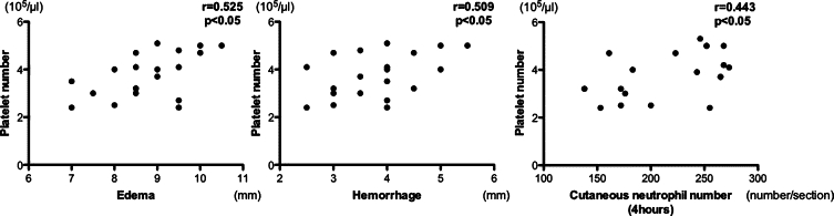
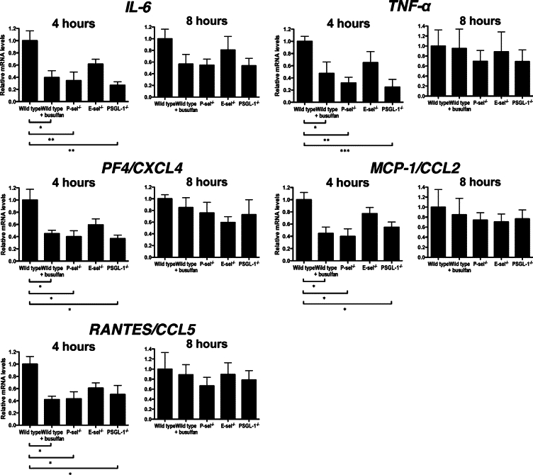
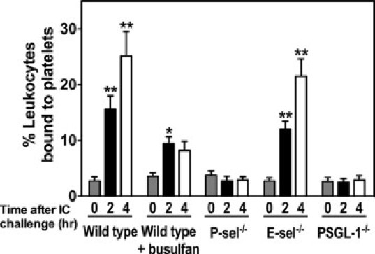
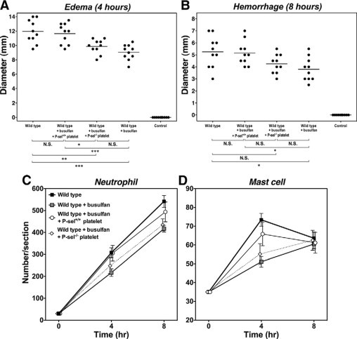
Platelets have been shown to be important in inflammation, but their role in the cutaneous Arthus reaction remains unclear. To assess the role of platelets in this pathogenetic process, the cutaneous Arthus reaction was examined in wild-type mice and mice lacking E-selectin, P-selectin, or P-selectin glycoprotein ligand-1 (PSGL-1) with or without platelet depletion by busulfan, a bone marrow precursor cell-specific toxin. Edema and hemorrhage induced by immune complex challenge significantly decreased in busulfan-treated wild-type mice compared with untreated mice. Busulfan treatment did not affect edema and hemorrhage in P-selectin- or PSGL-1-deficient mice, suggesting that the effect by busulfan is dependent on P-selectin and PSGL-1 expression. The inhibited edema and hemorrhage paralleled reduced infiltration of neutrophils and mast cells and reduced levels of circulating platelets. Increased cutaneous production of interleukin-6, tumor necrosis factor-alpha, and platelet-derived chemokines during Arthus reaction was inhibited in busulfan-treated wild-type mice relative to untreated mice, which paralleled the reduction in cutaneous inflammation. Flow cytometric analysis showed that immune complex challenge generated blood platelet-leukocyte aggregates that decreased by busulfan treatment. In thrombocytopenic mice, the cutaneous inflammation after immune complex challenge was restored by platelet infusion. These results suggest that platelets induce leukocyte recruitment into skin by forming platelet-leukocyte aggregates and secreting chemokines at inflamed sites, mainly through the interaction of P-selectin on platelets with PSGL-1 on leukocytes.


Figures

Figure 1
Figure 1
The effect of platelet depletion on edema and hemorrhage in the cutaneous reverse passive Arthus reaction. Mice were injected intradermally with rabbit IgG anti-chicken egg albumin Ab, then intravenously with chicken egg albumin and 1% Evans blue dye. After 4 or 8 hours, dorsal skins were assessed from wild-type, P-selectin (P-sel)−/−, E-selectin (E-sel)−/−, and PSGL-1−/− mice with or without busulfan treatment. Edema was evaluated after 4 hours as the diameter of extravasated Evans blue spot (A). Hemorrhage after 8 hours was assessed as the diameter of the purpuric spot (B). Wild-type mice that received an intradermal injection of polyclonal rabbit IgG followed by intravenous installation of chicken egg albumin served as controls (control). Horizontal bars indicate mean values of each group. N.S. = not significant. *P < 0.05, **P < 0.01, ***P < 0.001.
Figure 2
Figure 2
Arthus reaction-induced recruitment of neutrophils and mast cells in the skin from wild-type mice, wild-type mice treated with busulfan, E-selectin (E-sel)−/− mice, P-selectin (P-sel)−/− mice, and PSGL-1−/− mice at 4 and 8 hours after IC challenge. Numbers of neutrophils and mast cells per skin section were examined in H&E- and toluidine blue-stained skin sections, respectively. All values represent the means ± SEM calculated on the results obtained from 5 to 10 mice in each group. Statistical analysis is provided in the Results section.
Figure 3
Figure 3
Representative inflammatory cell recruitment in the skin from wild-type mice, wild-type mice treated with busulfan (wild-type + busulfan), P-selectin (P-sel)−/− mice, E-selectin (E-sel)−/− mice, and PSGL-1−/− mice. A, B: Neutrophils were detected by H&E staining at 4 and 8 hours after IC challenge. Original magnification, ×100. Scale bars = 50 μm. C, D: Mast cells (arrows) were detected as cells with metachromatic staining of granules in toluidine blue-stained sections at 4 and 8 hours after IC challenge. Original magnifications, ×50. Scale bars = 100 μm.
Figure 4
Figure 4
Correlation of circulating platelet counts against edema, hemorrhage, and neutrophil recruitment in the skin during the cutaneous reverse passive Arthus reaction. Edema, hemorrhage, and neutrophil recruitment were induced and evaluated in wild-type mice treated with busulfan, as described in Figure 1.
Figure 5
Figure 5
Arthus reaction-induced expression of IL-6, TNF-α, PF4/CXCL4, MCP-1/CCL2, and RANTES/CCL5 in the skin from untreated wild-type mice, wild-type mice treated with busulfan (wild-type + busulfan), P-selectin (P-sel)−/− mice, E-selectin (E-sel)−/− mice, and PSGL-1−/− mice at 4 and 8 hours after IC challenge. Total RNA was isolated from frozen skin tissues, reverse transcribed into cDNA, and then amplified using primers. Relative mRNA levels of IL-6, TNF-α, PF4, MCP-1, and RANTES in the skin samples were measured by real-time PCR and normalized relative to that of GADPH as an endogenous control. All values represent the mean ± SEM of results obtained from 5 to 8 mice in each group. *P < 0.05, **P < 0.01, ***P < 0.001.
Figure 6
Figure 6
Arthus reaction-induced platelet-leukocyte aggregate formation in whole blood from wild-type mice, wild-type mice treated with busulfan (wild-type + busulfan), E-selectin (E-sel)−/− mice, P-selectin (P-sel)−/− mice, and PSGL-1−/− mice before and at 2 and 4 hours after IC challenge. The percentages of leukocytes bound to platelets were assessed by flow cytometry. Platelets were identified with an anti-CD 41 antibody. Leukocyte-sized events were selected on the forward and side scattering profiles and the accuracy of the gating confirmed by staining with CD45. Events staining positive for both CD45 and CD41 were considered to represent platelet-leukocyte aggregates and were distinguishable from events staining for CD45 alone. All values represent the mean ± SEM of results obtained from five mice in each group. *P < 0.05 and **P < 0.001, versus levels before IC challenge in each group of mice.
Figure 7
Figure 7
The effect of platelet restoration on edema and hemorrhage formation and leukocyte recruitment in the skin during the cutaneous reverse passive Arthus reaction. Washed platelets were isolated from the blood of wild-type or P-selectin−/− mice. The resulting platelet suspension was injected intravenously into busulfan-treated wild-type mice 20 minutes before the IC challenge. Edema (A) and hemorrhage (B) were induced and evaluated in wild-type mice, busulfan-treated wild-type mice (wild-type + busulfan), busulfan-treated wild-type mice restored with platelets obtained from wild-type mice (wild-type + busulfan + P-sel+/+ platelet), or busulfan-treated wild-type mice restored with platelets obtained from P-selectin−/− mice (wild-type + busulfan + P-sel−/− platelet), as described in Figure 1. Numbers of neutrophils (C) and mast cells (D) were also examined in H&E- and toluidine blue-stained skin sections, respectively. Horizontal bars indicate mean values of each group (A, B). All values represent the means ± SEM calculated on the results obtained from 5 to 10 mice in each group (C, D). N.S. = not significant. *P < 0.05, **P < 0.01, ***P < 0.001.

Similar articles

Cited by

References

    1. Kohl J, Gessner JE. On the role of complement and Fc gamma-receptors in the Arthus reaction. Mol Immunol. 1999;36:893–903. - PubMed
    1. Arthus M. Injections répetées de serum de cheval chez le lapin. VR SocBiol. 1903;55:817–820.
    1. Baumann U, Chouchakova N, Gewecke B, Kohl J, Carroll MC, Schmidt RE, Gessner JE. Distinct tissue site-specific requirements of mast cells and complement components C3/C5a receptor in IgG immune complex-induced injury of skin and lung. J Immunol. 2001;167:1022–1027. - PubMed
    1. Baumann U, Kohl J, Tschernig T, Schwerter-Strumpf K, Verbeek JS, Schmidt RE, Gessner JE. A codominant role of Fc gamma RI/III and C5aR in the reverse Arthus reaction. J Immunol. 2000;164:1065–1070. - PubMed
    1. Hopken UE, Lu B, Gerard NP, Gerard C. Impaired inflammatory responses in the reverse arthus reaction through genetic deletion of the C5a receptor. J Exp Med. 1997;186:749–756. - PMC - PubMed

Publication types

MeSH terms