. 2010 Jan;176(1):381-92.
doi: 10.2353/ajpath.2010.090056. Epub 2009 Dec 11.

Endometrial cancer side-population cells show prominent migration and have a potential to differentiate into the mesenchymal cell lineage

Affiliations

Endometrial cancer side-population cells show prominent migration and have a potential to differentiate into the mesenchymal cell lineage

Kiyoko Kato et al. Am J Pathol. 2010 Jan.

Abstract

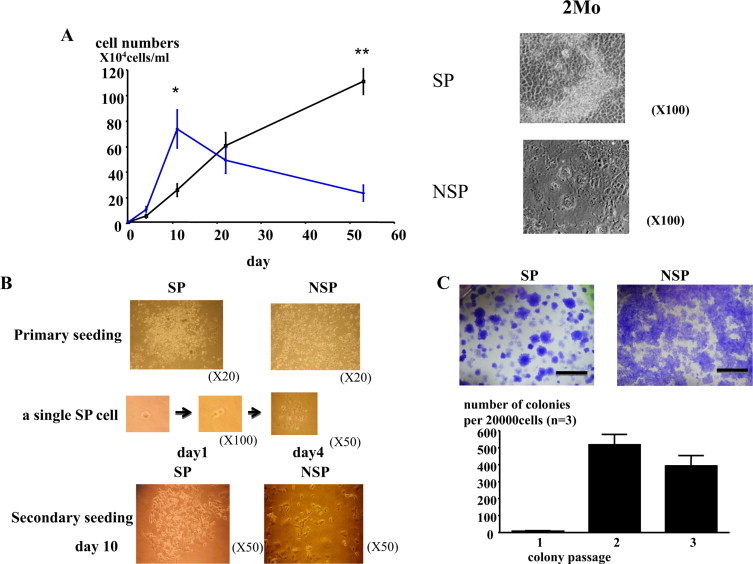
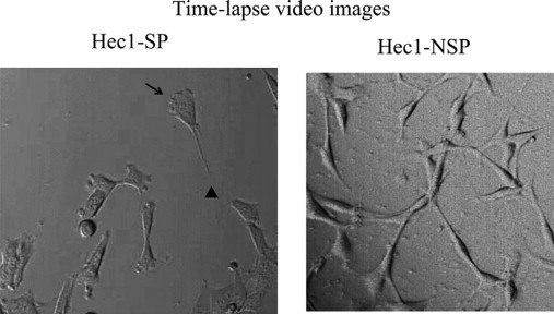
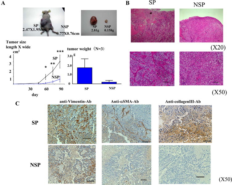
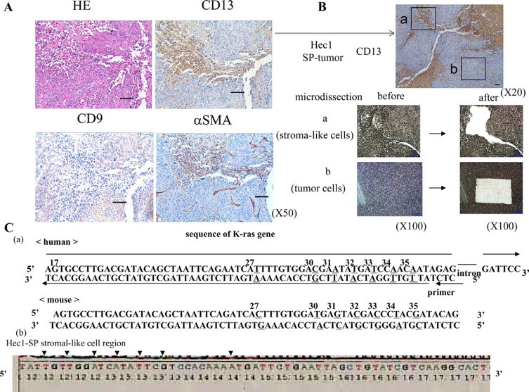
Cancer stem-like cell subpopulations, referred to as "side-population" (SP) cells, have been identified in several tumors based on their ability to efflux the fluorescent dye Hoechst 33342. Although SP cells have been identified in the normal human endometrium and endometrial cancer, little is known about their characteristics. In this study, we isolated and characterized the SP cells in human endometrial cancer cells and in rat endometrial cells expressing oncogenic human K-Ras protein. These SP cells showed i) reduction in the expression levels of differentiation markers; ii) long-term proliferative capacity of the cell cultures; iii) self-renewal capacity in vitro; iv) enhancement of migration, lamellipodia, and uropodia formation; and v) enhanced tumorigenicity. In nude mice, SP cells formed large, invasive tumors, which were composed of both tumor cells and stromal-like cells with enriched extracellular matrix. The expression levels of vimentin, alpha-smooth muscle actin, and collagen III were enhanced in SP tumors compared with the levels in non-SP tumors. In addition, analysis of microdissected samples and fluorescence in situ hybridization of Hec1-SP-tumors showed that the stromal-like cells with enriched extracellular matrix contained human DNA, confirming that the stromal-like cells were derived from the inoculated cells. Moreober, in a Matrigel assay, SP cells differentiated into alpha-smooth muscle actin-expressing cells. These findings demonstrate that SP cells have cancer stem-like cell features, including the potential to differentiate into the mesenchymal cell lineage.


Figures

Figure 1
Figure 1
Isolation of SP cells from human endometrial cancer cells. A: SP cells were present in primary endometrial cancer cells and Hec1 cells (0.20 ± 0.09% in primary endometrial cancer cells from seven cases and 0.63 ± 0.55% in Hec1 cells from 10 independent experiments). Verapamil treatment blocked the dye efflux, increased staining, and rendered the SP cells undetectable by FACS. B: Both Hec1-SP cells and -NSP cells were reanalyzed by FACS after 2 weeks in culture. SP cells from Hec1 cells generated both an SP and an NSP subpopulation. In contrast, NSP cells produced only NSP cells. The representative data of SP cells from one sorting sample were shown. Similar results were obtained from three independent experiments. C: The expression of CD9 and CD13 in SP and NSP cells of Hec1 cells were analyzed by immunohistochemistry. After 3 days of culture, the levels of both CD9 and CD13 expression were reduced in SP cells compared with those in NSP cells (magnification, ×100). The representative data were shown. Similar results were obtained from three independent experiments.
Figure 2
Figure 2
SP cells demonstrated the capacity for long-term proliferation and self-renewal. A: SP cells and NSP cells derived from Hec-1 cells were cultured on collagen-coated plates in mesenchymal stem cell maintenance medium (MF medium) for 2 months. Cell growth rate was analyzed for 2 months. SP cells overgrew, lost contact inhibition, and accumulated in colonies on top of the confluent cell layer. In contrast, NSP cells stopped growing after 2 weeks and became flat and enlarged. Data of cell numbers were represented as the mean ± SEM from three independent experiments. NSP>SP, P = 0.02; ∗∗SP>NSP, P = 0.0018. B: top, SP cells proliferated and formed colonies on the collagen-coated dish. NSP cells never formed colonies. Middle and bottom: The primary colonies were dissociated into single cells, which were individually cultured at 300 cells/cm2 in 100-mm collagen-coated dishes. Colonies were monitored to ensure that they were derived from a single cell. A single cell formed the secondary colony. C: The cloning plates were stained with crystal violet solution. The secondary cloning plates were shown. Hec1-SP cells, but not -NSP cells, formed well-separated colonies (scale bar, 10 mm). An average of 520 secondary colonies were formed per 2 × 10⁴ seeded single cells (2.6%), and an average of 390 tertiary colonies was formed per 2 × 10⁴ seeded single cells from the secondary colonies (1.9%). We counted the number of colonies in triplicate. Error bars represented SEM from the number of colonies in three independent experiments. (The number of colonies per 2 × 10⁴ seeded single cells: the primary colonies, 7.43 ± 3.09; the secondary colonies, 518 ± 60.4; the tertiary colonies, 393 ± 60.7.)
Figure 3
Figure 3
Hec1-SP cells showed podia formation and prominent migration. In time-lapse video imaging studies, the frequency of cell division was higher in Hec1-SP cells than in Hec1-NSP cells. Hec1-SP cells showed podia formation (lamellipodia at the leading edge, shown by arrow and uropodia at the trailing edge, shown by arrowhead). Hec1-NSP cells did not form podia.
Figure 4
Figure 4
Tumorigenicity was enhanced in Hec1-SP cells. A: SP cells or NSP cells were inoculated into the s.c. tissues of nude mice. Left: There was a trend to an increased size of tumor from SP cells compared with NSP cells, although it was masked by large variation between experiments. The tumor size was shown as the mean ± SEM from three independent experiments. Bottom left:y-axis: tumor size, length × wide (cm2); P = 0.045, ∗∗P = 0.056; ∗∗∗P = 0.074. Top right: Macroscopic appearances of the excised tumors after 12 weeks of inoculation were shown. Bottom right: The tumor weight after 12 weeks of inoculation was shown as the mean ± SEM from three independent experiments. y-axis: tumor weight (g). SP: 1.72 ± 0.54 g; NSP: 0.18 ± 0.10 g (P = 0.049). B: The tumors were stained with H&E. SP tumors not only were composed of tumor cells but also contained a markedly enriched stroma and ECM. In contrast, most NSP tumors were formed from the tumor cells, and their ECM was unremarkable (magnification, ×20, ×50; scale bar, 100 μm). C: The expression levels of vimentin, α-SMA, and collagen III were analyzed by immunohistochemistry. Both tumor cells and stromal-like cells in the SP-tumor were stained with vimentin. Stromal-like cells in the SP tumor expressed α-SMA. Tumor cells in the NSP tumor also expressed vimentin, but the level of expression was less than that in the SP tumors. α-SMA and collagen III were not expressed in the NSP tumor (magnification, ×20, ×50: scale bar, 100 μm; ×100: scale bar, 50 μm).
Figure 5
Figure 5
The developmental potential of SP cells to mesenchymal cell lineages. A and B: We microdissected CD9-positive tumor cells (b) and α-SMA-, CD13- positive, and CD9-negative surrounding stromal-like cells (a) in Hec1-SP tumors (magnification, ×20, ×50, ×100: scale bar, 100 μm;) C: (a) DNA sequence of exon 1 (codons 17 to 39) in the K-Ras gene is shown. The third base of codon 27 and codons 30 to 35 (underline) differ between the human and mouse. An intron is inserted between codons 37 and 38. (b) DNA sequences of microdissected tumor cells and stromal-like cells in the Hec1-SP tumor were analyzed. Both the tumor cells (data not shown) and stromal-like cells contained human K-ras DNA. Arrowhead: third base of codon 27, codons 31 to 35.
Figure 6
Figure 6
Most of stromal-like cells were derived from the inoculated Hec1-SP cells. FISH assay was performed on Hec1-SP tumor tissues using the spectrum orange-labeled CEP X (α satellite) DNA probe, which hybridizes to the centromere of human chromosome X, and the FITC-labeled DNA probe, which hybridizes to mouse pan-centromeric chromosome. In the area of tumor cells without ECM, only red signals (human chromosome) were detected. Both red signals and green signals (mouse chromosome) were detected in the area of stromal-like cells with enriched ECM. The ratio of cells with red signals was significantly more than that of cells with green signals (red: 76 ± 4%, green 24 ± 4% from three different regions of tumor cells or stromal-like cells with enriched ECM). Scale bar = 50 μm.
Figure 7
Figure 7
SP cells differentiated to α-SMA expressing cells. A total of 1 × 10⁵ Hec1-SP or -NSP cells were cultured in Matrigel with a mesenchymal stem cell maintenance medium (MF medium), a standard growth medium (DMEM containing 10% FCS), or smooth muscle cell differentiation medium. After 8 weeks, the level of α-SMA expression was analyzed in each Matrigel sample. SP cells cultured with either the standard growth medium (DMEM containing 10% FCS) or a smooth muscle cell differentiation medium expressed α-SMA. Hec1-NSP cells did not proliferate in the smooth muscle cell differentiation medium. Hec1-NSP cells did not express α-SMA in any culture condition. Scale bar = 20 μm.

Comment in

Similar articles

Cited by

References

    1. Bjerknes M, Cheng H. Clonal analysis of mouse intestinal epithelial progenitors. Gastroenterology. 1999;116:7–14. - PubMed
    1. Alonso L, Fuchs E. Stem cells of the skin epithelium. Proc Natl Acad Sci USA. 2003;100(Suppl 1):11830–11835. - PMC - PubMed
    1. Jankowski RJ, Deasy BM, Huard J. Muscle-derived stem cells. Gene Ther. 2002;9:642–647. - PubMed
    1. Spangrude GJ, Smith L, Uchida N, Ikuta K, Heimfeld S, Friedman J, Weissman IL. Mouse hematopoietic stem cells. Blood. 1991;78:1395–1402. - PubMed
    1. Morrison SJ, White PM, Zock C, Anderson DJ. Prospective identification, isolation by flow cytometry, and in vivo self-renewal of multipotent mammalian neural crest stem cells. Cell. 1999;96:737–749. - PubMed

Publication types

MeSH terms