. 2010 Jan 15;184(2):1031-40.
doi: 10.4049/jimmunol.0902773. Epub 2009 Dec 11.

CD8 T cell-initiated vascular endothelial growth factor expression promotes central nervous system vascular permeability under neuroinflammatory conditions

Affiliations

CD8 T cell-initiated vascular endothelial growth factor expression promotes central nervous system vascular permeability under neuroinflammatory conditions

Georgette L Suidan et al. J Immunol. .

Abstract

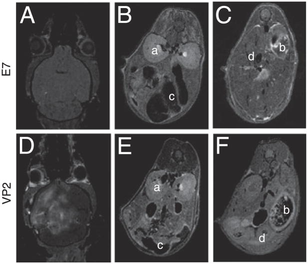
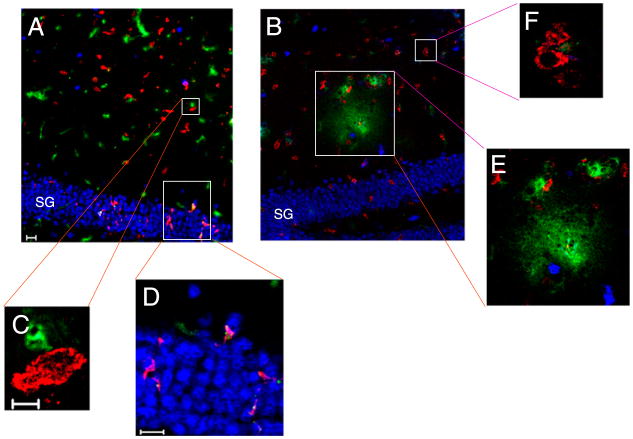
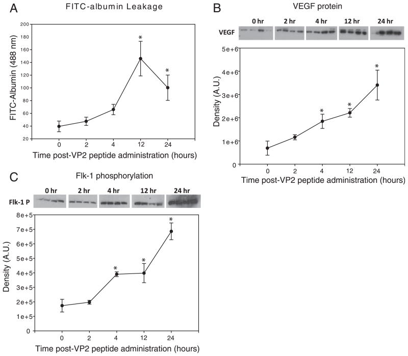
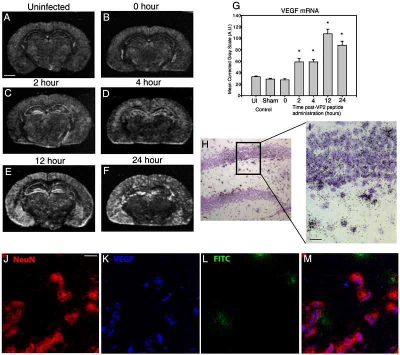
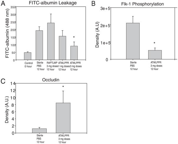
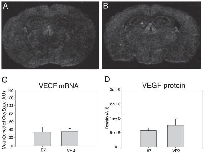
Dysregulation of the blood-brain barrier (BBB) is a hallmark feature of numerous neurologic disorders as diverse as multiple sclerosis, stroke, epilepsy, viral hemorrhagic fevers, cerebral malaria, and acute hemorrhagic leukoencephalitis. CD8 T cells are one immune cell type that have been implicated in promoting vascular permeability in these conditions. Our laboratory has created a murine model of CD8 T cell-mediated CNS vascular permeability using a variation of the Theiler's murine encephalomyelitis virus system traditionally used to study multiple sclerosis. Previously, we demonstrated that CD8 T cells have the capacity to initiate astrocyte activation, cerebral endothelial cell tight junction protein alterations and CNS vascular permeability through a perforin-dependent process. To address the downstream mechanism by which CD8 T cells promote BBB dysregulation, in this study, we assess the role of vascular endothelial growth factor (VEGF) expression in this model. We demonstrate that neuronal expression of VEGF is significantly upregulated prior to, and coinciding with, CNS vascular permeability. Phosphorylation of fetal liver kinase-1 is significantly increased early in this process indicating activation of this receptor. Specific inhibition of neuropilin-1 significantly reduced CNS vascular permeability and fetal liver kinase-1 activation, and preserved levels of the cerebral endothelial cell tight junction protein occludin. Our data demonstrate that CD8 T cells initiate neuronal expression of VEGF in the CNS under neuroinflammatory conditions, and that VEGF may be a viable therapeutic target in neurologic disease characterized by inflammation-induced BBB disruption.


Conflict of interest statement

Disclosures

The authors have no financial conflicts of interest.

Figures

FIGURE 1
FIGURE 1
CD8 T cell-mediated vascular permeability is specific to the CNS. At 24 h postadministration of VP2121–130 or mock E7 peptide, vascular integrity was assessed in the whole animal using gadolinium-enhanced T1-weighted MRI. At 24 h, there is gadolinium leakage into the brain of (D) VP2121–130 peptide-administered but not (A) E7 peptide-administered animals. Body scans demonstrate a lack of i.v.-injected gadolinium leakage into the peripheral organs with either (B and C) E7 peptide or (E and F) VP2121–130 peptide-administration (n = 4 per treatment group). Examples of major organs include (a) kidney, (b) spleen, (c) intestine, and (d) liver. All are negative for gadolinium leakage.
FIGURE 2
FIGURE 2
Brain-infiltrating CD8+ cells are in close proximity to neurons and blood vessels during early CNS vascular permeability. Mouse brains isolated from 7-d TMEV-infected mice were immunostained for NeuN (blue) and CD8a protein (red). FITC-albumin (green) was administered i.v. 1 h prior to brain harvest and denotes vessels as well as vascular leakage. A, At 0 h, CD8+ cells are detected in the hippocampus (original magnification ×20) in close proximity to (C) vessels (original magnification ×100), and (D) neurons of the SG of the hippocampal dentate gyrus (original magnification ×40). B, FITC-albumin leakage is dorsal to the SG 4 h postadministration of VP2121–130 (original magnification ×20). In this region, CD8+ cells are found near (F) intact vessels (original magnification ×100) and (E) areas with FITC-albumin leakage (original magnification ×40). Shown is a section from a representative mouse of four mice analyzed in each group. Scale bars are as follows: in A, 20 μm for A and B; in C, 5 μm for C and F; in D, 20 μm for D and E.
FIGURE 3
FIGURE 3
Increased brain VEGF protein and phosphorylation of VEGF receptor flk-1 occur prior to peak levels of CD8 T cell-mediated CNS vascular permeability. At 0, 2, 4, 12, and 24 h postadministration of VP2121–130 peptide, C57BL/6 mouse brains were assessed for (A) i.v.-injected FITC-albumin leakage into the CNS, (B) CNS VEGF protein levels, and (C) VEGF receptor, flk-1, phosphorylation. Four animals were assessed at each time point. *Denotes statistical significance with p < 0.05 when compared with 0 h baseline controls.
FIGURE 4
FIGURE 4
Neurons are a major source of VEGF expression in the CNS after induction of CD8 T cell-mediated CNS vascular permeability. Shown are representative coronal sections of mice at 0, 2, 4, 12, and 24 h postadministration of VP2121–130 peptide (n = 4 animals per time point). All sections were analyzed for VEGF mRNA and protein expression. In situ hybridization reveals areas of the brain that express VEGF mRNA in (A) uninfected, (B) 0 h, (C) 2 h, (D) 4 h, (E) 12 h, and (F) 24 h post-VP2121–130 peptide administration. G, VEGF mRNA labeling was quantified in the ipsilateral SG of the hippocampal dentate gyrus. H and I, Emulsion autoradiographs with cresyl violet counterstaining of the hippocampal dentate gyrus demonstrate substantial VEGF mRNA patchy neuronal labeling in the SG and subjacent hilar region (H, original magnification ×20; I, original magnification ×40). Also shown is confocal microscopy of (J) NeuN, (K) VEGF, (L) FITC-albumin in hippocampus 12 h postadministration of VP2121–130 peptide (original magnification ×100). NeuN immunostaining colocalizes VEGF cytokine as shown merged in (M). Scale bars are as follows: (A) 1300 μm for A–F, (H) 20 μm, (I) 20 μm, and (J) 10 μm for J–M. *Denotes statistical significance with p < 0.05 when compared with 0 h.
FIGURE 5
FIGURE 5
Inhibition of VEGF coreceptor, NRP-1, reduces flk-1 phosphorylation and preserves BBB integrity. At 12 h postadministration of VP2121–130 to initiate CNS vascular permeability, C57BL/6 mouse brains were assessed for i.v.-administered FITC-albumin leakage. Mice were treated to 1- or 3-mg dose regimens of NRP-1 inhibitor ATWLPPR, 3 mg doses of mock scrambled peptide RAPTLWP, or sterile PBS (n = 6 animals per treatment group). TMEV-infected mock control E7 peptide-injected animals served as an additional negative control. A, Treatment with 3 mg doses of ATWLPPR resulted in significantly less FITC-albumin leakage into the CNS when compared to treatment with RAPTLWP or PBS-administered controls. Treatment with 3 mg doses of ATWLPPR (n = 9 mice) resulted in a significant decrease in (B) phosphorylation of flk-1 and preservation of (C) CEC BBB tight junction protein occludin levels when compared with PBS-treated animals (n = 6 mice). *Denotes statistical significance with p < 0.05 when compared with sterile PBS group.
FIGURE 6
FIGURE 6
Perforin is required for upregulation of VEGF expression during CD8 T cell-mediated CNS vascular permeability. At 24 h post-administration of (A) mock E7 control peptide or (B) VP2121–130 peptide, C57BL/6 perforin-deficient mouse brains were analyzed for VEGF mRNA using in situ hybridization and VEGF protein using Western blot analysis (n = 4 animals for each analysis). C, VEGF mRNA in the SG of the hippocampal dentate gyrus and (D) VEGF protein levels in the whole brain reveal no changes in VEGF expression in perforin-deficient mice consistent with this strain being resistant to CNS vascular permeability.

References

    1. Medana IM, Turner GD. Human cerebral malaria and the blood-brain barrier. Int J Parasitol. 2006;36:555–568. - PubMed
    1. Suidan GL, Pirko I, Johnson AJ. A potential role for CD8+ T-cells as regulators of CNS vascular permeability. Neurol Res. 2006;28:250–255. - PubMed
    1. Huber JD, Egleton RD, Davis TP. Molecular physiology and pathophysiology of tight junctions in the blood-brain barrier. Trends Neurosci. 2001;24:719–725. - PubMed
    1. Abbott NJ. Astrocyte-endothelial interactions and blood-brain barrier permeability. J Anat. 2002;200:629–638. - PMC - PubMed
    1. Kabakus N, Gurgoze MK, Yildirim H, Godekmerdan A, Aydin M. Acute hemorrhagic leukoencephalitis manifesting as intracerebral hemorrhage associated with herpes simplex virus type I. J Trop Pediatr. 2005;51:245–249. - PubMed

Publication types

MeSH terms

Substances